
Mycomya minutata Edwards, 1931
|
publication ID |
https://doi.org/ 10.11646/zootaxa.3815.4.4 |
|
publication LSID |
lsid:zoobank.org:pub:172C594B-7321-4F1C-B8CC-158A195A7D73 |
|
DOI |
https://doi.org/10.5281/zenodo.6134635 |
|
persistent identifier |
https://treatment.plazi.org/id/03998794-FFF1-F708-FF40-675AFAF2F8C7 |
|
treatment provided by |
Plazi |
|
scientific name |
Mycomya minutata Edwards, 1931 |
| status |
|
Mycomya minutata Edwards, 1931 View in CoL
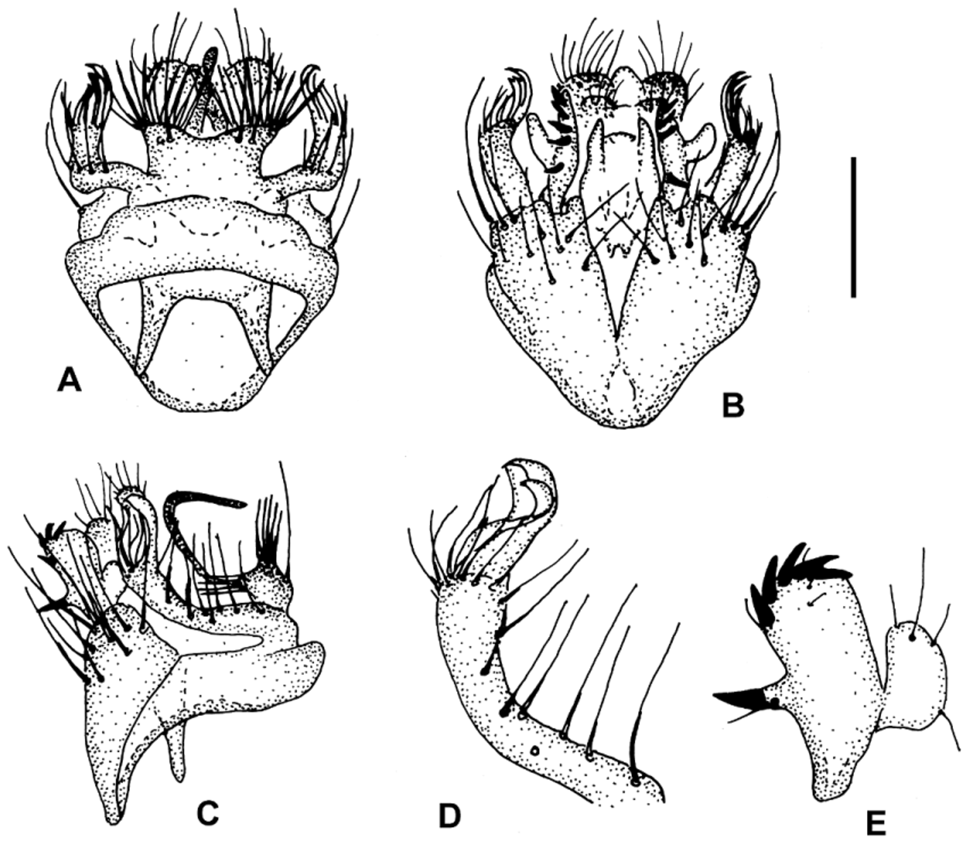
Figs. 3 View FIGURE 3 A–E
Mycomya minutata Edwards, 1931 View in CoL . Edwards 1931b: 266.
Material studied. Lectotype, by present designation. ♂. INDONESIA, Sumatra Fort de Kock, 1926, E. Jacobson B. M. 1930-538 (NHM). Paralectotypes. Same data, 1 ♂, 2 ♀♀ (NHM).
Description. ♂. Head. Palp, other mouthparts and face yellow, posterior parts of head brownish to brown. Antenna brownish, scape, pedicel and base of 1st flagellomere yellow. 1st flagellomere about 3x its width, 2nd flagellomere 2x its width. Thorax. Pronotum yellow, with 2–3 long setae. Scutum yellow with 3 brownish, posteriorly fused longitudinal stripes. Anepisternum yellow. Preepisternum yellow to yellowish. Scutellum brownish, with 4 long setae. Laterotergite yellowish to brownish. Mediotergite brownish, bare. Wing. Length 2.8–2.9 mm. Wing hyaline. Sc ending in R1 proximad of middle of small cell, Sc1 missing. Apical part of Sc bearing 1–2 small setae. Small cell about 2x as long as wide. Cu fork distinctly distal to M fork. M ratios: 1.01–1.13, 1.36–1.53. Cu ratios: 1.00–1.15, 1.56–1.88. Small setae: M petiole: 0; M1: 0; M2: 0; Cu petiole: 0; Cu1: 0; Cu2: 0. Halter pale yellowish. Legs. Coxae and femora yellow, tibiae and tarsi brownish. Coxa 2 without spur. Leg ratios: bt1:t1 = 0.77–0.79, bt2:t2 = 0.69, bt3:t3 = 0.57–0.58. Abdomen. Tergites brownish, sternites yellow. Hypopygium. Figs. 3 View FIGURE 3 A–E, yellow. Tergal part with two pairs of combs, outer combs widely separated from each other, both with about 10 spines ( Fig. 3 View FIGURE 3 A). Tergal lateral appendage long, slender, about 7x as long as its width, setose, with some curved, flattened, apical setae ( Fig. 3 View FIGURE 3 D). Sternal synsclerite with several setae on both sides, no sternal submedian filament ( Fig. 3 View FIGURE 3 B). Gonostylus short, about 2.5x as long as its width, with 5–6 apical tooth, 1 dark spur in its middle part, and membranous lateral lobe about 0.5x as long as main branch of gonostylus ( Fig. 3 View FIGURE 3 F). Aedeagus with lateral lobes, extending slightly beyond its apex ( Fig. 3 View FIGURE 3 E). Female. Wing length 2.9–3.0 mm. Thorax and abdomen similar to those of ♂. Leg ratios: bt1:t1 = 0.77–0.80, bt2:t2 = 0.66–0.68, bt3:t3 = 0.53–0.56. Sc with 4 small setae. Terminalia yellow. Apical segment of cercus relatively long and narrow.
Discussion. Mycomya minutata belongs to the subgenus Mycomyopsis (as discussed for M. apoensis above). Edwards (1931b: Fig. 5 View FIGURE 5 ) already described the male hypopygium of this small species. He stated that M. minutata ”belongs to the same group as M. fimbriata Mg. (Europe and N. India), M. unipectinata Edw. ( Ceylon) and M. plagiata Tonn. ( New Zealand, etc.)”. However, M. fimbriata ( Meigen, 1818) belongs to another subgenus ( Neomycomya , see Väisänen, 1984a), whereas both M. minutata and M. unipectinata Edwards, 1927 clearly belong to subgenus Mycomyopsis . Mycomya minutata differs from the other species of the subgenus in the shape and setosity of the tergal lateral appendage ( Fig. 3 View FIGURE 3 D) and the gonostylus ( Fig. 3 View FIGURE 3 F), as well as in the absence of the sternal submedian filament. The tergal lateral appendage resembles those in M. ducula Väisänen (2013c: fig. 8C) and M. jeti Väisänen (2013c: fig.10C) . The latter species differs from M. minutata in having a short sternal submedian filament ( Väisänen 2013c: figs. 10B, 10D). M. ducula is similar to M. minutata in having a short gonostylus with a spur, and lacking the sternal submedian filament ( Väisänen 2013c: figs. 8B, 8D). However, the setae of the tergal lateral appendage of M. ducula are in two separate groups, i.e. in the basal half and the apex ( Väisänen 2013c: fig. 8C), whereas in M. minutata the whole tergal lateral appendage is sparsely covered by setae ( Fig. 3 View FIGURE 3 D). The type locality of M. minutata is Fort de Kock (= Bukittinggi), which is about 920 m above the sea level, in the Sumatran Minangkabau highlands near the volcanoes of Mount Singgalah and Mount Marapi.

No known copyright restrictions apply. See Agosti, D., Egloff, W., 2009. Taxonomic information exchange and copyright: the Plazi approach. BMC Research Notes 2009, 2:53 for further explanation.
|
Kingdom |
|
|
Phylum |
|
|
Class |
|
|
Order |
|
|
Family |
|
|
Genus |
Mycomya minutata Edwards, 1931
| Väisänen, Rauno 2014 |
Mycomya minutata
| Edwards 1931: 266 |