
Neogreenia sophorica Wu
|
publication ID |
https://doi.org/ 10.11646/zootaxa.5418.5.1 |
|
publication LSID |
lsid:zoobank.org:pub:0DDD7278-0E9C-4979-ACB9-1915D4209282 |
|
DOI |
https://doi.org/10.5281/zenodo.10787181 |
|
persistent identifier |
https://treatment.plazi.org/id/03C24061-FFD9-D260-FF0F-255CFB3CF89D |
|
treatment provided by |
Plazi |
|
scientific name |
Neogreenia sophorica Wu |
| status |
|
Neogreenia sophorica Wu in Wu & Cheng, 2006
Neogreenia sophorica Wu in Wu & Cheng 2006: 62. Type data: CHINA: Beijing, Dongcheng District , on Sophora japonica View in CoL . Holotype, female, type depository: Beijing Forestry University, Beijing, China.
Host plants: Styphnolobium japonicum (L.) Schott ( Fabaceae ).
Distribution: China (Beijing).
Remarks. Wu & Cheng (2006) described and illustrated the adult female and first-instar and second-instar nymphs. However, the description of the second-instar nymph of N. sophorica was based on specimens mixed with third-instar female nymphs. After examining these cysts, we noticed that the second-instar nymphs had (i) far fewer large simple pores and (ii) the five anterior abdominal spiracles each had sieve-like disc-pores in the atrium, whereas the third-instar female nymphs had (i) groups of large simple pores on the dorsum and (ii) the six anterior abdominal spiracles each had sieve-like disc-pores in the atrium. We therefore describe the second-instar and third-instar female nymphs of N. sophorica below.
Material examined. Paratypes: 3 second-instar nymphs, CHINA: collection data same as for holotype, mounted together on 1 slide; 6 third-instar ♀♀ nymphs, collection data same as for holotype, 24.iv.2004, mounted together on 1 slide .
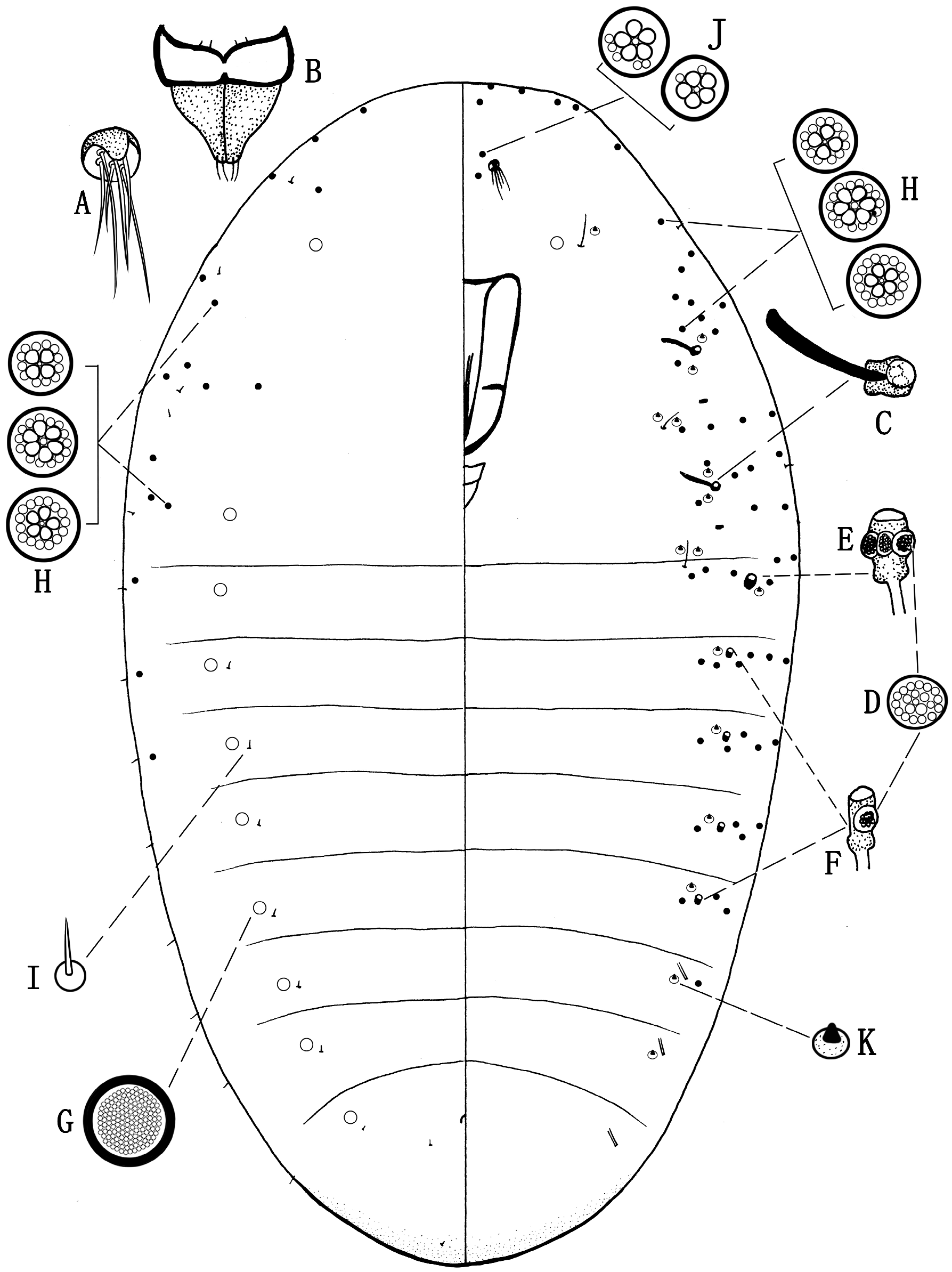
Second-instar nymph (cyst) (sexes indistinguishable)
Slide-mounted material (n=3) ( Fig. 6 View FIGURE 6 ). Body oval, 1.0– 1.6 mm long and 0.6–0.9 mm wide; derm entirely membranous or with posterior part slightly sclerotized. Antennae ( Fig. 6A View FIGURE 6 ) reduced and plate-like, each 10–13 μm in diameter, with margin sclerotized and protruding anteriorly, bearing 6 or 7 setae, each 15–25 μm long. Eyes absent. Labium ( Fig. 6B View FIGURE 6 ) 2 segmented, about 80 μm long and 68–80 μm wide; segment I with a pair of short setae (each about 5 μm long) on each side; segment II with 2 apical hair-like setae (each about 23 μm long) on each side. Clypeolabral shield longer than labium, 235–260 μm long and 130–150 μm wide; stylets present. Legs absent. Thoracic spiracles ( Fig. 6C View FIGURE 6 ) sclerotized, each opening about 10 μm in diameter, with a sclerotized bar and a group of 5 sieve-like disc-pores ( Fig. 6D View FIGURE 6 ) in atrium. Abdominal spiracles numbering 8 pairs, with 1 pair situated on each abdominal segment, anterior 5 pairs sclerotized; anteriormost pair ( Fig. 6E View FIGURE 6 ) with each opening about 10 μm in diameter, and 2 or 3 sieve-like disc-pores ( Fig. 6D View FIGURE 6 ) within atrium; spiracles ( Fig. 6F View FIGURE 6 ) on segments II– V smaller, each with opening about 7.5 μm in diameter, and with only 1 sieve-like disc-pore ( Fig. 6D View FIGURE 6 ) within atrium; posteriormost 3 pairs unsclerotized and tube-like, with each opening about 2.5 μm in diameter, and lacking pores within atrium. Anal opening about 10 μm wide, with U-shaped sclerotized partial ring lacking pores and setae, located medially on posteriormost dorsal segment. Cicatrices absent.
Dorsum. With disc-pores of 2 types: (i) large simple pores ( Fig. 6G View FIGURE 6 ), each about 10 µm in diameter, with sclerotized rim, numbering 0–2 pores (mostly 1) submarginally on each segment; (ii) compound multilocular disc-pores ( Fig. 6H View FIGURE 6 ), each about 7.5 µm in diameter, with 3–7 (mostly 4 or 5) subcentral loculi and with outer ring of distinct peripheral loculi, present on margins from head to abdominal segment III. Setae of only 1 type: spine-like setae ( Fig. 6I View FIGURE 6 ), each 6–8 μm long, present in submarginal and submedial areas.
Venter. With disc-pores of 3 main types: (i) compound multilocular disc-pores ( Fig. 6H View FIGURE 6 ), same size and structure as on dorsum; forming group around each thoracic spiracle and present on margins except for 2 posteriormost segments; also smaller ones ( Fig. 6J View FIGURE 6 ), each about 6 µm in diameter, with 3–5 subcentral loculi, numbering 2 or 3 between antennae; (ii) sieve-like disc-pores ( Fig. 6D View FIGURE 6 ), each about 5 µm in diameter, with many irregularly distributed loculi, present within atria of thoracic spiracles and anterior 5 pairs of abdominal spiracles; and (iii) large simple pores ( Fig. 6G View FIGURE 6 ), same size and structure as on dorsum; with 1 pore near each long hair-like seta on prothorax. Setae of 3 types present: (i) hair-like setae, each 18–28 μm long, situated submedially on prothorax, below each mesothoracic spiracle and submarginally on abdominal segment I; (ii) spine-like setae ( Fig. 6I View FIGURE 6 ), same size as on dorsum, a few on margins; and (iii) short conical spine-like setae ( Fig. 6K View FIGURE 6 ) with sclerotized basal sockets, each about 5 μm long; with 1 or 2 near each hair-like seta, 2 or 3 near each thoracic spiracle and 0 or 1 near each abdominal spiracle except for posteriormost pair, where absent.
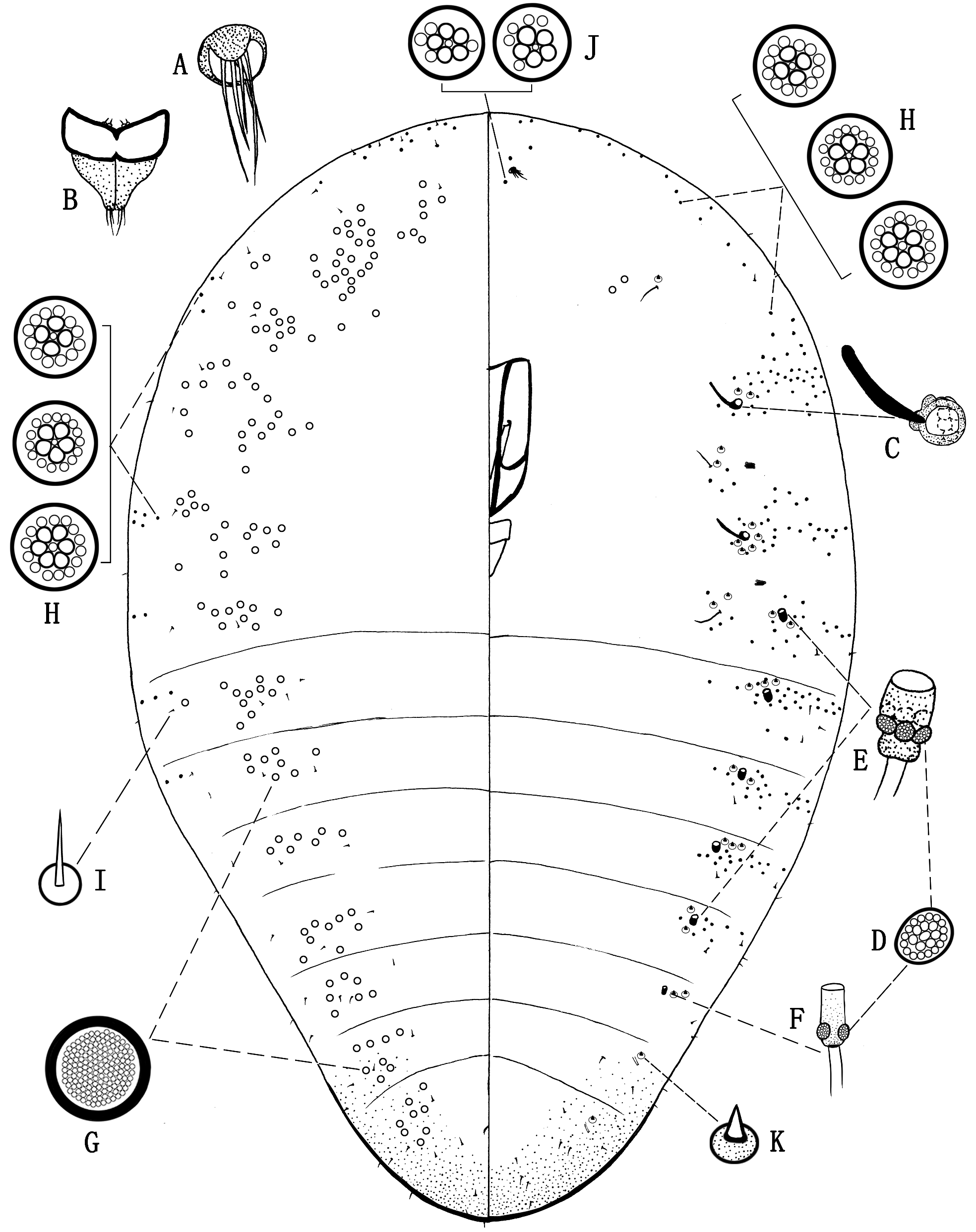
Third-instar female nymph (cyst)
Slide-mounted material (n=6) ( Fig. 7 View FIGURE 7 ). Body pyriform, rounded anteriorly and tapering posteriorly, 2.8–3.9 mm long and 1.8–2.0 mm wide; derm mostly membranous but abdominal apex sclerotized. Antennae ( Fig. 7A View FIGURE 7 ) reduced to small oval plates, each 20–25 μm in diameter, with margin sclerotized and protruding anteriorly, bearing 5 or 6 setae, each 12–40 μm long; situated medially on head. Eyes absent. Labium ( Fig. 7B View FIGURE 7 ) 2 segmented, 130–150 μm long and 100–110 μm wide; segment I 53–65 μm long, with margins sclerotized but centre membranous, bearing 1 pair of short setae (each 30–33 μm long) on each side; segment II 78–85 μm long, sclerotized, with 3 apical hair-like setae (each 20–25 μm long) on each side. Clypeolabral shield longer than labium, 370–385 μm long and 160–190 μm wide; stylets present. Legs absent. Thoracic spiracles ( Fig. 7C View FIGURE 7 ) sclerotized, each opening 12–18 μm in diameter, with sclerotized bar and group of 12–14 sieve-like disc-pores ( Fig. 7D View FIGURE 7 ) at inner end of atrium. Abdominal spiracles numbering 8 pairs, with anterior 6 pairs sclerotized; 5 anteriormost pairs ( Fig. 7E View FIGURE 7 ) each with opening about 20 μm in diameter and 4–7 sieve-like disc-pores in atrium; spiracles ( Fig. 7F View FIGURE 7 ) on abdominal segment VI much smaller, each with opening 9–10 μm in diameter and atrium containing 2 sieve-like disc-pores ( Fig. 7D View FIGURE 7 ); posteriormost 2 pairs of spiracles unsclerotized, small and tube-like, each with opening about 5 μm in diameter, lacking pores within atrium. Anal opening with U-shaped sclerotized ring, 18–20 μm wide, without pores or setae, located medially on posteriormost dorsal segment. Cicatrices absent.
Dorsum. With disc-pores of 2 types: (i) large simple pores ( Fig. 7G View FIGURE 7 ), each 13–15 µm in diameter, with sclerotized rim, forming group of 3–20 pores on submargin of each segment, most numerous on prothorax; (ii) compound multilocular disc-pores ( Fig. 7H View FIGURE 7 ), each about 10 µm in diameter, with 4–6 subcentral loculi and an outer ring of 12–14 peripheral loculi, present on margins from head to abdominal segment II. Setae of only 1 type present: spine-like setae ( Fig. 7I View FIGURE 7 ), each 8–10 μm long, present in submarginal and marginal areas.
Venter. With disc-pores of 4 main types: (i) large simple pores ( Fig. 7G View FIGURE 7 ), same size and structure as on dorsum, numbering 1–4 near each hair-like seta on prothorax; (ii) compound multilocular disc-pores ( Fig. 7H View FIGURE 7 ), same size and structure as on dorsum; forming group around each thoracic spiracle and present on margins except absent from 2 posterior abdominal segments; also smaller ones ( Fig. 7J View FIGURE 7 ), each 8–9 μm in diameter, with 5 subcentral loculi and 4–6 peripheral loculi irregularly distributed, these smaller pores numbering 5–7 between antennae; and (iv) sieve-like disc-pores ( Fig. 7D View FIGURE 7 ), each about 7.5 µm in diameter, some pores slightly polygonal, each with many irregularly distributed loculi, present within atria of thoracic spiracles and anterior 6 pairs of abdominal spiracles. Setae of 3 types: (i) hair-like setae, each 23–25 μm long, situated submedially on prothorax, and submarginally on mesothorax and abdominal segment I; (ii) spine-like setae ( Fig. 7I View FIGURE 7 ), same size as on dorsum, few on margin; and (iii) short conical spine-like setae ( Fig. 7K View FIGURE 7 ) with sclerotized basal sockets, each about 5 μm long; with 1 or 2 near each hair-like seta, 3–5 around each thoracic spiracle and 1–4 around each abdominal spiracle.
| V |
Royal British Columbia Museum - Herbarium |
| VI |
Mykotektet, National Veterinary Institute |
No known copyright restrictions apply. See Agosti, D., Egloff, W., 2009. Taxonomic information exchange and copyright: the Plazi approach. BMC Research Notes 2009, 2:53 for further explanation.
|
Kingdom |
|
|
Phylum |
|
|
Class |
|
|
Order |
|
|
Family |
|
|
Genus |
Neogreenia sophorica Wu
| Zheng, Xinyi, Watson, Gillian W., Zhang, Jiangtao, Tan, Zhixiang & Wu, San’An 2024 |
Neogreenia sophorica
| Wu, S. A. & Cheng, G. F. 2006: 62 |