
Oncocephala quadrilobata (Guérin, 1844), Guerin, 1844
|
publication ID |
https://doi.org/ 10.5281/zenodo.174422 |
|
DOI |
https://doi.org/10.5281/zenodo.6256924 |
|
persistent identifier |
https://treatment.plazi.org/id/03CF9F47-FF9F-FF9F-FEC9-FA9BFDD21A43 |
|
treatment provided by |
Plazi |
|
scientific name |
Oncocephala quadrilobata (Guérin, 1844) |
| status |
|
Oncocephala quadrilobata (Guérin, 1844) View in CoL
( Figs 3 View FIGURE 3 , 4 View FIGURE 4 , 14–20 View FIGURES 14 – 20 , 22 View FIGURES 21, 22 , 25, 26 View FIGURES 23 – 26 , 31–34 View FIGURES 31 – 34 , 36 View FIGURES 35, 36 )
Mature larva
Measurements of body (n=1): length (except head, from base of pronotum to end of body): 7.9 mm; width across mesonotum: 2.5 mm.
Body strongly flattened dorsoventrally, elongated, parallelsided ( Figs 3 View FIGURE 3 , 4 View FIGURE 4 , 31, 32 View FIGURES 31 – 34 ).
Colour of alcohol preserved larva, on dorsal as well as ventral side, yellow but with tips of abdominal lateral scoli and pulvilli of legs reddishbrown; without any distinct pattern ( Figs 31, 32 View FIGURES 31 – 34 ).
Segments of thorax and of abdomen transverse. Lateral margin of pronotum broadly rounded. Mesonotum and metanotum shorter and wider than pronotum. Abdominal segments 1–7 more or less as wide as meso and metanotum. Because of fusion of eighth and ninth abdominal segments, the last segment different in shape from the remaining ones: it has a pair of lateral scoli and forms broadly rounded end of body ( Figs 3 View FIGURE 3 , 4 View FIGURE 4 , 31, 32 View FIGURES 31 – 34 ).
Segments of thorax without lateral scoli. Abdominal segments with one lateral scolus on each side. Each scolus with one pointed seta apically, and the second one, visible from dorsal view, slightly below the top. One pointed seta dorsally and one ventrally midway along the length of abdominal scoli 1–7. Two setae dorsally and one ventrally midway along the length of scolus of the last abdominal segment. One or two campaniform sensilla on ventral side, at the base of each scolus ( Figs 3 View FIGURE 3 , 4 View FIGURE 4 ).
Nine pairs of spiracles: one pair on mesonotum and eight pairs on abdominal segments 1 to 8.
Dorsal and ventral side of body with short pointed setae and slightly shorter minute setae placed at the anterior margin of segments. Meso, matanotum and first 7 abdominal segments each has a transverse fold across the middle (plical area).
Pronotum with 14 pointed setae and 9 or 10 campaniform sensilla on each side, distributed along the margin. Anterior border of mesonotum, metanotum and the first abdominal segment with a pair of very minute setae medially and two minute setae laterally on each side; remaining abdominal segments with a pair of very minute setae medially and one minute seta laterally on each side. Moreover, mesonotum with a row of 8 pointed setae and two campaniform sensilla close to posterior margin; and 6 pointed setae and one campaniform sensillum on each side laterally. One pointed seta and a campaniform sensillum extremely laterally, on each side. Metanotum with a pair of campaniform sensilla close to anterior margin, a row of 10 pointed setae and two campaniform sensilla close to posterior margin; and 6 pointed setae and two campaniform sensilla on each side laterally. On each side, extremely laterally, one pointed seta and a campaniform sensillum. Dorsal side of first abdominal segment with two pairs of pointed setae and a pair of campaniform sensilla close to anterior margin, a row of 10 pointed setae and two campaniform sensilla close to posterior margin, and two pointed setae and two campaniform sensilla on each side close to spiracle. Dorsal side of abdominal segments 2–7 with two pairs of pointed setae and one or two pairs of campaniform sensilla close to anterior margin, a row of 13–16 pointed setae and two campaniform sensilla close to posterior margin, and two pointed setae and two or three campaniform sensilla on each side close to spiracle. Eighth abdominal segment fused with ninth without distinct border. The last segment with two pairs of pointed setae and a pair of campaniform sensilla close to anterior margin, 4 campaniform sensilla on each side laterally, two pairs of pointed setae and a pair of campaniform sensilla in area between spiracles, and two setae close to each spiracle. Moreover, behind spiracle on each side laterally, two small setae and one campaniform sensillum, a pair of setae and two pairs of campaniform sensilla in the middle, and 8 setae along extreme margin of segment. Chaetotaxy of dorsal side of body as in Figure 3 View FIGURE 3 .
Ventral surface of pro, meso, and metathorax with two pairs of minute setae at anterior margin, 4 pointed setae and 4 campaniform sensilla in the middle, and two setae at the base of each coxa. Abdominal segments 1–7 with a pair of minute setae at anterior border, a row of 6–12 pointed setae and 2 to 5 campaniform sensilla in the middle, and three setae on lateral side. The last abdominal segment with a pair of minutae setae at anterior border, 6 pointed setae in row and a pair of campaniform sensilla close to anterior border, a pair of setae and one campaniform sensillum on each side, and ten setae at extreme margin of body. In the middle of this segment (which is a connection of fused 8th and 9th abdominal segments) there is the small structure (10th abdominal segment?) consisting of two tubercles each bearing three pointed setae. Chaetotaxy of ventral side of body as in Figure 4 View FIGURE 4 .
Head well sclerotized, prognathous, partially retracted into pronotum ( Figs 31. 32 View FIGURES 31 – 34 ). Epicranial stem absent ( Figs 14, 15 View FIGURES 14 – 20 ). Median endocarina well developed extending between frontal arms. Frontal arms Ushaped. Frontoclypeal suture absent. Clypeal area with a pair of setae and a pair of campaniform sensilla. Frons, on each side, with two short setae placed anteriorly and three or two short setae and one campaniform sensillum placed medially close to frontal arm. Vertex (epicranium) on each side with five short setae and one campaniform sensillum. On each side of head, close to stemmata, four long pointed setae and one short seta and one campaniform sensillum. Temporal side with four campaniform sensilla and three setae: two as long as setae placed close to stemmata, and one slightly shorter than the first one.
Stemmata black, six on each side of head.
Antennae 3 segmented. Basal segment transverse, distinctly wider than remainder. Median segment stout. Third segment slightly elongated.
Labrum free, wider than long, without emarginate anterior margin ( Figs 16, 17 View FIGURES 14 – 20 ). Mid part of ventral surface (epipharyngeal area) with a pair of pointed setae, two campaniform sensilla and two groups of a few sensilla (campaniform or peg like). Central and anterior parts of ventral side of labrum armed with numerous long spines. Anterior ventral margin, on each side, with six stout long setae. Anterior dorsal margin with numerous (c. 19) stout long pointed setae. Dorsal side of labrum with four setae and four campaniform sensilla.
Mandibles heavily sclerotized, 3dentate ( Figs 18, 19 View FIGURES 14 – 20 ). Teeth blunt at apex. Two setae and a campaniform sensillum at the base on dorsal side, and a campaniform sensillum on dorsal side in the middle.
Maxillae and labium connate ( Fig. 20 View FIGURES 14 – 20 ). Each stipes with two short pointed setae and one campaniform sensillum. Palpiger distinct, heavily sclerotized in basal part, with one seta and a campaniform sensillum ventrally. Maxillary palpi twosegmented. First segment of maxillary palpi with one seta, second with a group of sensilla at apex, and with a campaniform sensillum below the apex. Galea fused with lacinia. Mala bears seven long setae and three short setae. Ligula covered with spines. Labial palpi onesegmented, with group of sensilla at apex. Prementum with two pairs of prominent sensilla, two long setae and six campaniform sensilla. Postmentum with 4 setae.
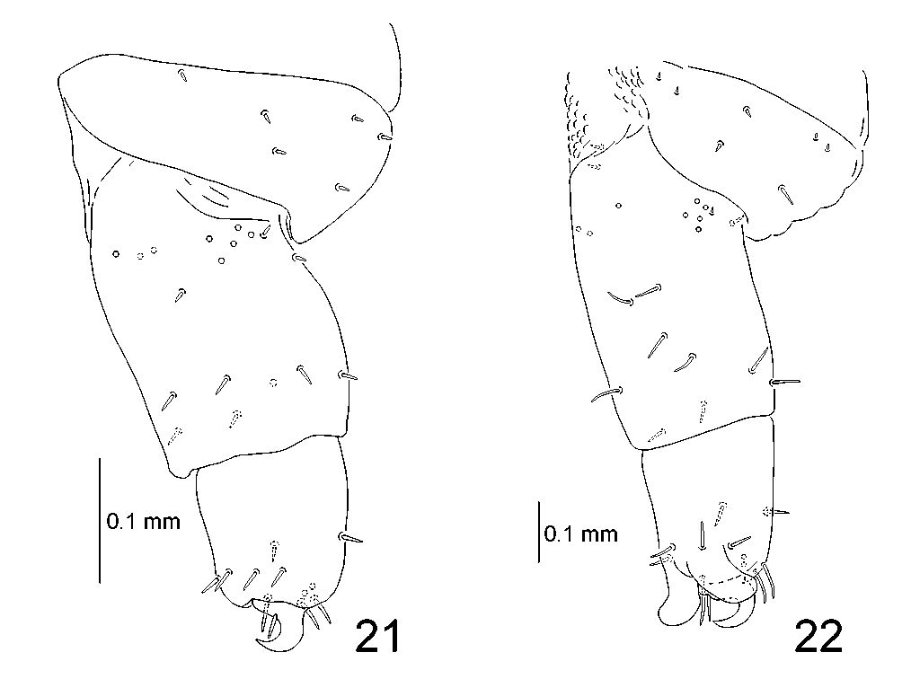
All legs stout, with similar chaetotaxy ( Figs 22 View FIGURES 21, 22 ), three segmented, built with: coxa, femur and tibiotarsus. Tibiotarsus apically with heavily sclerotized, short and curved, single and simple claw armed basally with a pointed seta. Above the claw two campaniform sensilla. Below the claw, on ventral side, two lobes — pulvilli. Tibiotarsus with 9 long pointed setae: complex of 7 setae surrounding pulvilli and claw, and two setae in dorsomedial part. Femur with 9 long setae and one short seta close to the base dorsally. Basally on internal side a group of 5 campaniform sensilla and one short pointed seta; one campaniform sensillum at the base, ventrally. Also basally, but on external side, two short pointed setae and two campaniform sensilla. Coxa with setae arranged in three groups: first with two short setae and one long pointed seta, second and third with a pair of short setae.
Pupa
Measurements (n = 1): length of body: 7.2 mm, width of fifth abdominal segment: 3.2 mm.
Body flattened dorsoventrally.
Alcohol preserved pupa appears light brown with brown to dark brown spiracles. Dorsal side of 2–5 abdominal segments with brown tubercles arranged in two rows: first row close to anterior border with 4 tubercles, second placed close to posterior border with 12–14 tubercles ( Figs 25, 26 View FIGURES 23 – 26 ).
Pro, meso and metanotum without lateral scoli. Head visible from dorsal view. Abdominal segments 1–7 with simple lateral scoli on each side. Last abdominal segment with two simple scoli and four flattened processes ( Figs 25, 26 View FIGURES 23 – 26 , 33, 34 View FIGURES 31 – 34 ). Each simple scolus armed apically with seta, and with one or two setae close to base.
Eight spiracles: one pair on each abdominal segment. Spiracles of first 4 segments of similar diameter. Diameter of spiracles of 6th to 8th segments slightly smaller than that of segments 1st–4th. Spiracle of 5th segment the most prominent, elongated, distinctly longer than lateral scoli ( Figs 25 View FIGURES 23 – 26 , 33 View FIGURES 31 – 34 ).


No known copyright restrictions apply. See Agosti, D., Egloff, W., 2009. Taxonomic information exchange and copyright: the Plazi approach. BMC Research Notes 2009, 2:53 for further explanation.