
Paranaitis bowersi (Benham, 1927)
|
publication ID |
https://doi.org/ 10.1046/j.1096-3642.2003.00069.x |
|
persistent identifier |
https://treatment.plazi.org/id/793A8787-5C04-600F-FF0F-FEEDFD66CC1C |
|
treatment provided by |
Carolina |
|
scientific name |
Paranaitis bowersi |
| status |
|
PARANAITIS BOWERSI View in CoL ( BENHAM, 1927)
Phyllodoce bowersi Benham, 1927: 77 , pl. 1, figs 27– 31; Monro, 1936: 111; Hartman, 1959: 160; Wesenberg-Lund, 1962: 40.
Phyllodoce (Anaitis) bowersi: Monro, 1930: 72–73 .
Anaitides bowersi: Hartman, 1964: 49 View in CoL , pl. 15, figs 1, 2.
Anaitis bowersi: Uschakov, 1962: 140–141 , pl. 1, figs B–V.
Paranaitis bowersi: Averincev, 1972: 106 View in CoL ; Uschakov, 1975: 148, fig. 1.
Material examined
Holotype ( BMNH 1928.2.29.13), Ross Sea, 284 m.
Description
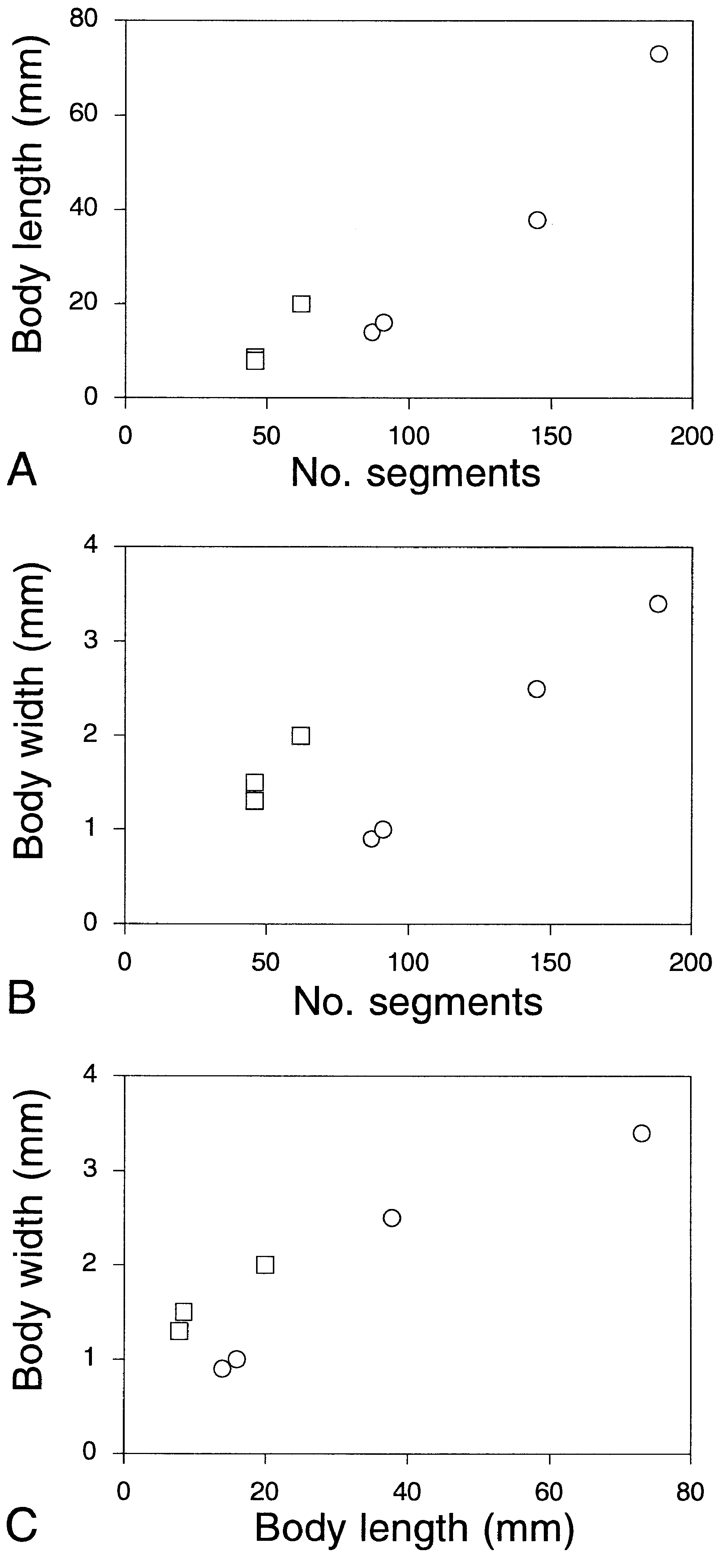
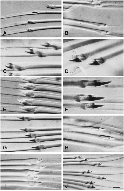
Holotype complete specimen, c. 60 mm long, 4.7 mm wide, for 117 segments. Preserved specimen uniformly pale; live specimen pale pink ( Benham, 1927). Body dorso-ventrally flattened, of uniform width, with tapering posterior end. Prostomium anteriorly rounded, wider than long, with shallow but distinct ligula. Paired antennae and palps short, conical, directed laterally. Eyes absent. Nuchal papilla longer than diameter, situated posteriorly in ligula. Nuchal organs probably fully retracted in holotype, situated in lateral slit between prostomium and segment 1; shape unknown. Distal part of proboscis with six rows of rounded tubercles. Proximal part (less than 1/10 of total length) dorsally smooth, laterally and ventrally with c. 30 conical large papillae. Chitinous papillae probably absent. Terminal ring with c. 15 rounded well defined papillae. Paired large papillae present laterally inside ring.
Segments 1 and 2 fused. Cirri of segment 1, dorsal and ventral cirri of segment 2, and dorsal cirri of segment 3 cylindrical, long and tapered. Cirri of segment 1 and ventral cirri of segment 2 reaching c. segment 5. Dorsal cirri of segment 2 reaching c. segment 7. Dorsal cirri of segment 3 reaching c. segment 8. Segment 2 with small neuropodial lobes with c. 7 chaetae, lobes partly fused to ventral cirrophores. Segment 3 with small neuropodia with c. ten chaetae, with ventral cirri of similar size and shape as following segments. Aciculae in segments 2 and 3 unknown.
Dorsal cirri of median segments reniform, wider than long. Dorsal cirrophores of median and posterior segments with narrow extension on dorsal side of cirri; extension absent from anterior segments. Neuropodium with supra-acicular lobes longer than subacicular lobes, with c. 25 chaetae. Dorsal and ventral chaetae similar within single fascicle. Rostrum of chaetal shaft asymmetrical, with single main tooth on anterior side. Ventral cirri with rounded ends, c. 1.5 times as long as wide, slightly shorter than neuropodial lobes. Pygidial cirri cylindrical, with rounded ends, c. 1.5 times as long as wide. Pygidial papilla very short, knob-like.
Habitat
219–1837 m.
Distribution
Known from Ross Sea and eastern sector of Antarctica ( Monro, 1930), and Gulf de Ancud, Chile (Wesenberg- Lund, 1962).
Remarks
Paranaitis bowersi differs from other Paranaitis in the character combination dorsally elongated dorsal cirrophores of median and posterior segments, and proboscis with long distal part covered by six rows of tubercles, and with short proximal part with large lateral and ventral papillae.
PARANAITIS CAECA ( MOORE, 1903) View in CoL
( FIGS 6–9 View Figure 6 View Figure 7 View Figure 8 View Figure 9 , 37 View Figure 37 )
Eumida caeca Moore, 1903: 426–428 View in CoL , pl. 23, fig. 1 (misspelled as Eumidia caeca View in CoL ); Izuka, 1912: 203, pl. 21, fig. 5; Hartman, 1959: 152.
Paranaitis caeca: Eibye-Jacobsen, 1991: 129 View in CoL .
Material examined
Holotype (USNM-15716), Sagami Bay, Japan, 74– 78 m; 2 specimens (ZIHU-2015), SW off Miura Peninsula, Sagami Bay , 35∞08.13¢N, 139∞36.21¢E, shell sand, 39 m, 12 May 1998, coll. F.P.; 1 specimen (ZIHU-1919), Otsuchi Bay , Iwate Honshu , Japan, 39∞20.7¢N, 141∞57.7¢E, sandy mud, 49 m, 7 May 1997, coll. T.K.
Description
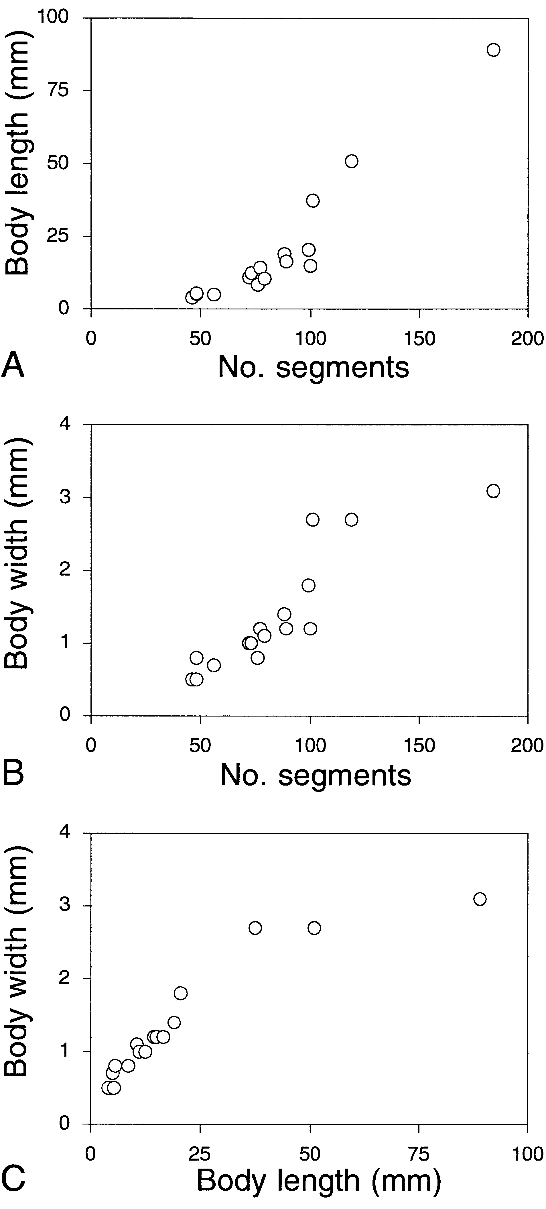
Holotype complete ovigerous female, 73 mm long, 3.4 mm wide, for 188 segments; see Figure 9 View Figure 9 for measurements of other specimens. Live animal white. Dorsum of segments 8–11 with brown pigmentation, forming transverse band. Dorsum of following segments with brown spot medially on each segment ( Fig. 6A View Figure 6 ), and inner part of dorsal cirri with single large rounded brown spots; medial and lateral spots together forming three longitudinal lines. Colour well retained after preservation. Body long, dorso-ventrally flattened, anteriorly and posteriorly tapered. Prostomium rounded pentagonal, with shallow but distinct ligula. Paired antennae and palps stout, conical, laterally directed ( Fig. 6B, C View Figure 6 ). Eyes absent. Nuchal papilla large, similar in shape to paired antenna, situated in shallow ligula. Nuchal organs rounded, large, situated laterally between prostomium and segment 1, visible dorsally and ventrally when everted ( Fig. 6B, C View Figure 6 ). Proboscis not divided into proximal and distal parts, with lateral row of rounded papillae each side; dorsal surface entirely covered by pointed papillae, ventral surface by rounded small papillae ( Fig. 7 View Figure 7 ). Terminal ring with c. 12 papillae. Paired large papillae present laterally inside ring.
Segments 1 and 2 fused. Cirri of segment 1, dorsal and ventral cirri of segment 2, and dorsal cirri of segment 3 stout, cylindrical, distally tapered ( Fig. 8A–C View Figure 8 ). Cirri of segment 1 reaching c. segment 5. Dorsal cirri of segment 2 with acicula, reaching c. segment 9 ( Fig. 8B View Figure 8 ). Ventral cirri of segment 2 reaching c. segment 6. Dorsal cirri of segment 3 with acicula, reaching c. segment 10 ( Fig. 8C View Figure 8 ). Segment 2 with dorsal and ventral aciculae, and c. 4 chaetae arising from cirrophores of ventral cirri ( Fig. 8B View Figure 8 ). Segment 3 with dorsal aciculae, small neuropodial lobes with ventral aciculae and c. ten chaetae, and ventral cirri.
Dorsal cirri of median segments kidney-shaped, wider than long ( Fig. 8F–I View Figure 8 ). Dorsal cirrophores distally enlarged. Neuropodium with supra-acicular lobes longer than subacicular lobes, with 25–30 chaetae. Dorsal and ventral chaetae similar within single fascicle. Rostrum of chaetal shaft symmetrical, with single short main tooth both sides ( Fig. 37B View Figure 37 ). Ventral cirri oval with rounded to weakly pointed ends, longer than neuropodia ( Fig. 8G,I View Figure 8 ). Pygidial cirri cylindrical, with rounded ends, 2–3 times as long as wide ( Fig. 6D View Figure 6 ). Pygidial papilla present.
Habitat
Sandy mud, 49 m. Paranaitis uschakovi and P. caeca , two poorly known species from Japan, are similarly in lacking eyes and in having white body with brown pigmentation. Eibye-Jacobsen (1991), based on an examination of the holotype of P. caeca and, presumably, the original description of P. uschakovi , considered the two species as distinct based on the following differences: (1) shape of prostomium, (2) length of dorsal cirri of segments 2 and 3 (those of segment 3 distinctly longer in P. uschakovi , of equal length in P. caeca ), (3) asymmetrical dorsal cirri in P. uschakovi (supposedly considered symmetrical in P. caeca ), and (4) length of ventral cirri (shorter than neuropodium in P. uschakovi , longer in P. caeca ). We examined the newly collected specimens of both species from Otsuchi Bay, as well as the holotypes of P. caeca and P. uschakovi , and concluded that most characters above are not suited to differentiate the two taxa. Instead, they may be separated by the following features: (1) shape of nuchal papilla (long and pointed in P. caeca ; short and rounded in P. uschakovi ), (2) position of nuchal organs (laterally in P. caeca ; ventro-laterally in P. uschakovi ), (3) shape of paired antennae, palps and anterior cirri (narrower paired antennae and palps, and thinner cirri of segments 1–3 in P. uschakovi ), (4) presence of proboscis papillae in P. caeca , (5) presence of ciliated furrows on the dorsal cirrophores of P. uschakovi , (6) presence of a dorsal longitudinal line in P. caeca , (7) morphology of the rostrum of chaetal shaft (symmetrical without main tooth in P. caeca ; asymmetrical with single main tooth in P. uschakovi ), and (8) different growth patterns ( Fig. 9 View Figure 9 ).
Distribution
Known from Sagami Bay, Uraga Channel ( Izuka, 1912) and Otsuchi Bay, Japan.
Remarks
Paranaitis caeca differs from other Paranaitis in the character combination of dorsally papillated proboscis, absence of eyes, elongated and pointed nuchal papilla, symmetrical rostrum of chaetal shaft, and unique pigmentation pattern.
PARANAITIS GARDINERI PERKINS, 1984 View in CoL
( FIGS 10–12 View Figure 10 View Figure 11 View Figure 12 )
Paranaitis gardineri Perkins, 1984: 563–565 View in CoL , fig. 4.
Paranaitis polynoides: Gardiner, 1976: 110 View in CoL , fig. 6m –p; Gathof, 1984: 19-21 to 19-23, figs 19-17 to 19-18. Not Anaitis polynoides Moore, 1909 View in CoL .
Material examined
Holotype (USNM-52876), off Cape Lookout, North Carolina, USA; 2 paratypes (USNM-52877), Wrightsville Beach, North Carolina; 1 paratype (HZM-P- 17600), Hutchinson Island, Florida, USA; 1 paratype ( FSBC I 30424), Hutchinson Island, 27∞20.24¢N, 80∞13.04¢W, 10.9 m; 1 paratype ( FSBC I 30425), Hutchinson Island , 27∞21.23¢N, 80∞13.24¢W, 10 m; 1 paratype ( FSBC I 30426), Hutchinson Island , 27∞22.08¢N, 80∞13.46¢W, 10.6 m; 1 specimen (mounted for SEM, not kept), Hutchinson Island , 27∞21.6¢N, 80∞13.2¢W, 11 m, 4 April 1997, coll. F.P.; 6 specimens (USNM-45527), Tampa Bay , Florida; 1 specimen (USNM-56127), Gulf of Mexico , 29∞36¢N, 87∞48¢W, 37 m; 1 specimen (USNM-51022), Beaufort, North Carolina, 34∞04¢N, 75∞25¢W, 20 m; 1 specimen (USNM-59331), off Georgia, Gulf of Mexico , USA, 31∞01¢N, 80∞17¢W, 40 m; 1 specimen (USNM- 59332), off Georgia, 31∞01¢N, 80∞17¢W, 40 m; 1 specimen (USNM-59333), off Georgia, 30∞57¢N, 79∞58¢W, 183 m; 1 specimen (USNM-59334), off Georgia, 31∞03¢N, 80∞26¢W, 34 m; 1 specimen (USNM-75587), off Florida, Gulf of Mexico , 29∞34¢N, 80∞22¢W, 44 m; 1 specimen (USNM-59335), Mississippi Sound, Alabama, Gulf of Mexico , 30∞03.12¢N, 88∞14.24¢W, 21 m; 1 specimen (USNM-75589), Mississippi Sound, 29∞59.20¢N, 88∞18.49¢W, 28.5 m; 1 specimen (USNM- 108159), off South Carolina, 31∞44.06¢N, 80∞13.06¢W, 33 m.
Description
Holotype complete with regenerated posterior end, proboscis everted, 37.5 mm long (excluding proboscis), 2.7 mm wide, for 101 segments. Up to 89 mm most animals from Florida lacking colour pattern ( Perkins, 1984). Preserved specimens pale yellow with brown spots on dorsum and dorsal cirri. Eyes blackish. Body long and slender, anteriorly and posteriorly tapered. Prostomium anteriorly rounded, with distinct deep ligula ( Fig. 10A View Figure 10 ). Paired antennae and palps conical, antero-laterally to laterally directed. Eyes large, rounded, with lenses, situated at posterior margin of prostomium. Short rounded nuchal papilla inserted posteriorly in ligula, often partly covered by segment 1. Nuchal organs retractile, rounded with horseshoe-shaped bands of cilia, ventro-laterally situated ( Fig. 10B View Figure 10 ). Proximal part of proboscis lacking tubercles, with gradual transition to distal part with tubercles ( Fig. 11A View Figure 11 ). Rounded chitinous papillae covering dorsal, lateral and ventral surfaces of both proximal and distal part ( Figs 11A,B View Figure 11 ). Papillae of terminal ring poorly defined. Paired large papillae present laterally inside ring. Segments 1 and 2 fused. Cirri of segment 1, dorsal and ventral cirri of segment 2, and dorsal cirri of segment 3 cylindrical, long and tapered. Cirri of segment 1 reaching c. segment 7. Dorsal cirri of segment 2 reaching c. segment 9. Ventral cirri of segment 2 reaching c. segment 7. Dorsal cirri of segment 3 reaching c. segment 11. Segment 2 without neuropodial lobes and chaetae. Segment 3 with small neuropodial lobes with c. 7 chaetae and small ventral cirri. Aciculae in segments 2 and 3 unknown. Dorsal cirri of median segments oval, slightly longer than wide in small specimens ( Fig. 10C View Figure 10 ); reniform, wider than long in large specimens. Dorsal cirrophores distinct, wide. Neuropodium with supra-acicular lobes longer than subacicular lobes, with c. 35 chaetae. Dorsal and ventral chaetae similar within single fascicle. Rostrum of chaetal shaft asymmetrical, with single large main tooth on anterior side ( Fig. 10E View Figure 10 ). Ventral cirri with rounded ends, as long as or slightly shorter than neuropodial lobes ( Fig. 10D View Figure 10 ). Pygidial cirri c. 7 times as long as wide, tapering to pointed ends ( Fig. 11C View Figure 11 ). Pygidial papilla present ( Fig. 10F View Figure 10 ).
long, 3.1 mm wide, for 184 segments (paratype, USNM-52877); see Figure 12 View Figure 12 for measurements of other specimens. Live animals from North Carolina reported as having diffuse purple spots on dorsum, ventrum, and digitate and flattened dorsal cirri; Habitat
Sand mixed with gravel and shell fragments, muddy sand and coarse calcareous sand, intertidally to 11 m.
Distribution
Known only from south-east US Atlantic coast.
Remarks
Paranaitis gardineri differs from other Paranaitis in the character combination proboscis with gradual transition between proximal and distal parts, long pygidial cirri with pointed ends, and specific body pigmentation. Paranaitis misakiensis is similar in proboscis features, but differs in having oval dorsal cirri with narrow dorsal cirrophores, pygidial cirri with rounded ends, and different body pigmentation. Based on re-examination of Gathof’s (1984) specimens (USNM-56127), her records of P. polynoides from the US Atlantic coast are referred to P. gardineri .


| T |
Tavera, Department of Geology and Geophysics |
| US |
University of Stellenbosch |
No known copyright restrictions apply. See Agosti, D., Egloff, W., 2009. Taxonomic information exchange and copyright: the Plazi approach. BMC Research Notes 2009, 2:53 for further explanation.
|
Kingdom |
|
|
Phylum |
|
|
Class |
|
|
Order |
|
|
Family |
|
|
Genus |
Paranaitis bowersi
| Kato, Tetsuya & Pleijel, Fredrik 2003 |
Paranaitis caeca: Eibye-Jacobsen, 1991: 129
| Eibye-Jacobsen D 1991: 129 |
Paranaitis gardineri
| Perkins TH 1984: 565 |
Paranaitis polynoides:
| Gathof JM 1984: 19 |
| Gardiner SL 1976: 110 |
Paranaitis bowersi: Averincev, 1972: 106
| Uschakov PV 1975: 148 |
| Averincev VG 1972: 106 |
Anaitides bowersi:
| Hartman O 1964: 49 |
Anaitis bowersi:
| Uschakov PV 1962: 141 |
Phyllodoce (Anaitis) bowersi: Monro, 1930: 72–73
| Monro CCA 1930: 73 |
Phyllodoce bowersi
| Wesenberg-Lund E 1962: 40 |
| Hartman O 1959: 160 |
| Monro CCA 1936: 111 |
| Benham WB 1927: 77 |
Eumida caeca Moore, 1903: 426–428
| Hartman O 1959: 152 |
| Izuka A 1912: 203 |
| Moore JP 1903: 428 |