
Paraxantia hubeiensis Liu & Kang
|
publication ID |
https://doi.org/ 10.5281/zenodo.186270 |
|
DOI |
https://doi.org/10.5281/zenodo.6216336 |
|
persistent identifier |
https://treatment.plazi.org/id/03D387A6-FFC8-8A3C-0BB1-F9C8FDA2F894 |
|
treatment provided by |
Plazi |
|
scientific name |
Paraxantia hubeiensis Liu & Kang |
| status |
sp. nov. |
Paraxantia hubeiensis Liu & Kang sp. nov.
( Figs. 3 View FIGURES 1 – 5 , 8 View FIGURES 6 – 10 , 13 View FIGURES 11 – 15 , 18 View FIGURES 16 – 25 , 23, 23, 32, 33, 41, 45, 52, 57, 62)
Holotype: male, CHINA: Hubei: Hefeng, Fenshuiling Forestry Spot, 1200m, 30.v.1989, no. 1344465, Coll. Sun Baowen ( IZAS).
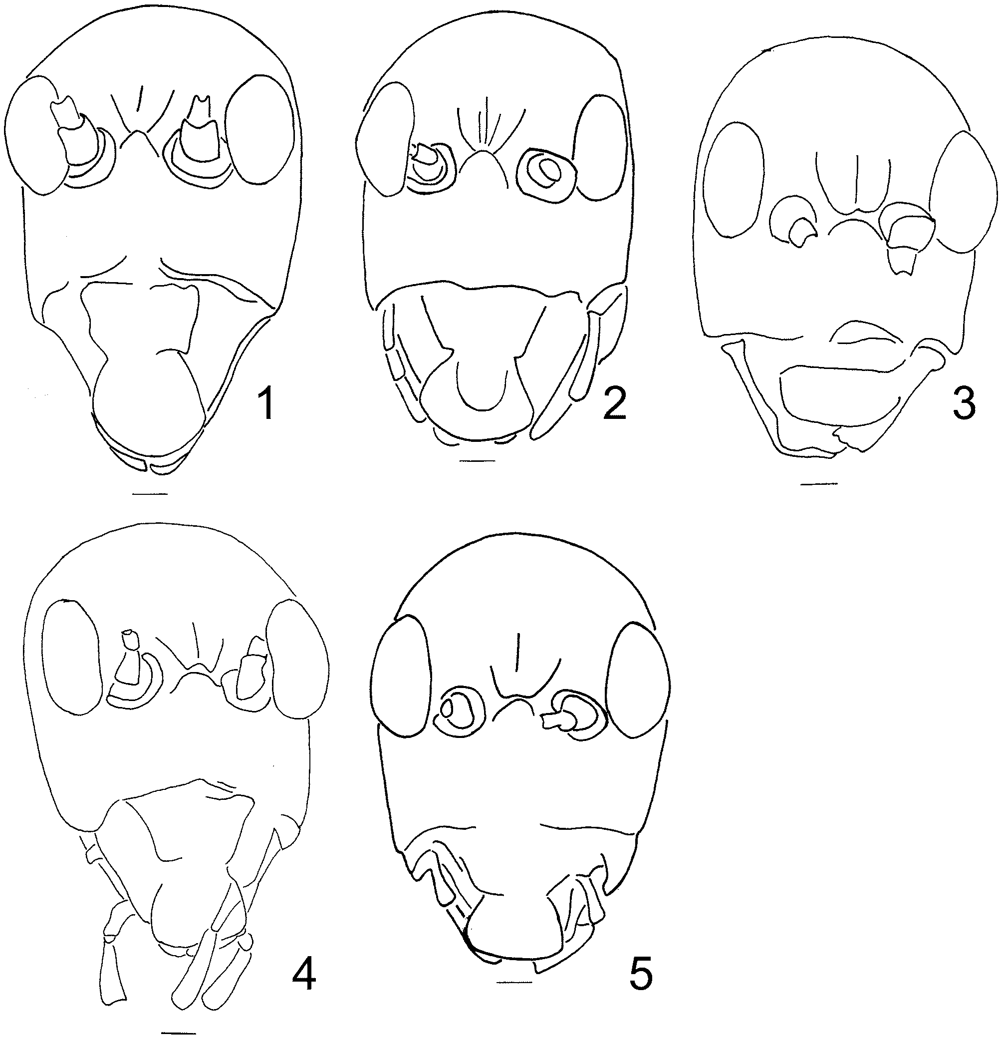
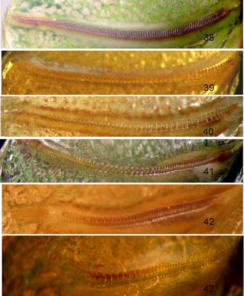
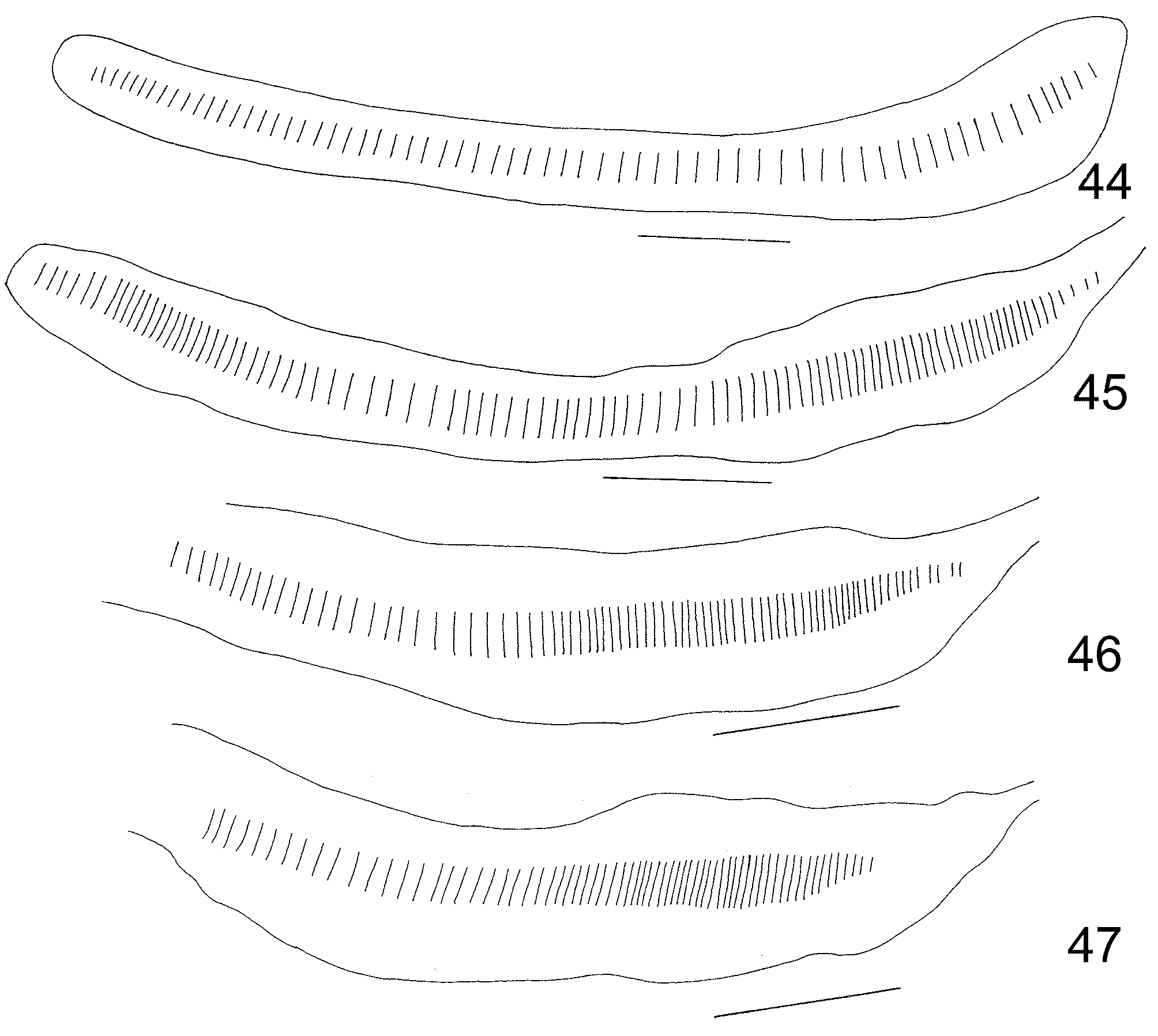
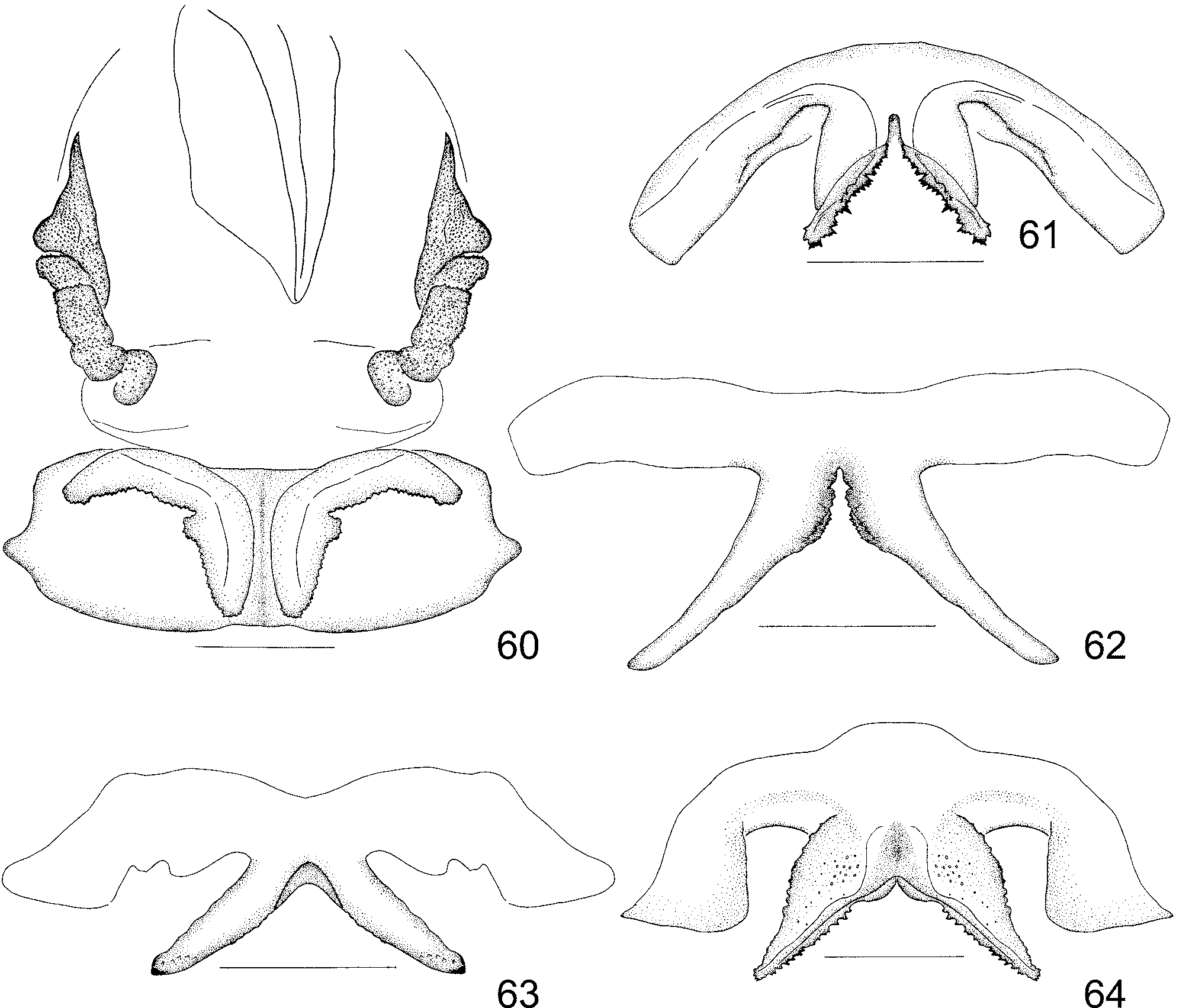
Description: Male (holotype). Size distinctly large for typical phaneropterine insects, and distinctly smaller than Paraxantia tibetensis . Pronotal disk ( Figs. 8 View FIGURES 6 – 10 , 13 View FIGURES 11 – 15 ) with deeply engraved first transverse groove, lying at basal fifth, and distinct middle transverse groove, lying slightly before middle; one oblique slightly granular line beginning in middle of each lateral carina, then ending in middle of posterior margin. Anterior femur armed with 6–8 small spines on ventro-anterior margin; median femur armed with 7–10 small spines on ventro-anterior margin; posterior femur with 24–26 anterior and 0–2 subapical posterior spines on ventral margins. Anterior tibiae only with 1 dorsal spine and 1 ventral apical spine on posterior margin, and 4 ventral spines as well as 1 ventral apical spine on anterior margin; median tibiae also only with 1 dorsal spine and 1 ventral apical spine on posterior margin, and 4–5 ventral spines as well as 1 ventral apical spine on anterior margin; posterior tibiae with 23 anterior and 23–24 posterior dorsal spines. Tegmen: Wings developed well. Hind wing longer than tegmen. Tegmen extending beyond apex of hind femur. Radial vein of tegmen with one oblique branch reaching posterior margin after radial sector vein. Left stridulatory area (Fig. 32) with posterior margin slightly sharply angular, greatest width between CuM vein and posterior margin rather large, about 5.5 millimeters. Stridulatory vein ( Figs. 41 View FIGURES 38 – 43 , 45 View FIGURES 44 – 47 ) rather long, with camber fine stridulatory file composed of about 73 widely arranged teeth, among which 22 widely arranged large teeth occupying middle third part, remaining teeth gradually becoming smaller towards both ends. Right stridulatory area with distinct irregular quadrangular mirror (Fig. 33).
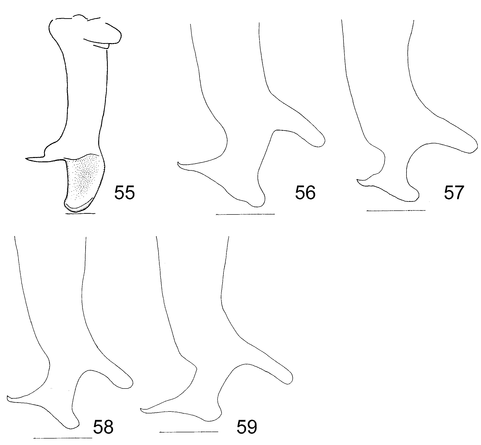
Epiproct sharp triangular. Cerci robust, bifurcate at apical fourth, dorsal one conical, produced inwards and upwards, with apex rounded; ventral one produced and horizontally inwards, abruptly tapering into a long sharp spine ( Fig. 57 View FIGURES 55 – 59 ). Subgenital plate wider than long, with distinct middle carina; apical margin with a wide triangular notch at middle; styli small, just slightly longer than notch (Fig. 52). unpaired lower sclerite of phallus with distinct shorter upper arm than sheet-like spinous lower one, notch between lower lateral arm sharply angular ( Fig. 62 View FIGURES 60 – 64 ).
Female unknown.
Color: Green, just compound eyes, tip of tibial spines, spines of internal cercal fork and sclerites of genitalia brown.
Measurements of male (mm): Length of body 31.2; length of pronotum 8.1; length of tegmen 60.1; width of tegmen 20.8; greatest width of tegminal dorsal part 5.5; length of hind wing 65.9; length of anterior femur 7.1; length of middle femur 10.9; length of posterior femur 26.5.
Etymology: The name shows the type locality.
Discussion: The new species resembles Paraxantia sinica ( Liu, 1993) in the general view, including the structure of head, common characteristics of pronotum, wings, legs and abdominal apex in Paraxantia . These two species just differ in details of stridulatory area of tegmen, and male abdominal apex. Paraxantia hubeiensis differs from P. s i n e n s i s by obtusely angular stridulatory area, stridulatory file, cerci and genitalia.
Distribution: China: Hubei: Hefeng.


| IZAS |
Institut Zoologii Akademii Nauk Ukraini - Institute of Zoology of the Academy of Sciences of Ukraine |
No known copyright restrictions apply. See Agosti, D., Egloff, W., 2009. Taxonomic information exchange and copyright: the Plazi approach. BMC Research Notes 2009, 2:53 for further explanation.