
Parvidrilus gianii, Martínez-Ansemil & Châtelliers & Martin & Sambugar, 2012
|
publication ID |
https://doi.org/ 10.1111/j.1096-3642.2012.00857.x |
|
DOI |
https://doi.org/10.5281/zenodo.5480038 |
|
persistent identifier |
https://treatment.plazi.org/id/039ADA53-1F51-6975-FC8C-252B60F6FBEC |
|
treatment provided by |
Marcus |
|
scientific name |
Parvidrilus gianii |
| status |
SP. NOV. |
PARVIDRILUS GIANII MARTÍNEZ- ANSEMIL & SAMBUGAR SP. NOV. ( FIG. 5 View Figure 5 )
Types: Holotype. MNCN 16.03 View Materials /3071, mature specimen, stained in paracarmine and whole-mounted in Canada balsam. Seldesuto cave (43°18′21.0′′N, 3°37′34.5′′W, 192 m asl), Matienzo, Cantabria, Spain, 2.ix.2002, leg. Ana Camacho. GoogleMaps
Paratype. MNCN 16.03 View Materials /3072, immature specimen from type locality, 2.ix.2002, leg. Ana Camacho, stained in paracarmine and whole-mounted in Canada balsam .
Etymology: Named after Narcisse Giani ‘maître et ami’, to whom many European oligochaetologists are very much indebted.
Description: Entire mature worm, length 1.2 mm, 26 segments, width 40 Mm at V, 42 Mm XII. Prostomium rounded, 15 Mm long, 25 Mm wide at base. Body wall thin (especially in dorsal part), unpapillated; foreign particles adhering to cuticle here and there along the body. Numerous transversal rows of thin cutaneous glands per segment. Clitellum not elevated, occupying at least XII- XIII.
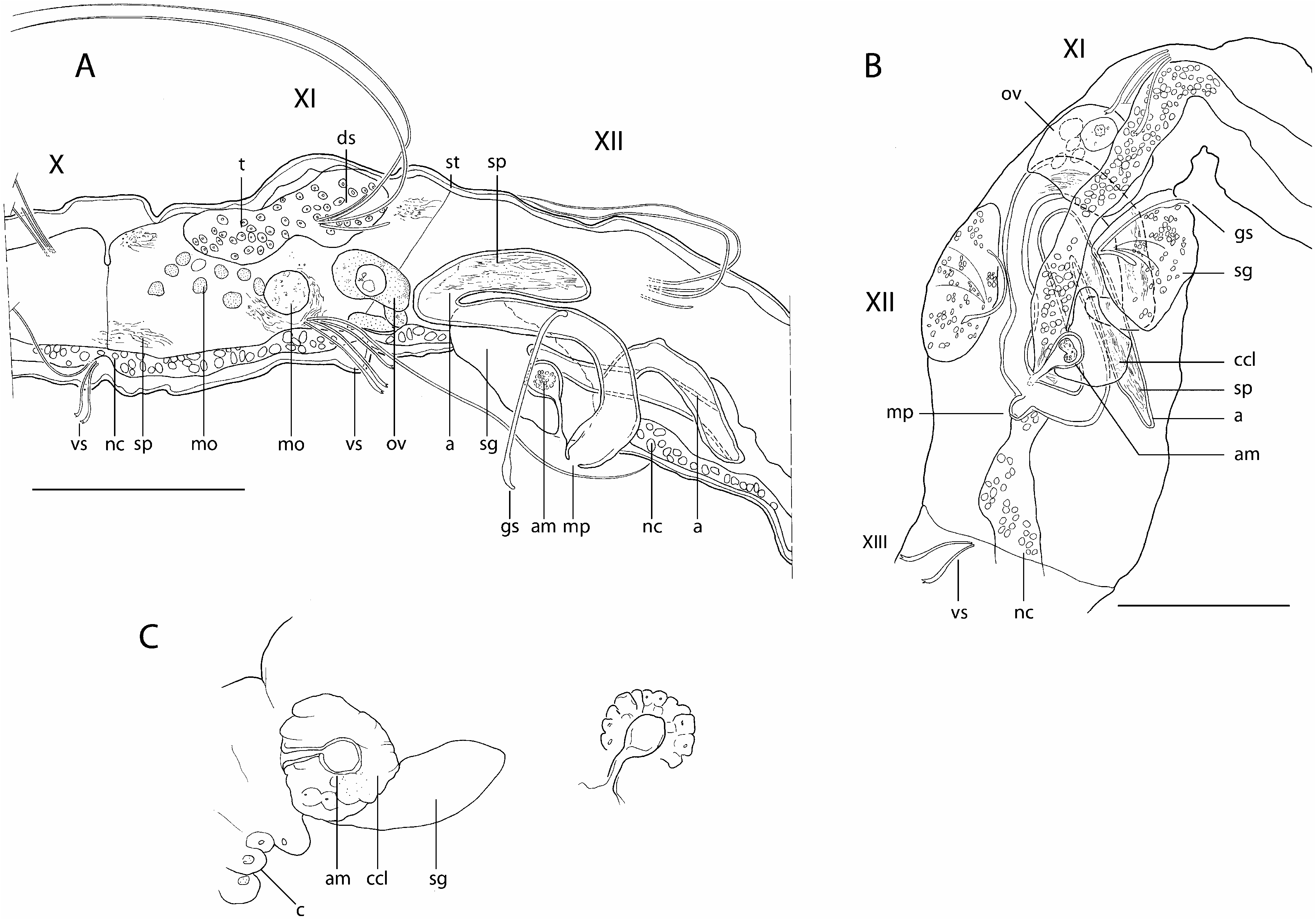
Dorsal (dorsolateral) and ventral (ventrolateral) setae present from III and posteriorly situated in each segment. Dorsal bundles with two (three) straight, thin single-pointed needles (20–28 Mm long) and one or two long, thin, and flexible hair setae (105–190 Mm long) ( Fig. 5A, B, h, n View Figure 5 ). Ventral bundles composed of (one) two–three (four) strongly curved crotchets, 20–26 Mm long, with enlarged distal half and doublepronged tip with minute distal tooth ( Fig. 5A–C View Figure 5 , cr); one thin hair seta (60–90 Mm long) in preclitellar ventral bundles of III–VI. No modified genital setae; a single bifid crochet in ventral bundles of segment XII.
No eyes. Brain long, extending into segment IV, deeply incised posteriorly. Ventral nerve cord in contact with epidermis, beneath muscles of the body wall. Most setigeral segments have a mid-dorsal glandular pouch opening posteriorly on each segment, at about the transversal setal line; glandular pouches absent from IV–VI.
Digestive tract complete, entirely ciliated and ending in a terminal anus. An eversible pharynx, with small dorsal pad set off from oesophagus, followed by a narrow, thick walled winding tube extending into IX, with thick muscular walls at about VII–IX; alimentary canal completed by a gut clearly enlarged and filled in with sediment posteriorly from segment XIII ( Fig. 5E, F, g View Figure 5 ). Compact pharyngeal glands present in IV–VI. Digestive tract surrounded from V by a well-developed layer of chloragogen cells. Coelomocytes and nephridia not observed.
No compact testes attached to septum but free germ cells and morulae floating in the coelomic cavity of segment XI ( Fig. 5A View Figure 5 , mo). Two small sperm funnels opening in ventral part of septum XI/XII and continuing into very thin vasa deferentia (1.5-2 Mm wide), very likely to be long and tightly folded, entering atria basally ( Fig. 5A, F, f View Figure 5 , vd). Atria elongate (about 150 Mm long, 14–16 Mm wide), extending in segments XII- XIII, curling anteriad ( Fig. 5A, C–F, a View Figure 5 1 View Figure 1 , a 2 View Figure 2 ), merging below the nerve cord and a thick muscular arch ( Fig. 5A, E View Figure 5 , ma, nc) into a common ejaculatory duct with cuticular walls, and opening on tip of a mid-ventral porophore located somewhat anterior to the transversal setal line of segment XII ( Fig. 5A, F, c View Figure 5 , mp, p). Atria made up by outer thin muscular layer (<1 Mm thick) and thick lining of vacuolated tissue, with indistinct lumen, except at the most basal portion, where the lumen is large and epithelial cells are finely granulated. Sparse unordered sperm embedded into vacuolated cells all along atria. Prostate glands absent. Two small ovaries with maturing eggs attached to septum 11/12 ( Fig. 5A, F, o View Figure 5 ). Two small oviducts seemingly attached to septum 12/13 ( Fig. 5A View Figure 5 , od). A single spermatheca present in XIII. Spermathecal ampulla (empty) ovoid (30 Mm long, 9 Mm high), with a nucleated epithelial wall surrounded by a thick muscular lining; spermathecal duct about 13 Mm long, as a loosely defined structure, without lumen, basally enlarged, in continuity with the ventral body wall at the most anterior part of segment XIII, in a somewhat lateral position, at the left side of the worm ( Fig. 5A, D–E View Figure 5 , sa, sd).
Remarks: The combination of long atria, a single spermatheca in segment XIII, and the cuticular wall of the common ejaculatory duct characterize P. gianii sp. nov. Amongst the three Parvidrilus species that have a single spermatheca in segment XIII, P. gianii is easily distinguishable from the others by its ovoid spermathecal ampulla surrounded by a thick muscular lining, and elongated atria (ten times longer than wide). The cuticular wall of the ejaculatory duct of this new species is unique amongst the genus.
Distribution and habitat: Parvidrilus gianii is known only from the type locality, the Seldesuto cave, Matienzo, Cantabria, Spain. The species was sampled at 100 m from the entrance, along the shore of the lake, by stirring up rocks and sand covered by about 25 cm of water. The depth of the lake deepens very quickly a short distance from the shore; the gallery ends in a siphon.


No known copyright restrictions apply. See Agosti, D., Egloff, W., 2009. Taxonomic information exchange and copyright: the Plazi approach. BMC Research Notes 2009, 2:53 for further explanation.
|
Kingdom |
|
|
Phylum |
|
|
Class |
|
|
Order |
|
|
Family |
|
|
Genus |