
Parvidrilus stochi, 2012
|
publication ID |
https://doi.org/ 10.1111/j.1096-3642.2012.00857.x |
|
DOI |
https://doi.org/10.5281/zenodo.5480040 |
|
persistent identifier |
https://treatment.plazi.org/id/039ADA53-1F57-6977-FC7E-24876705FA3F |
|
treatment provided by |
Marcus |
|
scientific name |
Parvidrilus stochi |
| status |
SP. NOV. |
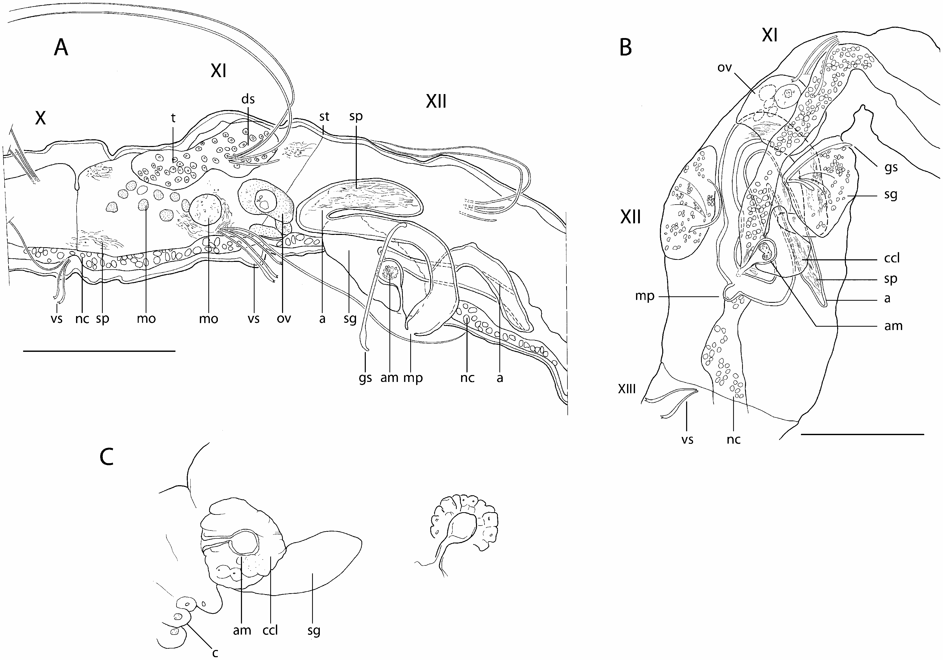
PARVIDRILUS STOCHI SAMBUGAR & MARTÍNEZ- ANSEMIL SP. NOV. ( FIG. 6 View Figure 6 )
Types: Holotype. VRO1003, mature specimen unstained, whole-mounted in Canada balsam. Monte Majore cave (40°30′47′′N, 8°36′33′′E), Thiesi , Sardinia, Italy, 26.vi.2008, leg. Fabio Stoch, Gianfranco Tomasin, Beatrice Sambugar, Paolo Marcia. GoogleMaps
Other material examined: One partially mature specimen, stained in paracarmine and whole-mounted in Canada balsam, from type locality, 17.iii.2005, leg. Fabio Stoch, Gianfranco Tomasin, Jos Notenboom. Two immature specimens, stained in paracarmine and whole-mounted in Canada balsam, from type locality, 8.ix.2006, leg. Fabio Stoch, Paolo Marcia.
Etymology: Named after our friend Fabio Stoch, for his important contributions to the knowledge of European subterranean fauna.
Description: Entire mature worms, length 1.2 mm, 28 segments, width 45 Mm at V, 55 Mm at XII. Prostomium rounded, 15 Mm long, 29 Mm wide at base. Body wall thin (especially in dorsal part), unpapillated; foreign particles adhering to cuticle along the body, but neither dense nor continuous. Numerous transversal rows of thin cutaneous glands per segment. Clitellum weakly developed (about XI- XIII), but some large cells observed in XII and XIII ( Fig 6C View Figure 6 , cl).
Dorsal (dorsolateral) and ventral (ventrolateral) setae present from III, posteriorly situated in each segment. Dorsal bundles with two (three) straight, thin single-pointed needles (20–28 Mm long) and one (two) long, thin and flexible hair setae (115–200 Mm long) ( Fig. 6A–C, h, n View Figure 6 ). Ventral bundles with (one) two–three (four) strongly curved crotchets, 20–27 Mm long, with enlarged distal half and double-pronged tip with minute distal tooth ( Fig. 6A–C View Figure 6 , cr), and accompanied by one thin hair seta (95–110 Mm long) only present in preclitellar ventral bundles III- VIII. No modified genital setae; ventral bundles of segment XII with three bifid crochets.
No eyes. Brain long, extending into segment IV, deeply incised posteriorly. Ventral nerve cord in contact with epidermis, beneath muscles of the body wall. Most setigeral segments with a mid-dorsal glandular pouch opening posteriorly on each segment, on or immediately adjacent to the transversal setal line; glandular pouches absent from IV- VI.
Digestive tract complete, entirely ciliated, ending in a terminal anus. An eversible pharynx, with small dorsal pad set off from oesophagus, followed by a narrow, winding tube, extending into IX, with thick muscular walls at about VII- IX; alimentary canal completed by a gut beginning in X, enlarged and filled in with sediment from segment XV ( Fig. 6D–F, g View Figure 6 ). Compact pharyngeal glands present in IV- VI. Digestive tract surrounded by a well-developed layer of chloragogen cells from V. Coelomocytes and nephridia not observed.
No compact testes attached to septum, but many free germ cells and morulae floating in the coelomic cavity of segment XI and in the most posterior and anterior parts of X and XII, respectively ( Fig. 6A, E View Figure 6 , mo). Sperm funnels and vasa deferentia not observed. Atria very elongate (about 250 Mm long), twisted, tubular, with a diameter slightly decreasing from the proximal to the distal end (20–15 Mm), extending in segments XII –XIV ( Fig. 6A, D–F, a View Figure 6 1 View Figure 1 , a 2 View Figure 2 ), merging below the nerve cord and opening on the tip of a mid-ventral porophore located between the two ventral setal bundles of segment XII ( Fig. 6A, D, E View Figure 6 , ed, ma, mp, p). Atria made up by extremely thin (muscular?) outer layer and thick lining of vacuolated tissue, with indistinct lumen, except at the most basal portion, where the lumen is large and the epithelial cells are finely granulated. Space occupied by vacuolated cells seemingly progressively replaced by sperm (about two thirds of atrial length of the holotype and only a very small part of atria of the maturing specimen collected in March 2005). Prostate glands absent. Two ovaries attached to septum 11/12 (poorly visible) ( Fig. 6A, o View Figure 6 ). A single egg sac containing mature eggs extending into segment XV ( Fig. 6A, e View Figure 6 ). Two small oviducts seemingly attached to septum 12/13. A single spermatheca present in XII. Spermathecal ampulla ovoid (33 Mm long, 10 Mm high), thin walled, followed by a conical, bent duct (about 20 Mm long), without clear cut off from ampulla, thin walled distally, and proximally constituted by a loosely defined tissue ending close to male pore, in an anterior and somewhat lateral position, on left side of the worm. Ampulla and distal part of spermathecal duct full of spermatozoids ( Fig. 6A, D View Figure 6 , sa, sd).
Remarks: Parvidrilus stochi sp. nov. is one of the three Parvidrilus species known so far that have a single spermatheca in segment XII and are devoid of genital setae. The most outstanding features of P. stochi sp. nov. are the very long, wide, and twisted atria (15 times longer than wide), combined with a spermatheca in the atrial segment that has a large spermathecal duct distally constituted by a loosely defined tissue. The ovoid shape of the spermatheca of this new species is only comparable to that of P. gianii .
Distribution and habitat: Parvidrilus stochi is known only from the type locality in the Monte Majore cave (Sardinia, Italy). The cave opens in a small, isolated Miocene limestone outcrop rising from a volcanic plateau dating from the Oligocene- Miocene volcanism. The upper level of the cave is fossilized and percolating waters are collected in pools on clay and rock; the lower gallery is crossed by a small brook, which collects the waters sinking from a small valley at the entrance of the cave.


| V |
Royal British Columbia Museum - Herbarium |
| VI |
Mykotektet, National Veterinary Institute |
No known copyright restrictions apply. See Agosti, D., Egloff, W., 2009. Taxonomic information exchange and copyright: the Plazi approach. BMC Research Notes 2009, 2:53 for further explanation.
|
Kingdom |
|
|
Phylum |
|
|
Class |
|
|
Order |
|
|
Family |
|
|
Genus |