
Pediopsoides (Pediopsoides) Matsumura
|
publication ID |
https://doi.org/ 10.11646/zootaxa.4150.3.5 |
|
publication LSID |
lsid:zoobank.org:pub:9A230549-A9DF-47E6-BD92-6B599F0ACAFE |
|
DOI |
https://doi.org/10.5281/zenodo.6087717 |
|
persistent identifier |
https://treatment.plazi.org/id/A333EE78-F85C-FFAC-26FF-1C6CD783FD78 |
|
treatment provided by |
Plazi |
|
scientific name |
Pediopsoides (Pediopsoides) Matsumura |
| status |
|
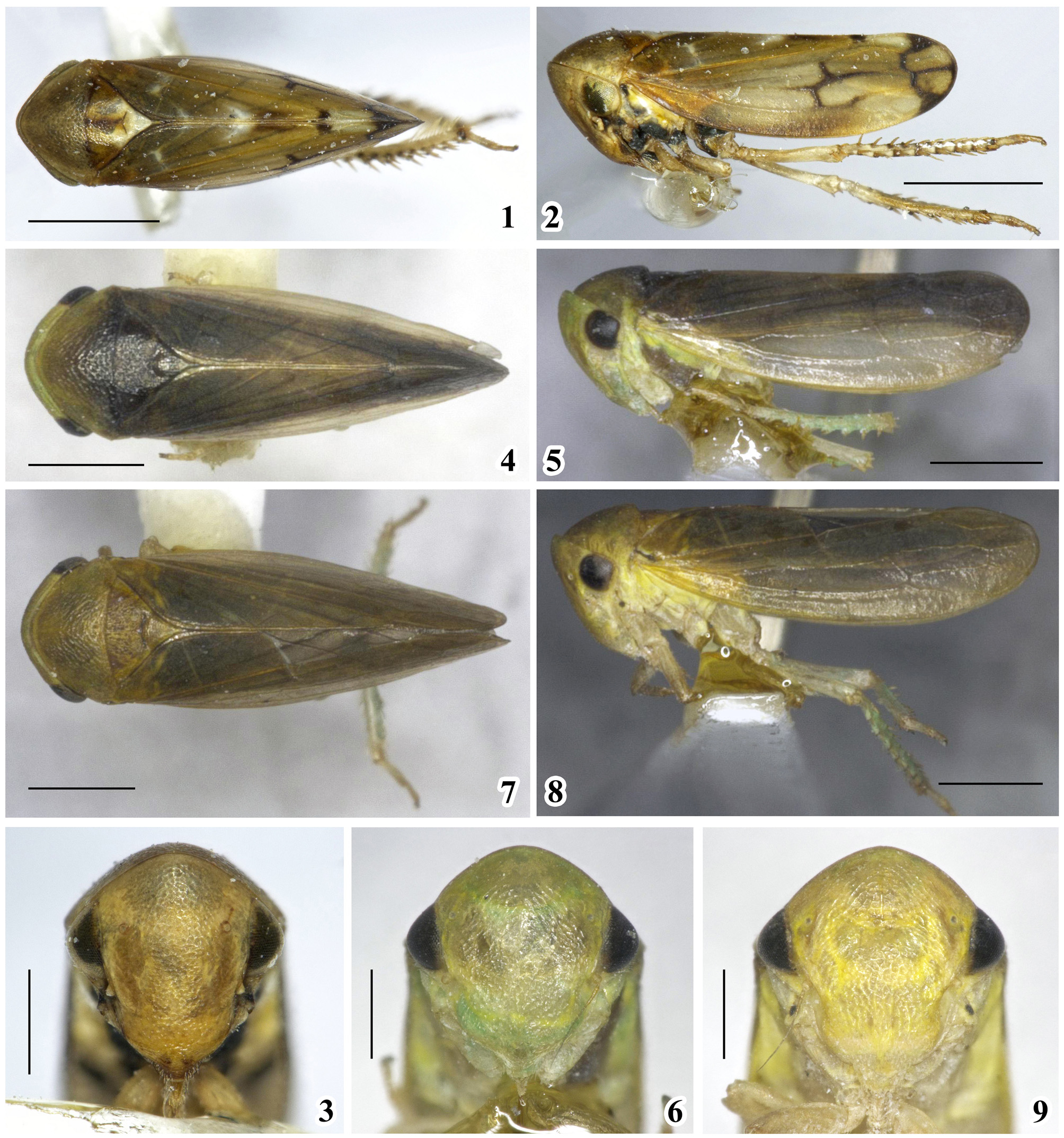
Key to species (³) of the subgenus Pediopsoides (Pediopsoides) Matsumura View in CoL
P. (P.) formosanus (Matsumura) distributed in Taiwan, known only from the female, is excluded.
1. Clypellus black ( Li et al. 2012: 536, Fig. 7 View FIGURES 1 – 9 )....................................... P. (P.) nigorolabium Li, Dai & Li
- Clypellus not black....................................................................................2
2. Aedeagal shaft with pair of processes...................................................................... 3
- Aedeagal shaft without pair of processes................................................................... 6
3. Shaft processes ( Zhang 2010: 61, Figs 30–31 View FIGURES 28 – 35 ) on apex of aedeagus, hooklike................. P. (P.) jingdongensis Zhang View in CoL
- Shaft processes at base or middle of aedeagus, spinelike....................................................... 4
4. Shaft processes at base of aedeagus ( Figs 16–17 View FIGURES 10 – 19 )................................... P. (P.) alba Li, Dai & Li View in CoL sp. nov.
- Shaft processes at middle of aedeagus..................................................................... 5
5. Aedeagal shaft processes ( Huang & Viraktamath 1993: 367, Fig. 28 View FIGURES 28 – 35 ) pointed basally; dorsal connective ( Huang & Viraktamath 1993: 367, Fig. 27 View FIGURES 20 – 27 ) with apophysis twisted caudoventrally at middle........................ P. (P.) femorata (Hamilton) View in CoL
- Aedeagal shaft processes ( Viraktamath 1996: 188, Figs 32–33 View FIGURES 28 – 35 ) pointed dorsally; dorsal connective ( Viraktamath 1996: 188, Figs 34–35 View FIGURES 28 – 35 ) without apophysis at middle............................................. P. (P.) kodaiana Viraktamath View in CoL
6. Style with subapical toothlike process ( Li et al. 2013: 20, Fig. 16 View FIGURES 10 – 19 ); dorsal connective ( Li et al. 2013: 20, Fig. 21 View FIGURES 20 – 27 ) with furcated
process at middle, like species in genus Oncopsis View in CoL .................................... P. (P.) tishetshkini Li, Dai & Li View in CoL - Style without subapical toothlike process; dorsal connective without process, or with unbranched process at middle........ 7
7. Aedeagal shaft ( Figs 24–25 View FIGURES 20 – 27 ) slender, in ventral view apex strongly expanded laterally, lamelliform; pygofer ventral margin ( Fig. 21 View FIGURES 20 – 27 ) with 5 small teeth............................................... P. (P.) amplificata Li, Dai & Li View in CoL sp. nov.
- Aedeagal shaft relatively stout, in ventral view apex not expanded laterally; pygofer ventral margin serrated, or with spines, or with wide process..................................................................................... 8
8. Pygofer ventral margin ( Yang & Zhang 2013: 588, Figs 3 View FIGURES 1 – 9 –A, 3–H) with single process widened at base and slanting inwards; apical half of aedeagal shaft ( Yang & Zhang 2013: 588, Fig. 3 View FIGURES 1 – 9 –E) strongly twisted dorsally, rectangular............................................................................................ P. (P.) anchorides Yang & Zhang View in CoL
- Pygofer ventral margin serrated or with 2 small spines........................................................ 9
9. Dorsal connective ( Fig. 31 View FIGURES 28 – 35 ) with long process at middle twisted ventrally; apex of ventral margin of pygofer ( Fig. 29 View FIGURES 28 – 35 ) with 2– 3 small teeth....................................................... P. (P.) longiapophysis Li, Dai & Li View in CoL sp. nov.
- Dorsal connective without process at middle; apex of ventral margin of pygofer with 2 spines, or not.................. 10
10. Ventral margin of pygofer with 2 spines................................................................... 11
- Ventral margin of pygofer ( Li et al. 2013: 19, Fig. 7 View FIGURES 1 – 9 ) without spines................... P. (P.) dainghanensis Li, Dai & Li
11. Dorsal connective ( Hamilton 1980: 898, Fig. 71) clearly S shaped; pygofer lobe ( Hamilton 1980: 898, Fig. 71) broad.................................................................................. P. (P.) satsumensis (Matsumura) View in CoL
- Dorsal connective ( Li et al. 2012: 537, Fig. 15 View FIGURES 10 – 19 ) geniculate; pygofer lobe ( Li et al. 2012: 537, Fig. 8 View FIGURES 1 – 9 ) narrow, elongate..................................................................................... P. (P.) bispinata Li, Dai & Li View in CoL


No known copyright restrictions apply. See Agosti, D., Egloff, W., 2009. Taxonomic information exchange and copyright: the Plazi approach. BMC Research Notes 2009, 2:53 for further explanation.