
Phatnoma mattijoae, Jepson, James E., Penney, David & Green, David I., 2011
|
publication ID |
https://doi.org/ 10.5281/zenodo.278259 |
|
DOI |
https://doi.org/10.5281/zenodo.6189834 |
|
persistent identifier |
https://treatment.plazi.org/id/03CB0053-BB28-6541-FF17-FEB6F3FCFCC7 |
|
treatment provided by |
Plazi |
|
scientific name |
Phatnoma mattijoae |
| status |
sp. nov. |
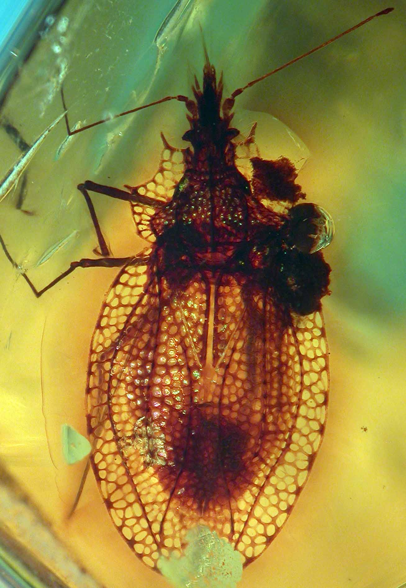
Phatnoma mattijoae sp. nov.
Fig. 1 View FIGURE 1 .
Etymology. Named after Matthew and Joanna Penney, nephew and niece of the second author. Material. NHM II 3047. Known only from the holotype. Complete adult specimen, female.
Distribution. Miocene Dominican amber forest.
Diagnosis. The relative position and structure (drawn out to a point) of the paranotal projections and the elongate cells on the outermost margin of the costal area serve to identify the new species.
Description. 3.88 mm long, 1.9 mm wide (at widest point, across hemelytra). Head porrect; extending in front of compound eyes. Eyes small, set close to pronotum. Antennal process (antennophores) projecting outwards at 45 degrees; antennae 4-segmented: basal segment 0.14 mm, widened distally, second segment 0.09 mm and of uniform thickness, third segment 1.09 mm with constricted base, distal segment 0.17 mm, expanded towards tip and with terminal setae. Head spines consist of pair of suberect curved occipitals, pair of erect frontals, pair of anteriorly directed jugals, and single, anteriorly directed long clypeal.
Thorax. Collar well developed with three rows of areolae. Pronotum with three raised longitudinal carinae (median and two laterals), each with single row of cells. Lateral margins expanded to form paranota consisting of two–three rows of cells, each with anterior and medial projection separated by five cells.
Hemelytra. 2.2 mm long, completely developed. Small scutellum. Large clavus; four areolae wide (at widest point). Clavus almost half size of hemelytra in length. Right hemelytron slightly overlapping left. Covered by large-rounded areolae, majority regular size, apart from outermost marginal cells of costal area, these noticeably more elongate. Costal area broad with three rows (occasionally four) of areolae. Subcostal area wide, with five rows of areolae (at its widest). Discoidal area slightly smaller than subcostal area with maximum of four rows of areolae; discoidal area subdivided by transverse veins. Sutural area four rows of areolae wide. Stenocostal area absent. Subcostal and discoidal areas higher than costal area. Hind wings not visible.
Legs. Long and slender, femora and tibiae approximately same length. All femora twice as thick as tibiae, tarsi two-segmented with two tarsal claws.
Abdomen. Obscured by wings, difficult to observe, shorter than hemelytra. Underside and terminalia difficult to observe because of cut and curve of amber; however, ovipositor is present.
Remarks. Phatnoma mattijoae sp. nov. is most similar to the extant Phatnoma ovatum Champion, 1897 from Guatemala; because both have their largest areolae in a row on the outer margin of the costal area on the hemelytra. They differ because P. ovatum does not have the medial paranotal projections drawn out to a point.
No known copyright restrictions apply. See Agosti, D., Egloff, W., 2009. Taxonomic information exchange and copyright: the Plazi approach. BMC Research Notes 2009, 2:53 for further explanation.