
Phlugis gracila Nickle, 2003
|
publication ID |
https://doi.org/ 10.11646/zootaxa.4311.4.7 |
|
publication LSID |
lsid:zoobank.org:pub:31838383-Dc41-47C5-B926-98Ced011B19C |
|
DOI |
https://doi.org/10.5281/zenodo.6023749 |
|
persistent identifier |
https://treatment.plazi.org/id/8355B653-900B-5F1A-BBEE-A237FB70F85C |
|
treatment provided by |
Plazi |
|
scientific name |
Phlugis gracila Nickle, 2003 |
| status |
|
Phlugis gracila Nickle, 2003 View in CoL
(Figs. 1–8)
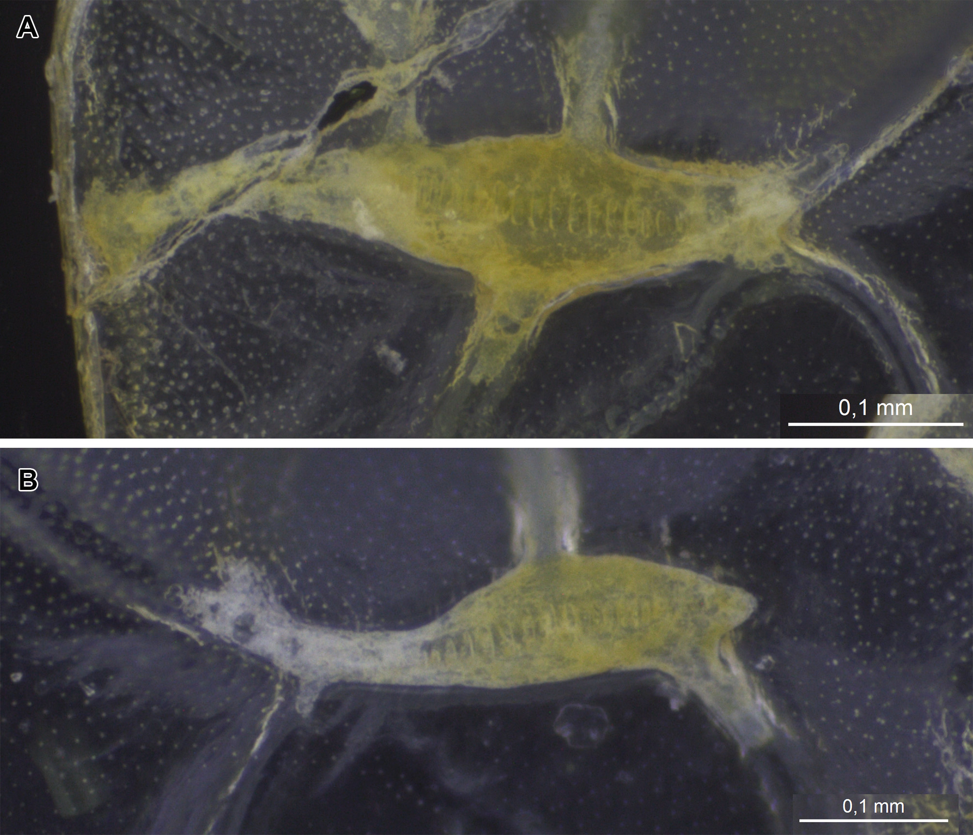
Diagnosis. Body narrow and elongate (Figs. 1A, 5A). Fore femur with 3 internal spines and 4 external ventral spines (Figs. 1F, 5G). Left stridulatory file slightly sinuous, medially thickened and narrowing gradually to the apex ( Fig. 3 View FIGURE 3 A). Right stridulatory file curved, medially thickened and narrowing abruptly at the beginning of distal region ( Fig. 3 View FIGURE 3 B). Subgenital plate sinuous, medially with dorsal margins making two triangular projections, posteriorly straight and distally rectangular in lateral view (Fig. 1I –K). Tenth tergite with two triangular projections posterolateral and convex posteromedial (Fig. 1I). Male cocealed genitalia elongate, with dorsal lobes laterally projected with blunt apex, which makes the genitalia similar to an anchor ( Fig. 4 View FIGURE 4 A–D). Titillator forming two slightly curved sclerotised triangular projections, with a blunt apex trespassing posterior margin of the lower folds of ventral lobe ( Fig. 4 View FIGURE 4 A–D). Posterior portion of genitalia with an aperture located medially between projections of titillator ( Fig. 4 View FIGURE 4 A–D).
Specimens examined. 4♂ - 6♀: BRASIL, Amazonas, Alvarães, Rio Solimões , próximo da Comunidade Caburini, 24–25.iii.2017, coleta manual em bancos de herbáceas aquáticas flutuantes, (D.M.M. Mendes & J.C. Oliveira leg.) [03°09'26"S / 64°45'48.6"W](3♂ - 5♀) GoogleMaps ; idem, Manaus, Ilha da Majantaria , 28.iv.1990, armadilha de luz, (M.F. Vieira & L. Aquino leg.)(1♂ - 1♀) .
Geographic distribution. Peru: Loreto; Brazil (new record): Amazonas (Fig. 08).
Description. Male. We include here only characteristics that were not used in the original description made by Nickle (2003).
Wings. Tegmen narrow and elongate, length 10 times longer than width, measued in medial region ( Fig. 2 View FIGURE 2 ). Costal margin of tegmen with the base briefly sinuous and posteriorly straight to the apex ( Fig. 2 View FIGURE 2 ). Anal margin of tegmen slightly sinuous from the base to the beginning of apical region, where it curves to the apex ( Fig. 2 View FIGURE 2 ). Left stridulatory file slightly sinuous, medially thickened and narrowing gradually to the apex ( Fig. 3 View FIGURE 3 A). Basal and apical teeth small, enhancing in length and width towards medial region of the row. Total length of the row: 0.3 mm; Maximum width of the vein: 0.1 mm; Total number of teeth: 39. Right stridulatory vein curved, medially thickened and narrowing abruptly at the beginning of distal region, with same width to the apex ( Fig. 3 View FIGURE 3 B). Total length of the row: 0.2 mm; Maximum width of the vein: 0.1 mm; Total of teeth: 21.
Male internal genitalia. Shape elongate ( Fig. 4 View FIGURE 4 A–D). Ejaculatory vesicles reniform with an ejaculatory duct shorter than half the length of ejaculatory vesicle ( Fig. 4 View FIGURE 4 A–D). Upper folds of ventral lobe narrow and elongate, trespassing lateral margin of the ventral lobe ( Fig. 4 View FIGURE 4 A–B). Dorsal lobes laterally projecting, blunt apex, which makes the genitalia similar to an anchor ( Fig. 4 View FIGURE 4 A–D). Dorsal folds elongate, slightly narrowing from the base to the apex and trespassing dorsal lobes margins. Titillator sclerite forming two slightly curved triangular projections, with a blunt apex trespassing posterior margin of the lower folds of ventral lobe ( Fig. 4 View FIGURE 4 A–D). Posterior portion of genitalia with an aperture located medially between projections of titillator ( Fig. 4 View FIGURE 4 A–D). Lower folds of ventral lobe short, wrinkled, with various small protuberances and posteriorly forming a projection with blunt apex ( Fig. 4 View FIGURE 4 C–D).
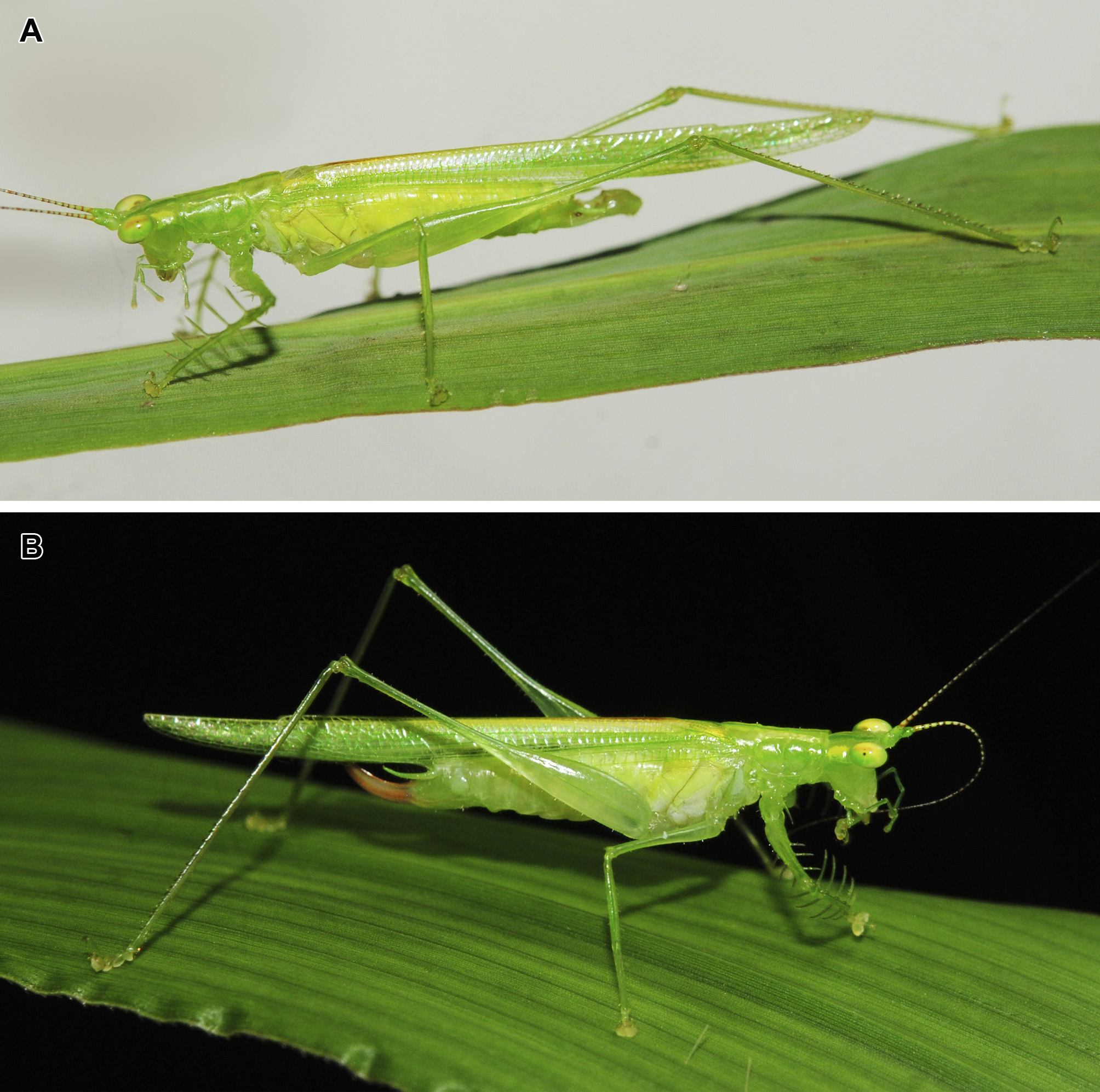
Coloration. Color described based on photos of living specimens ( Fig. 6 View FIGURE 6 A–B). Body light green with some dark-green areas. Antennae light green with the intersection between antennomeres brown spotted. Eyes anteriorly light green, medially yellow with an orange dorsal spot and posteriorly dark-green. Dorsal region of the gena with yellow spot behind the eyes. Pronotum dorsally with two yellow bands which are connected with the spots of the gena and follows parallel to the tegmen base. Tegmen when closed with yellow stripe on dorsal margin and anteriorly overlapped by a small brown stripe. Legs, cerci and subgenital plate light green.
Female. General. Morphology essentially equal to male ( Fig. 5 View FIGURE 5 A–K), except for the following characteristics:
Head. Head apex straight and without projections ( Fig. 5 View FIGURE 5 B). Compound eyes elliptical ( Fig. 5 View FIGURE 5 B–D). Frons, clypeus and labrum smooth, with medial height of the frons 1.5 times higher than medial height of the clypeus ( Fig. 5 View FIGURE 5 B).
Thorax. Pronotum dorsally straight, with posterior portion of pronotal disc slightly elevated in lateral view ( Fig. 5 View FIGURE 5 D). Dorsal carinae curved towards the lateral margins of pronotum in dorsal view ( Fig. 5 View FIGURE 5 C). Posterior portion of pronotal disc anteriorly bilobed and posteriorly concave in dorsal view ( Fig. 5 View FIGURE 5 C). Lateral lobes smooth, with ventral anteromedial margin concave and posteriorly straight in lateral view ( Fig. 5 View FIGURE 5 D). Maximum height of the lateral lobe less than 2 times than dorsal length of pronotum ( Fig. 5 View FIGURE 5 D). Mesobasisternum trapezoidal shaped, posteriorly with triangular concavity ( Fig. 5 View FIGURE 5 F). Metabasisternum diamond shaped, medially with small triangular concavity ( Fig. 5 View FIGURE 5 F).
Abdomen. Cerci slightly curved, conical, distally acuminate and with the apex reaching the medial portion of the ovipositor ( Fig. 5 View FIGURE 5 I–K). Subgenital plate triangular and lacking stiles ( Fig. 5 View FIGURE 5 J). Ovipositor slightly curved, with base expanded and progressively narrowing to distal region, ending in an acuminated apex ( Fig. 5 View FIGURE 5 K). Distal region of ovipositor with small teeth, larger teeth on ventral valve ( Fig. 5 View FIGURE 5 K).
Measurements (mm). TL: male 15.1–16 / female 17.7–18.1; TegL: male 20.8–19.8 / female 20.5–21.2; TegH: male 1.9–2.3 / female 2–2.1; WF: male 1.6–1.7 / female 1.9–2; PL: male 4.2–5 / female 3.7–3.9; FF: male 4.2–5.2 / female 3.8–4.3; FT: male 5.4–6 / female 5.5–5.6; MF: male 4.7–5.6 / female 4.8–5.5: MT: male 5,2–6.1 / female 5.7–5.9; HF: male 11.8–11.9 / female 12.2–12.7; HT: male 11.8–12.3 / female 12.4–12.6; Lplac: male 3.6–3.7 / female 1.4–1.6; LC: male 2.6–2.7 / female 2–2.3; OL: 4.6–4.8.
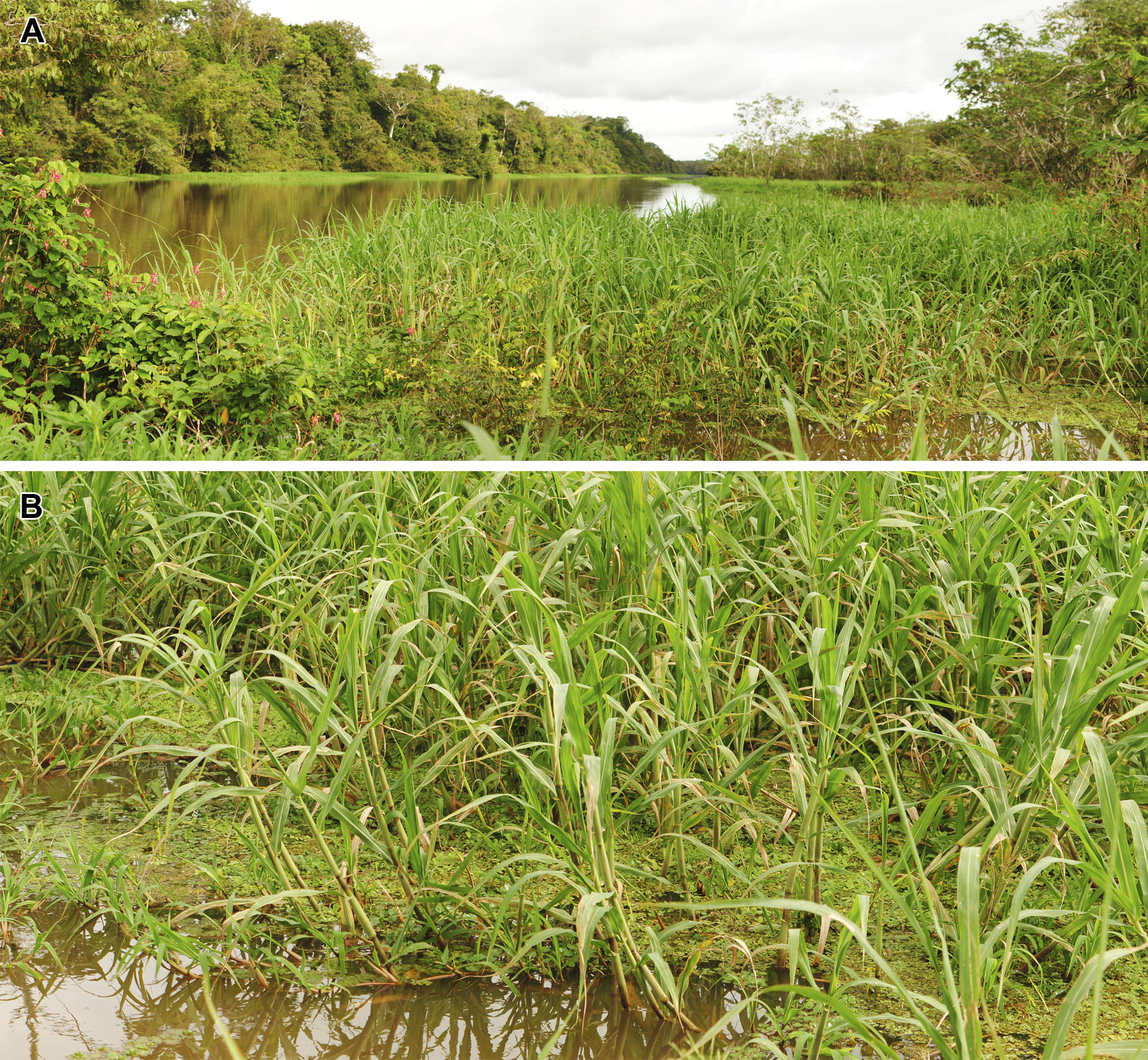
Natural history notes. The collecting was made in Amazonian floodplain forests, also known as várzeas, during flooding period, when the water of the rivers and tributaries spreads to the forest. In that habitat occurs great proliferation of various species of floating aquatic macrophytes, mainly some Poaceae (grasses) ( Fig. 7 View FIGURE 7 A). Specimens of P. gracila were found associated to Echinochloa polystachya (Poaceae) ( Fig. 7 View FIGURE 7 B).


No known copyright restrictions apply. See Agosti, D., Egloff, W., 2009. Taxonomic information exchange and copyright: the Plazi approach. BMC Research Notes 2009, 2:53 for further explanation.
|
Kingdom |
|
|
Phylum |
|
|
Class |
|
|
Order |
|
|
Family |
|
|
Genus |