
Pholcus lingguanensis Yao & Li
|
publication ID |
https://doi.org/ 10.11646/zootaxa.4170.1.1 |
|
publication LSID |
lsid:zoobank.org:pub:1BB4CDF3-C941-41CF-9987-CF9AFE0F71BD |
|
DOI |
https://doi.org/10.5281/zenodo.5328503 |
|
persistent identifier |
https://treatment.plazi.org/id/03F587F1-AF01-FFC9-0E94-F9C7FB710687 |
|
treatment provided by |
Plazi |
|
scientific name |
Pholcus lingguanensis Yao & Li |
| status |
sp. nov. |
Pholcus lingguanensis Yao & Li View in CoL sp. nov.
Figs 17–18 View FIGURE 17 View FIGURE 18
Type material. Holotype: male, Lingguan Cave (33°12.832′N, 112°11.377′E, elevation 336 m), Lingguandian Village , Erlong Town , Zhenping County, Nanyang, Henan, China, 22 May 2014, Y. Li and J. Liu leg GoogleMaps . Paratypes: 3 males 4 females, same data as holotype GoogleMaps .
Etymology. The specific name refers to the type locality; adjective.
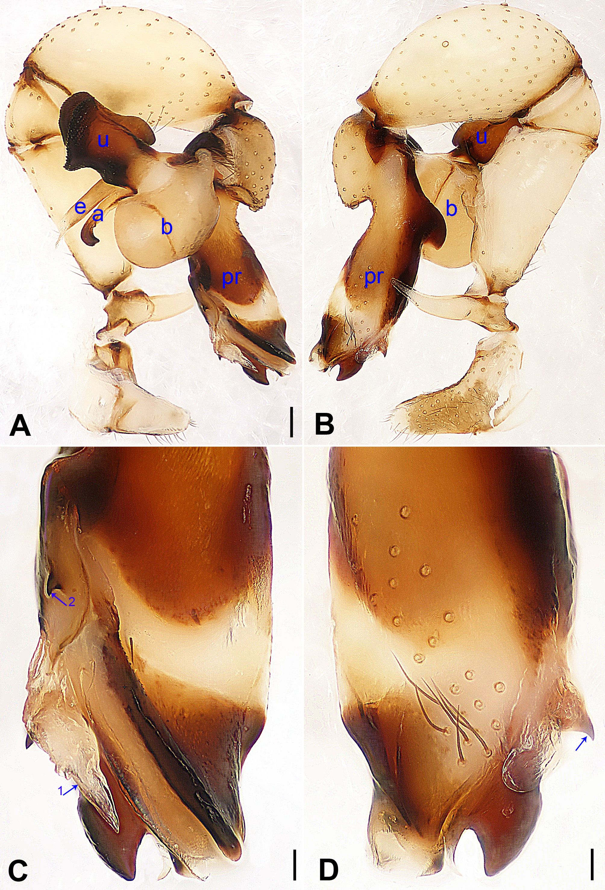
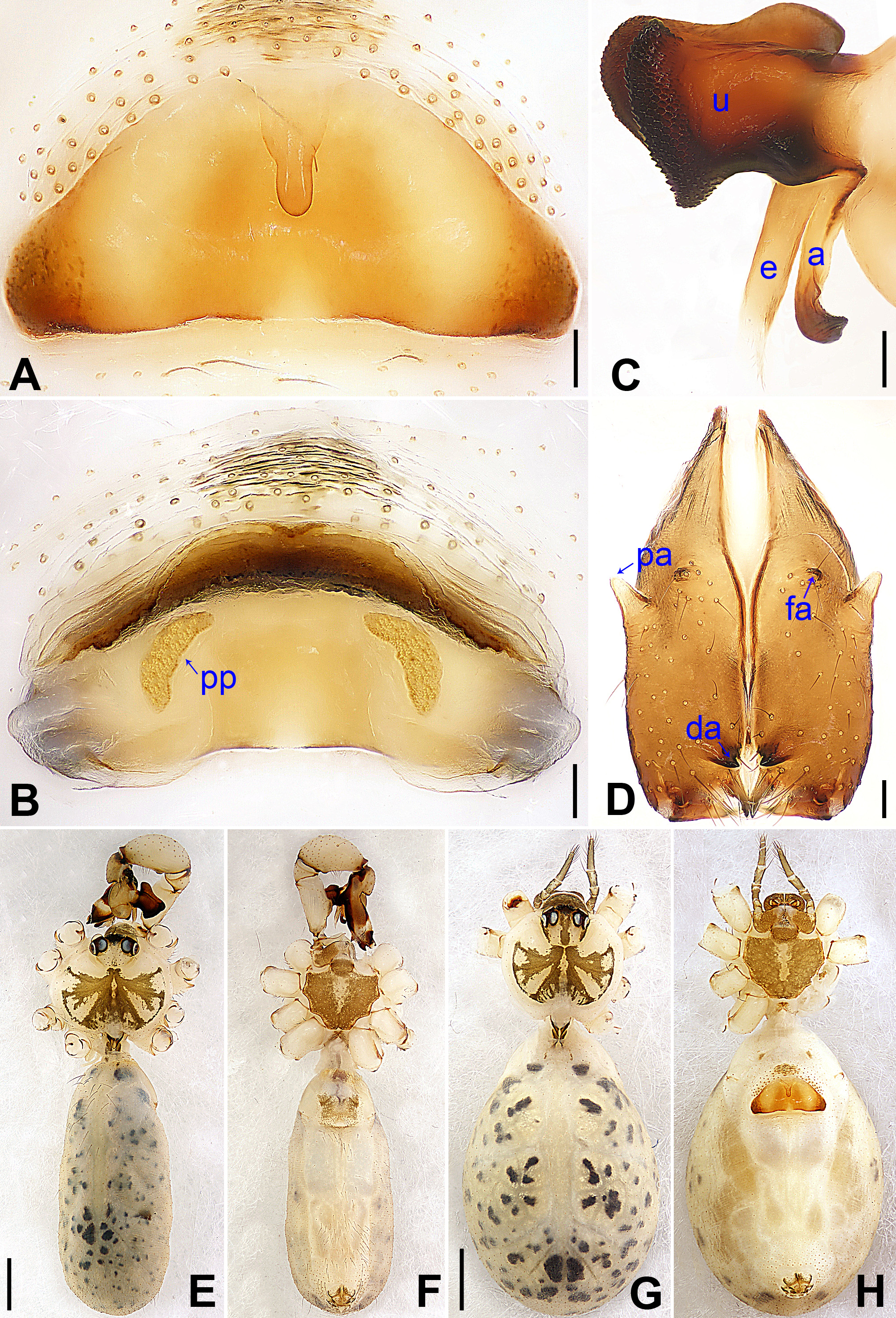
Diagnosis. This species resembles P. parayichengicus Zhang & Zhu, 2009 (see Zhang & Zhu 2009: 62, figs 33–34 and Yao & Li 2012: 29, figs 140–141) with similar male chelicerae ( Fig. 18 View FIGURE 18 D), bulbal apophyses ( Fig. 18 View FIGURE 18 C) and epigynum ( Fig. 18 View FIGURE 18 A) but can be distinguished by the absence of small retrolateral apophyses subdistally on the long male pedipalpal trochanteral apophyses ( Figs 17 View FIGURE 17 A–B), by the small, membranous prolatero-distal process (arrow 1 in Fig. 17 View FIGURE 17 C) and the very small, sclerotized ventro-subdistal apophysis on the procursus (arrow in Fig. 17 View FIGURE 17 D) and by the curved vulval pore plates ( Fig. 18 View FIGURE 18 B).
Description. Male (holotype): Total length 5.52 (5.96 with clypeus), carapace 1.56 long, 1.90 wide, opisthosoma 3.96 long, 1.78 wide. Leg I: 42.98 (11.15 + 0.80 + 10.64 + 18.01 + 2.38), leg II: 29.58 (8.27 + 0.76 + 7.48 + 11.67 + 1.40), leg III: 22.25 (6.41 + 0.70 + 5.32 + 8.72 + 1.10), leg IV: 28.96 (8.46 + 0.68 + 7.24 + 11.22 + 1.36); tibia I L/d: 63. Distance PME-PME 0.23; diameter PME 0.14; distance PME-ALE 0.06; distance AME- AME 0.07; diameter AME 0.11. Sternum wider than long (1.25/0.91). Habitus as in Figs 18 View FIGURE 18 E–F. Carapace yellowish, with brown radiating marks extending to ocular area and brown bands marginally; ocular area yellowish; clypeus brown; sternum brown with yellowish marks medially. Legs yellowish, femora (subproximally and subdistally) and tibiae (subproximally and subdistally) with slightly darker rings. Opisthosoma yellowish, with small spots dorsally and laterally. Ocular area elevated, without eye stalks (as in P. gonggarensis sp. nov., cf. Fig. 27 View FIGURE 27 C). Thoracic furrow absent. Chelicerae as in Fig. 18 View FIGURE 18 D, with a pair of proximo-lateral apophyses, a pair of black distal apophyses, and a pair of frontal apophyses. Pedipalps as in Figs 17 View FIGURE 17 A–B; trochanter with a ventral apophysis; femur with a ventral ridge; tibia with a projection prolaterally; procursus simple proximally but complex distally, with a spine-like prolatero-ventral apophysis medially (arrow 2 in Fig. 17 View FIGURE 17 C), a small, membranous prolatero-distal process (arrow 1 in Fig. 17 View FIGURE 17 C) and a small, sclerotized ventro-subdistal apophysis (arrow in Fig. 17 View FIGURE 17 D); uncus with a scaly edge; appendix hooked, with a small branch subdistally; embolus weakly sclerotized. Retrolateral trichobothrium of tibia I at 8%; legs with short vertical setae on tibiae, metatarsi and tarsi; without spines and curved setae; tarsus I with 23 distinct pseudosegments.
Female: Similar to male, habitus as in Figs 18 View FIGURE 18 G–H. Total length 5.13 (5.51 with clypeus), carapace 1.41 long, 1.60 wide, opisthosoma 3.72 long, 2.64 wide; tibia I: 8.01; tibia I L/d: 57. Distance PME-PME 0.21; diameter PME 0.11; distance PME-ALE 0.06; distance AME-AME 0.06; diameter AME 0.09. Sternum wider than long (1.06/ 0.78). Ocular area with brown marks; Epigynum ( Fig. 18 View FIGURE 18 A) with a knob. Vulva ( Fig. 18 View FIGURE 18 B) with a sclerotized anterior arch and two curved pore plates.
Distribution. China (Hena, type locality; Fig. 28 View FIGURE 28 ).
Natural History. The species was found on the wall in the entrance zone of the cave.


No known copyright restrictions apply. See Agosti, D., Egloff, W., 2009. Taxonomic information exchange and copyright: the Plazi approach. BMC Research Notes 2009, 2:53 for further explanation.