
Phyllodesmium jakobsenae
|
publication ID |
https://doi.org/ 10.5281/zenodo.157307 |
|
publication LSID |
lsid:zoobank.org:pub:A0D72C03-B8AA-4521-893B-21569DA08F23 |
|
DOI |
https://doi.org/10.5281/zenodo.5622830 |
|
persistent identifier |
https://treatment.plazi.org/id/24644335-527B-FF89-0269-FD9D3972FD52 |
|
treatment provided by |
Plazi |
|
scientific name |
Phyllodesmium jakobsenae |
| status |
|
Phyllodesmium jakobsenae nov. sp.
Figures 1–5 View FIGURE 1 View FIGURE 2 View FIGURE 3 View FIGURE 4
Repository
ZSM ñ Zoologische Staatssammlung München, Germany (Zoological State Collection Munich):
Holotype: ZSM Moll 20040189, Paratype: ZSM Moll 20040190
Localities
Pulau Bunaken (Bunaken Island, Bunaken Islands National Park), North Sulawesi ( Indonesia). Seven specimens (including the holotype) found in the lagoon in front of “Papa Boa Bungalows” on the southern side of Bunaken Island (07/12/2003; Western end of Liang Beach, GPS: 01°37'3.1''N, 124°45'51.1''E; 0.3–0.5 m depth). One specimen found in front of the South West cape of Bunaken Island (07/20/2003; 0.5 m depth).
Ethymology
This species is dedicated to Mrs. Wera Jakobsen, a passionate diver, who supported alphataxonomy of marine slugs by a donation to BIOPAT (Patrons for Biodiversity).
Description
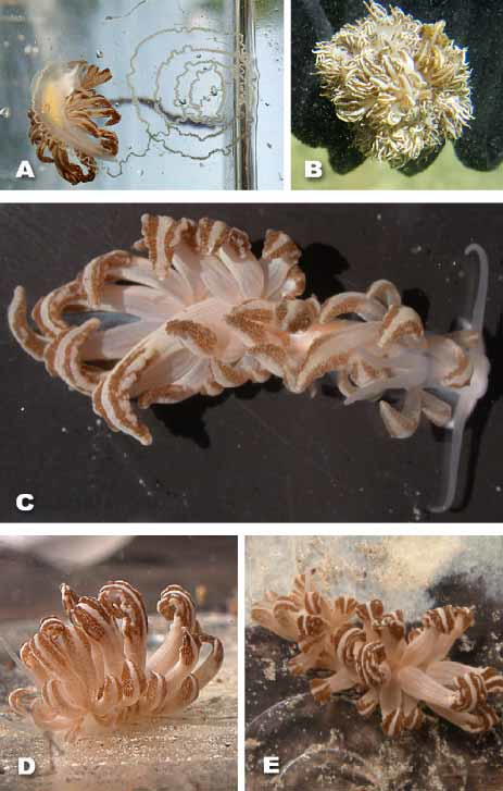
Color and external morphology of living animal: Body of living animals, including oral tentacles, rhinophores and foot translucent white ( Fig. 1 View FIGURE 1 A –E). Gonads shining through translucent epidermis in pale yellow ( Fig. 1 View FIGURE 1 A), buccal bulb in light orange.
Animals elongate, up to approximately 30 mm in length (without cerata), with a few short and several very long cerata (longer than rhinophores and oral tentacles), covering whole notum ( Fig. 1 View FIGURE 1 C–E). Smooth oral tentacles slightly longer than rhinophores, usually directed laterally ( Fig. 1 View FIGURE 1 C). Rhinophores similar in shape and surface texture to oral tentacles, standing close together. Both rhinophores and oral tentacles tapering. Anterior foot angular, slightly extended and with some white pigmentation mainly along anterior edge. Posterior end of foot tapering.
Cerata (25–35) arranged in clusters of up to 7 on each pad. Compared to more dorsal ones, lateral cerata in most specimens very small and short. Posterior cerata appearing longer than more anterior ones ( Fig. 1 View FIGURE 1 C). Upper third of elongate, dorsoventrally flattened ceras spatulate or rather arrowheadshaped. In middle of broadened part white longitudinal stripe present, extending from base of ceras to tip and running parallel to main digestive glandular duct. Lateral of this central whitish stripe one broad brownish longitudinal stripe on each side ( Fig. 1 View FIGURE 1 C–E), composed of very small singular brownish dots, probably representing clusters of zooxanthellae within digestive gland. In some specimens brownish stripes riddled with nodulose white markings (dots or longitudinal lines). Edge of ceras appearing whitish and slightly nodulose, tips blunt and rounded. Cerata usually partly curled distally. Basal whitish part of ceras more or less circular in crosssection, smooth with distinct raised ridge on ventral midline of proximal third of each ceras. Ridge continuing into white line of upper part of ceras. Basal end of ceras appearing clubbed, tapering slightly more distally before broadening abruptly into arrowheadshaped apical part. Thin longitudinal, creamy colored lines on this part of ceras.
Description of preserved animals
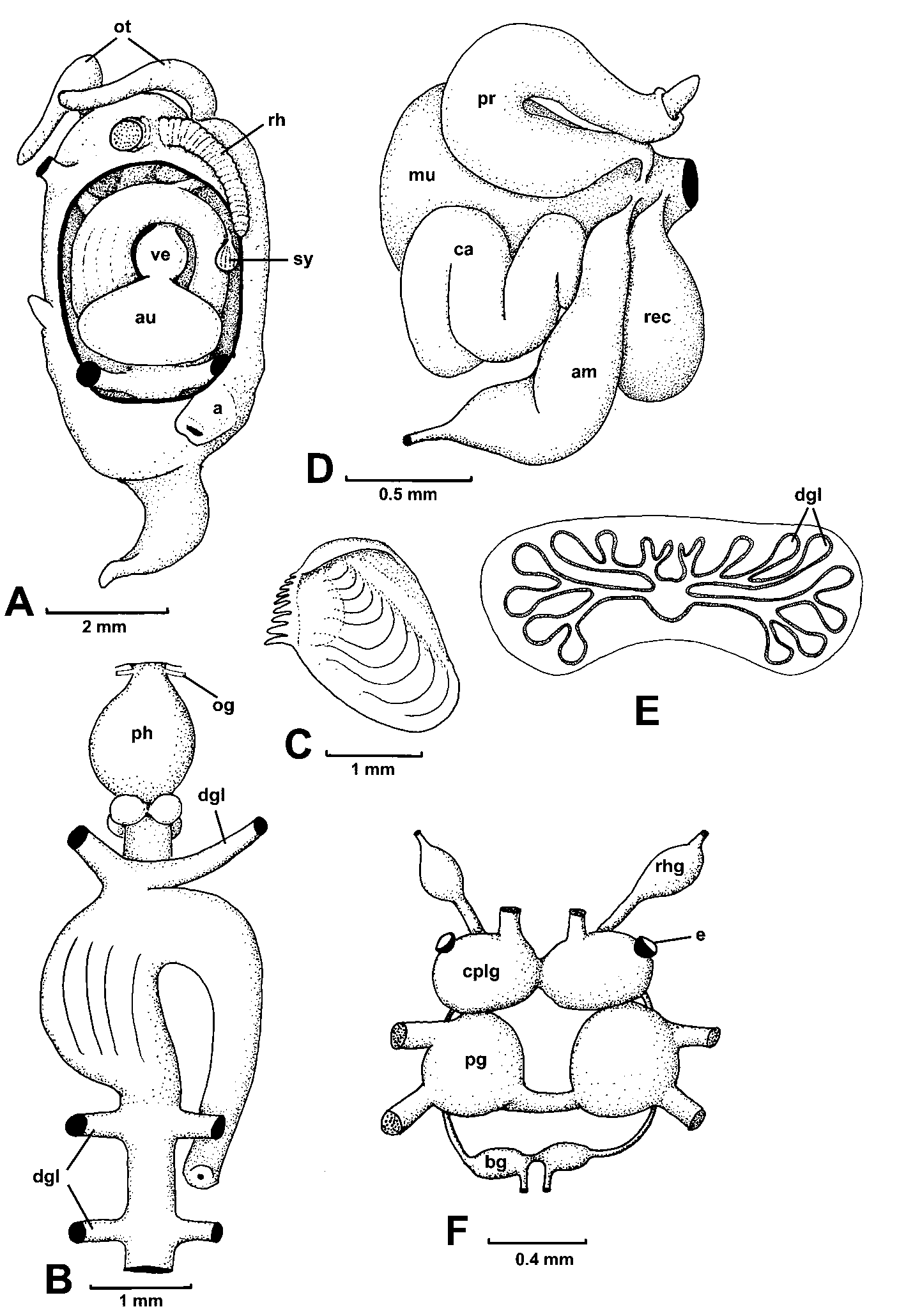
External morphology: Due to starving condition, many animals have shrunken considerably. Nearly all cerata repelled, only small ones still attached to body. No distinct notal rim present. Foot anteriorly without propodial tentacles, posterior part pointed. Oral tentacles wrinkled, slightly longer than rhinophores, with tiny groove on dorsal side. Rhinophores heavily wrinkled, giving the appearance of being lamellate, and standing close to each other ( Fig. 2 View FIGURE 2 A). Larger cerata of preserved animals nearly as long as body. Insertion of cerata difficult to see in preserved specimens, probably arranged in arches, but lying on distinct pads. Four pads on each side, lying opposite to each other. First cerata lateral of rhinophores. Anal papilla dorsally on right side behind second pad ( Fig. 2 View FIGURE 2 A). Genital opening below first cerata pad. Whole epidermis composed of specialized vacuolated cells ( Figs. 4 View FIGURE 4 C–E).
Central nervous system: Central nervous system located behind pharynx ( Fig. 2 View FIGURE 2 B). Cerebral and pleural ganglia completely fused ( Fig. 2 View FIGURE 2 F). Buccal ganglia lying beneath oesophagus at junction with pharynx. Large rhinophoral ganglion at base of each rhinophore, connected to cerebral ganglion by short connective. Eyes situated directly at cerebral ganglia ( Fig. 2 View FIGURE 2 F), of usual nudibranch arrangement with pigment and lense ( Fig. 4 View FIGURE 4 F). Statocysts lying between cerebropleural and pedal ganglia. Only one statolith present ( Fig. 4 View FIGURE 4 F)
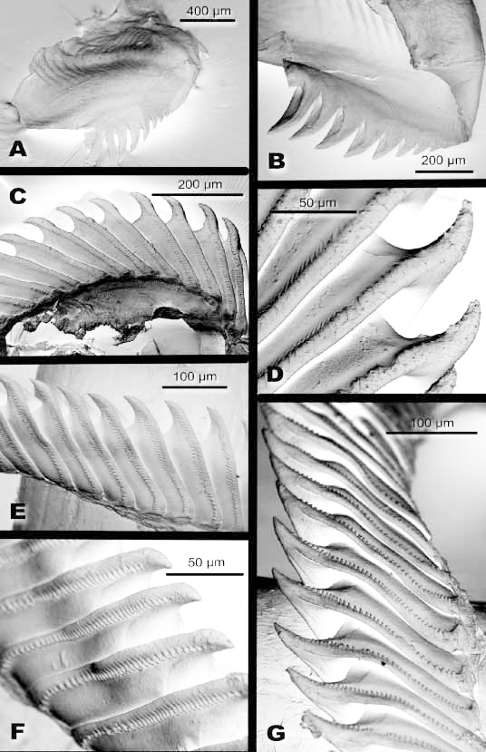
Digestive system: Oral tube short, leading into bulbous pharynx ( Fig. 2 View FIGURE 2 B). Huge oral glands present ( Fig. 4 View FIGURE 4 G), forming many lobes around pharynx. Glandular tissue composed of cells with blue to violet staining contents, indicating acid and neutral mucopolysaccharides. Paired outleading ducts from each side uniting beneath pharynx and opening into very short oral tube. Labial cuticle without any armature. Two jaw plates oval shaped ( Figs. 2 View FIGURE 2 C, 3A). Along cutting edge of jaw plates up to 9 somewhat hollowed and clogshaped denticles present, getting progressively longer in size ( Figs. 3 View FIGURE 3 A, B). Longest denticles about 300µm in length. Radular formula of two specimens 39 x 0.1.0 (No.3) and 40 x 0.1.0. (No.5). Each rhachidian tooth with large (50µm long) median pointed cusp ( Figs. 3 View FIGURE 3 C–G), clawshaped in lateral view ( Figs. 3 View FIGURE 3 C, D). Margin of rhachidians with about 38 to 43 relatively short (5 to 10µm long) denticles ( Figs. 3 View FIGURE 3 E–G). In anterior part of radula of specimen No. 5 denticles flattened, showing attrition ( Figs. 3 View FIGURE 3 C, D). Salivary glands could not be found either by dissection or in histological slides. Oesophagus short, entering stomach on ventral side ( Fig. 2 View FIGURE 2 B). Interior of oesophagus completely covered by specialized vacuolated cells. Stomach and posterior part of digestive gland not separated, characterized by many longitudinal folds. Two anterior digestive glandular branches starting above entrance of oesophagus and leading to first cerata pads on both sides. Right duct orange to brown and therefore differing in color compared to all other main digestive glandular ducts, which are translucent to whitish. One main duct leading to posterior part of body with opposite lying ducts leading into posterior cerata pads. Intestine starting next to oesophagus and anterior digestive glandular branches, leading on the right side to anus ( Fig. 2 View FIGURE 2 B). No typhlosolis in intestine observed in dissected animals, but small one present in histological slides.
Digestive gland in cerata: One central narrow digestive gland duct extending through each ceras. Especially in apical, broadened part, central duct ramifying in primary and secondary branches, the latter radiating towards ceratal wall ( Figs. 2 View FIGURE 2 E, 4B). Each secondary branch terminating in saclike structure. On side of flattened part of ceras not exposed to light, sacs concentrated only at margin of each ceras (lower area in Fig. 2 View FIGURE 2 E, Fig. 4 View FIGURE 4 B). A stripe free of these sacs parallel to central duct, with white pigmentation. Light orientated side of each ceras ( Fig. 2 View FIGURE 2 E upper part, Fig. 4 View FIGURE 4 A) densely packed with sacs directly beneath ceratal wall. Central stripe with secondary branchings not ending in sacs but in thickened terminations also present, but much smaller. Zooxanthellae present in digestive glandular cells of these sacs, in lumen of sacs, and in epithelium of branches ( Figs. 4 View FIGURE 4 C, D). Cnidosac of these large cerata with cells containing one large vacuole. No contents observed in these cells. Distinct muscular layer around cnidosac present.
Small cerata, which did not autotomize, showing different morphology and histology: central digestive glandular branch not ramifying, occupied by many glandular cells. Cnidosac cells filled with tiny nematocyst like structures, arranged around central lumen. Beneath ceras epithelium small glandular cells (“cellules spéciales” see Edmunds 1966: 34) present ( Fig. 4 View FIGURE 4 E), lacking in the larger cerata. Epithelial glandular mucous cells present in smaller cerata ( Fig.4 View FIGURE 4 E).
Excretory system: Syrinx large, inside highly folded, opening ventrally into pericard on anterior right side ( Fig. 2 View FIGURE 2 A).
Circulatory system: Ventricle mediodorsal between first and second cerata pad. Atrium lying behind ventricle, both arranged in longitudinal direction ( Fig. 2 View FIGURE 2 A).
Reproductive system: Gonad follicles not arranged into lobes but forming rather uniform layer lying beneath digestive gland. Gonad reaching into anterior third of ventral visceral cavity. Male and female follicles separate. Distal genital system occupying whole anterior part of visceral cavity. Gonoduct, with sausageshaped ampulla, dividing into vas deferens and oviduct next to atrium, separation lying inside of nidamental glands ( Fig. 2 View FIGURE 2 D). Vas deferens soon widening into sausageshaped prostate, forming one large coil, ending in one small papilliform penis. Receptaculum seminis slightly enlongate, entering atrium next to oviduct opening.
Biological notes
All specimens were found nestled into coral colonies of an unknown species of Xenia (Ehrenberg, 1831; Xeniidae, Alcyonaria, Octocorallia ). The bodies of the slugs were not visible, only the cerata were slightly surpassing the coral’s tentacles. The animals are very well camouflaged, the cerata mimicking parts of the tentacles of Xenia in color and shape. One to three specimens were found in each Xenia colony. Figure 1 View FIGURE 1 B shows one Xenia colony with two slugs sitting in the middle. Part of the colony is preserved in formalin/ seawater and kept together with the holotype.
The locality in front of “Papa Boa Bungalows” is a shallow lagoon fringed by mangroves. All specimens were found in the intertidal zone between 0.3 and 0.5 m depth. The substrate was a mixture of sand, mud, coral rubble and some living hard corals. On the coral rubble there were single colonies of Xenia . Most of the sandy patches around the Xenia colonies were covered with seagrass. Water temperature at locality was 31°C on average (July 2003).
The second locality close the South West cape of Bunaken Island is a coral reef with sandy patches in between. On these sandy patches the slugs were also sitting in Xenia attached to coral rubble.
With the exception of the holotype, all specimens were immediately put into aquaria in order to perform longterm starvation experiments. All specimens shrunk considerably after some days and autotomized most of their cerata. Especially the arrowheadshaped and broadened apical parts of the remaining cerata shrunk after some days. Observations in the aquarium showed an interesting diurnal behaviour: During daytime, the specimens mainly remained inactive. The long narrowed base of the cerata was often extremely contracted and shortened in most of the specimens. Additionally the apical, arrowheadshaped part of the cerata was extremely broadened. During nighttime the slugs became more active and moved through the aquarium. The bases of the cerata were prolonged again and the upper part of the cerata was less broad than before. The cerata easily autotomized when the animals were disturbed. Detached cerata exuded a sticky secretion and moved for some minutes. This secretion may have come from the few mucous cells found in the epidermis and probably not from the “cellules spéciales” ( Edmunds 1966: 34) since they have not been observed in the large cerata.
Some specimens laid spawn masses after one or two days of capture. The spawn mass is a whitish ribbonlike cord, with egg capsules arranged in a line. It is attached to the substrate in form of a spiral (3 to 4 cm in diameter) ( Fig. 1 View FIGURE 1 A). After 5 to 6 days kept in the aquarium (average temperature 29–31°C) the free swimming veliger larvae hatched.
Measurements of photosynthetic activity (PAM)
Figure 5 A shows the yield values of Phyllodesmium jakobsenae , P. crypticum Rudman, 1981 and the octocoral Xenia sp. plotted versus the irradiance. Both Phyllodesmium species were feeding on probably the same species of Xenia but in different localities: P.
jakobsenae was found close to Bunaken Island, whereas P. crypticum was found in Lembeh Strait, North Sulawesi. The yield values of both Phyllodesmium species decrease exponentially with increase of irradiance, and the values of P. crypticum are nearly identical to the ones of P. jakobsenae . In lower irradiances the values of Xenia sp. are only slightly higher than those of the slugs, but around 500 µmol and higher, values for the soft coral are significantly higher. In Figure 5 B, the yield values of one specimen of P. jakobsenae (No.8) are plotted versus the number of cultivation days under starving conditions in the aquarium. The yield values stay on a high level between 0.5 and 0.7 for the whole ten days of the experiments. In Figure 5 C, the ground fluorescence (F0) values of one specimen of P. jakobsenae (No.1) are plotted versus the number of cultivation days under starving conditions in the aquarium. The F0 values decrease quickly from an average of 350 in the beginning to approximately 100 after 8 days.

No known copyright restrictions apply. See Agosti, D., Egloff, W., 2009. Taxonomic information exchange and copyright: the Plazi approach. BMC Research Notes 2009, 2:53 for further explanation.