
Physocypria bullata Vávra, 1897
|
publication ID |
https://doi.org/ 10.11646/zootaxa.2820.1.1 |
|
DOI |
https://doi.org/10.5281/zenodo.5294274 |
|
persistent identifier |
https://treatment.plazi.org/id/03EB87C8-6F47-FFEA-FF30-FA7DA7637725 |
|
treatment provided by |
Felipe |
|
scientific name |
Physocypria bullata Vávra, 1897 |
| status |
|
Physocypria bullata Vávra, 1897 View in CoL
( Figures 25–30A, B View FIGURE 25 View FIGURE 26 View FIGURE 27 View FIGURE 28 View FIGURE 29 View FIGURE 30 )
1897 Physocypria bullata Vávra : 7, Figs 1–5 View FIGURE 1 View FIGURE 2 View FIGURE 3 View FIGURE 4 View FIGURE 5
1944 Physocypria bullata Vávra —Klie: 11, Fig. 2 View FIGURE 2 .
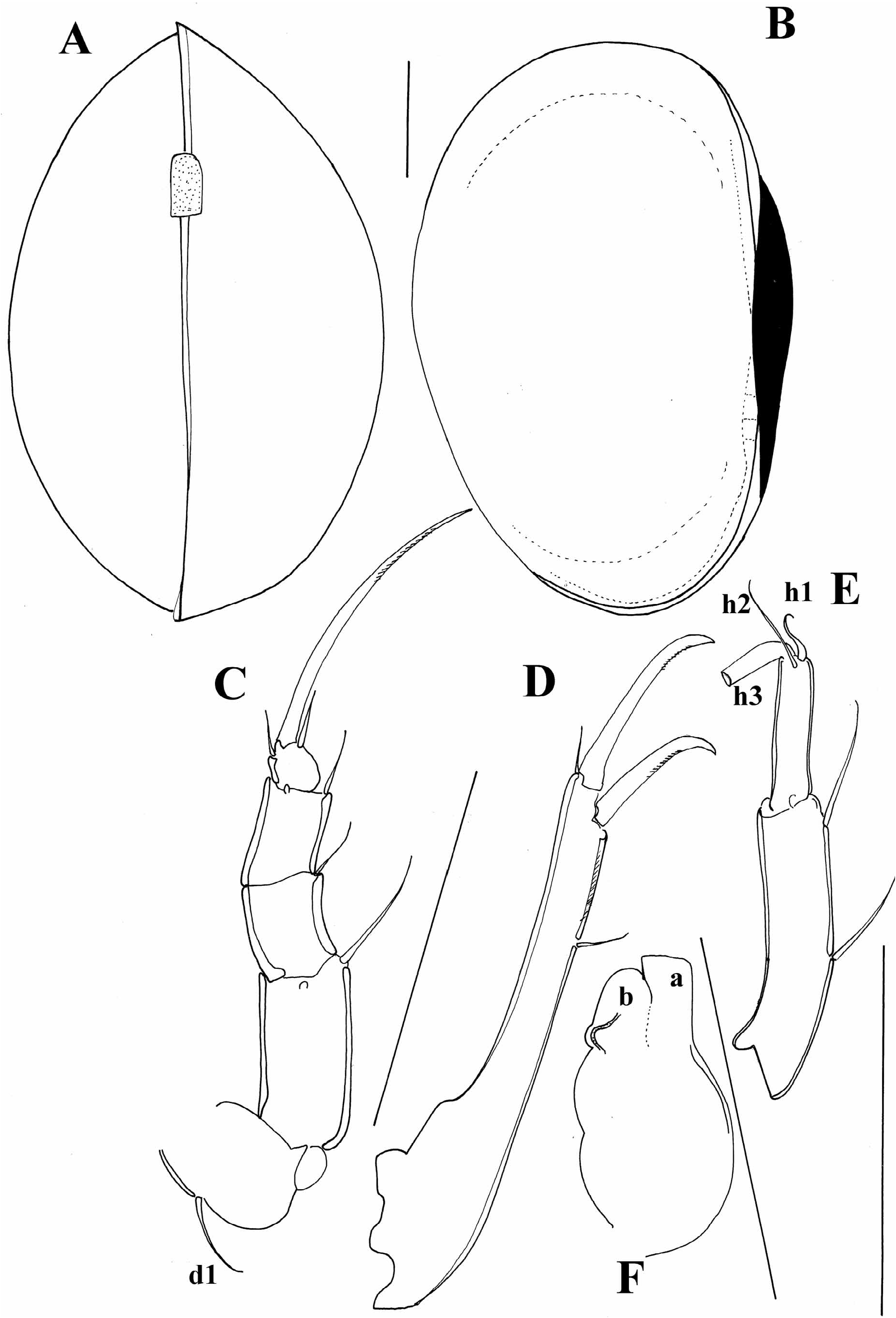
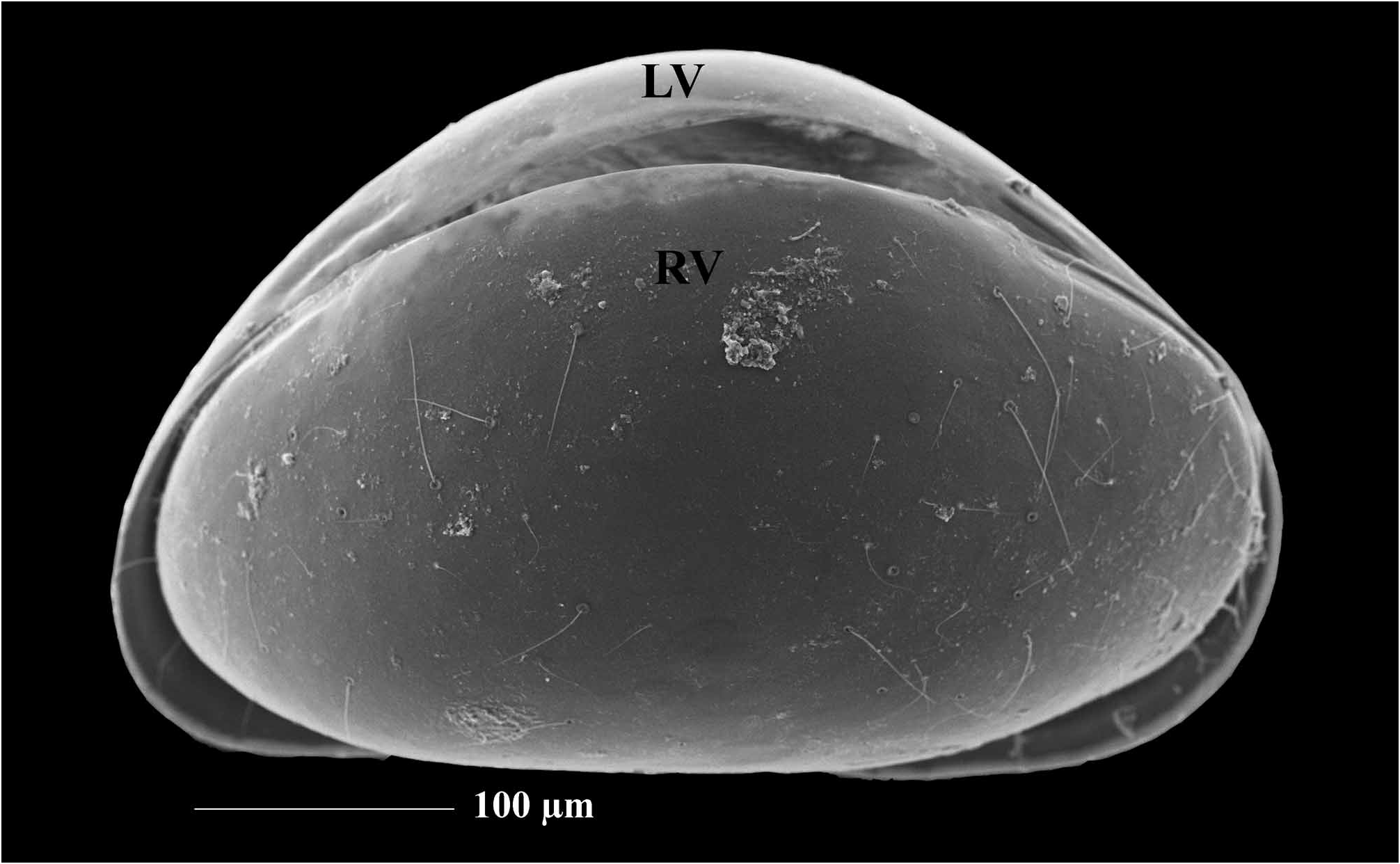
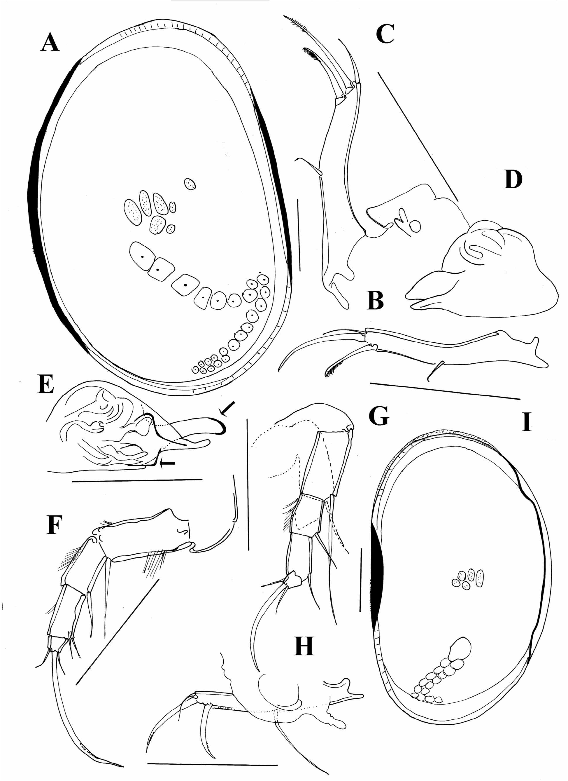
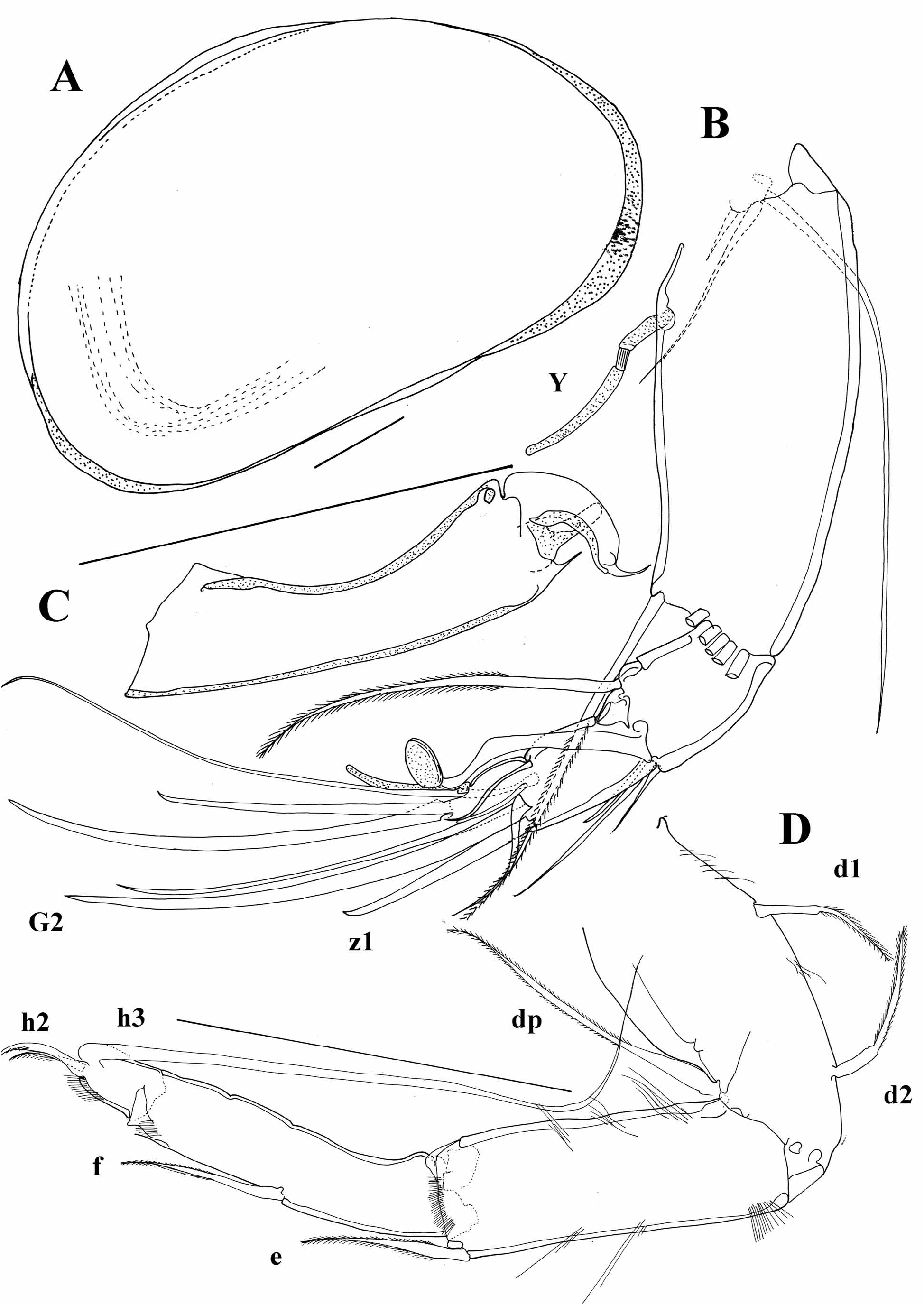
Redescription. Female: Carapace ovoid in lateral view ( Figure 26A View FIGURE 26 ), L = 0.4 mm. Dorsal margin rounded on RV and flat on LV. RV overlapping LV dorsally with flange. Greatest H of RV lying around mid L and equaling 68% of L, on LV 63%. Both anterior and posterior margins rounded, but posterior being narrower than anterior one. Marginal pore canals short, straight and denser anteriorly than posteriorly. Selvage peripheral anteriorly, while posteriorly inwardly displaced. Ventral margin convex. LV overlapping RV anteriorly, posteriorly and ventrally. Both LV and RV with marginal tubercles along free margin.
A1 ( Figure 29B, D View FIGURE 29 ): 7-segmented. First segment with two posterior and two anterior setae. Both posterior setae normally developed. Second segment with one anterior seta and a clearly visible Rome organ posteriorly. Third segment with one posterior and one anterior seta. Fourth and fifth segments with two short posterior and two long anterior setae. Penultimate segment with total of five distal setae, dorsal seta (alpha) claw like, other setae being very long. Terminal segment with four setae, most posterior one being transformed into a claw, and most anterior one being transformed into aesthetasc ya, which is twice as long as terminal segment.
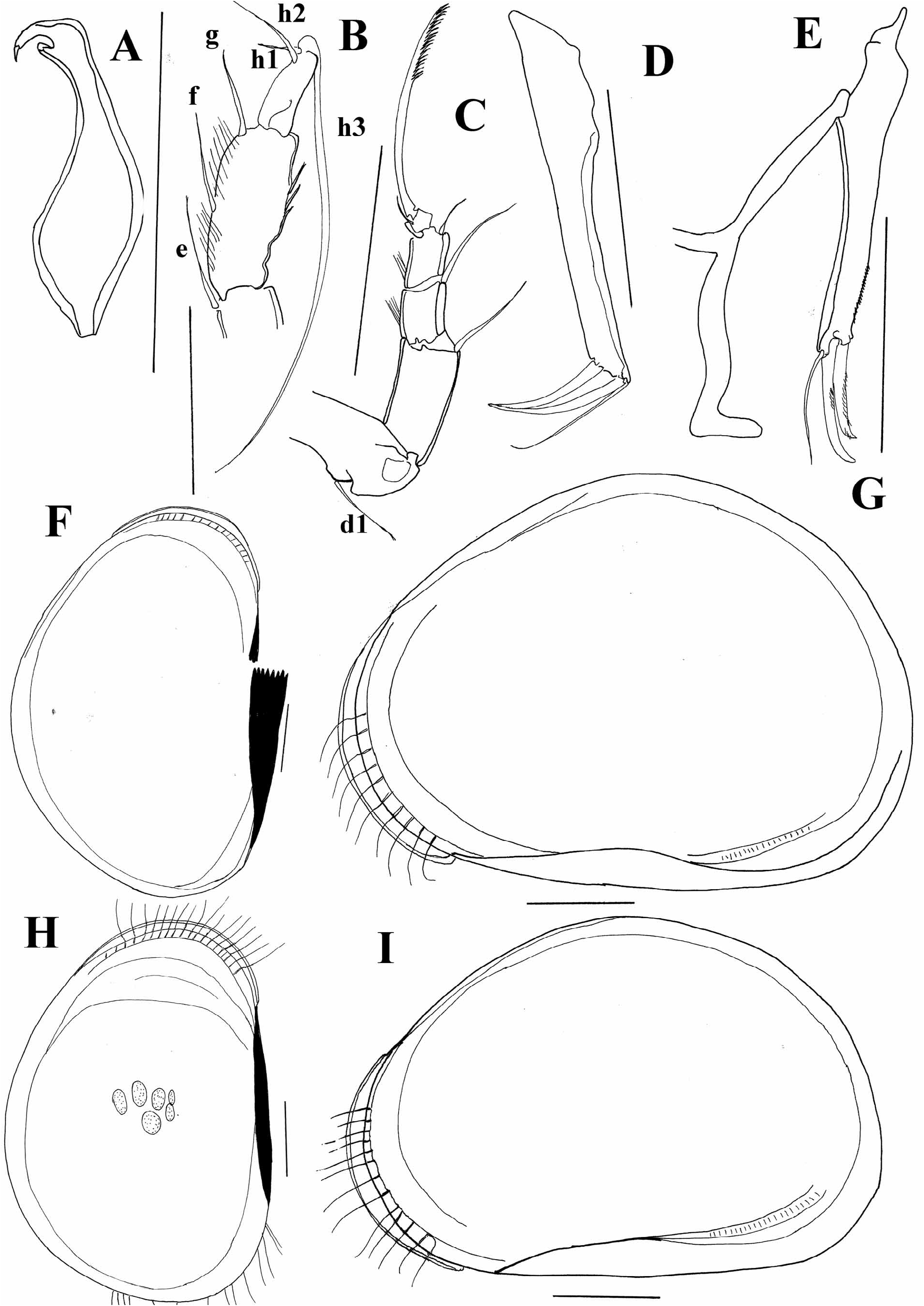
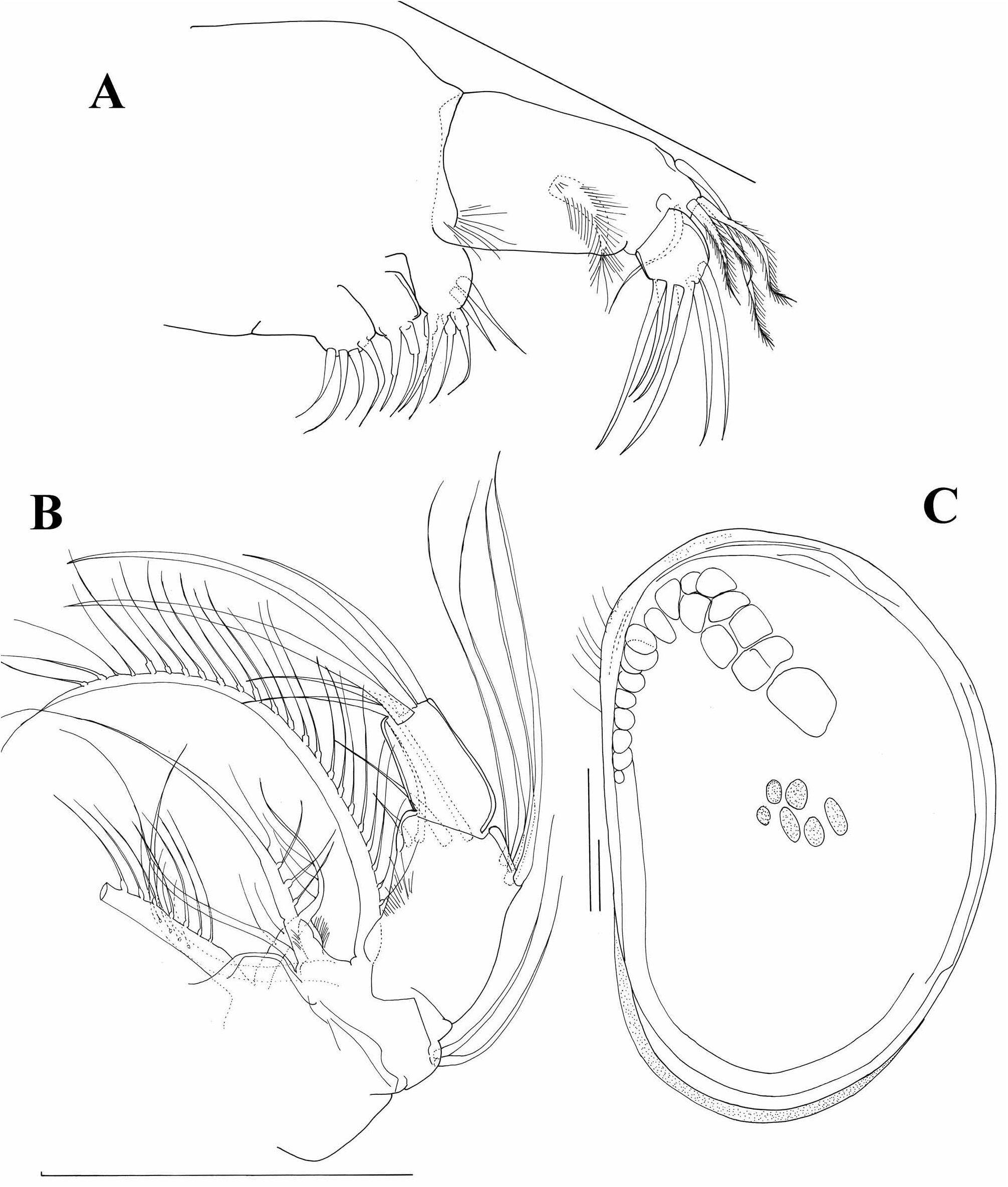
A2 ( Figure 26D View FIGURE 26 ): Exopod consisting of plate, one long and two short setae. First endopodal segment with one long, plumose, ventral seta, and five long swimming setae, and one short (most external one). Penultimate segment with two external setae and four internal “t” setae. Setae z1 transformed into a short claw, slightly longer than terminal segment, other z setae reaching 1/3 of terminal claws. Claws G1 and G3 subequally long. Claw G2 very half L of G1. GM claw reaching 2/3 of G1 and Gm being half L of GM.
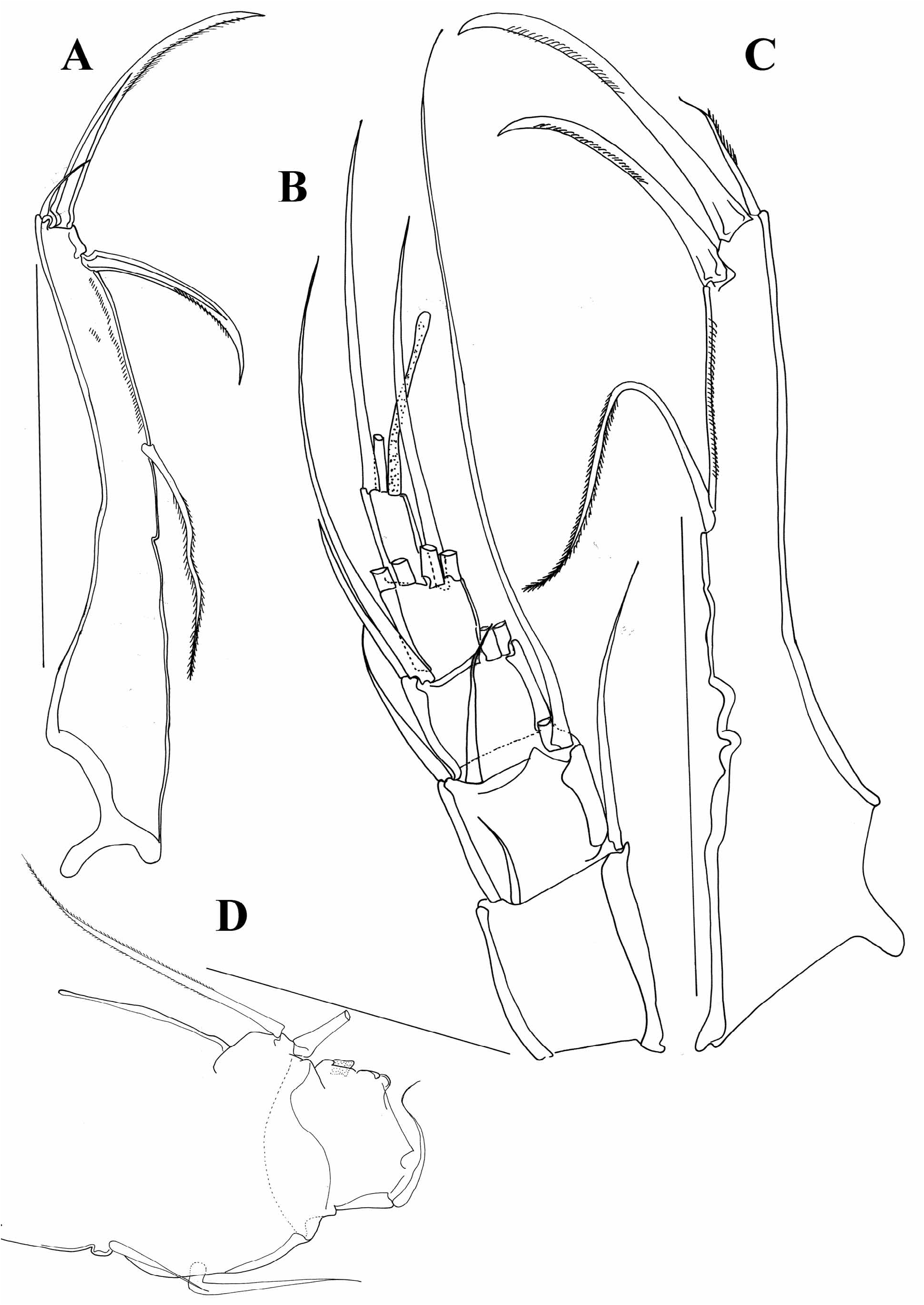
Md ( Figure 30B View FIGURE 30 ): First segment on the palp with two setae. Second segment externally with two setae, while internally with three long (plumose) setae, of which one clearly longer than other two; one smooth seta (as long as shorter plumose setae) and one thick, short plumose (beta seta). Penultimate segment with four setae extero-laterally, two setae distally, and two setae intero-distally. Terminal segment with three strong claws and two setae. None of the claws fused with segment. Terminal segment only two times longer than wide.
Mxl ( Fig 30A View FIGURE 30 ): Palp positioned quite distant from endites. First segment of palp wide with five setae anteriorly and one thick seta posteriorly. Terminal segment with four claws and two setae. Terminal segment square shaped.
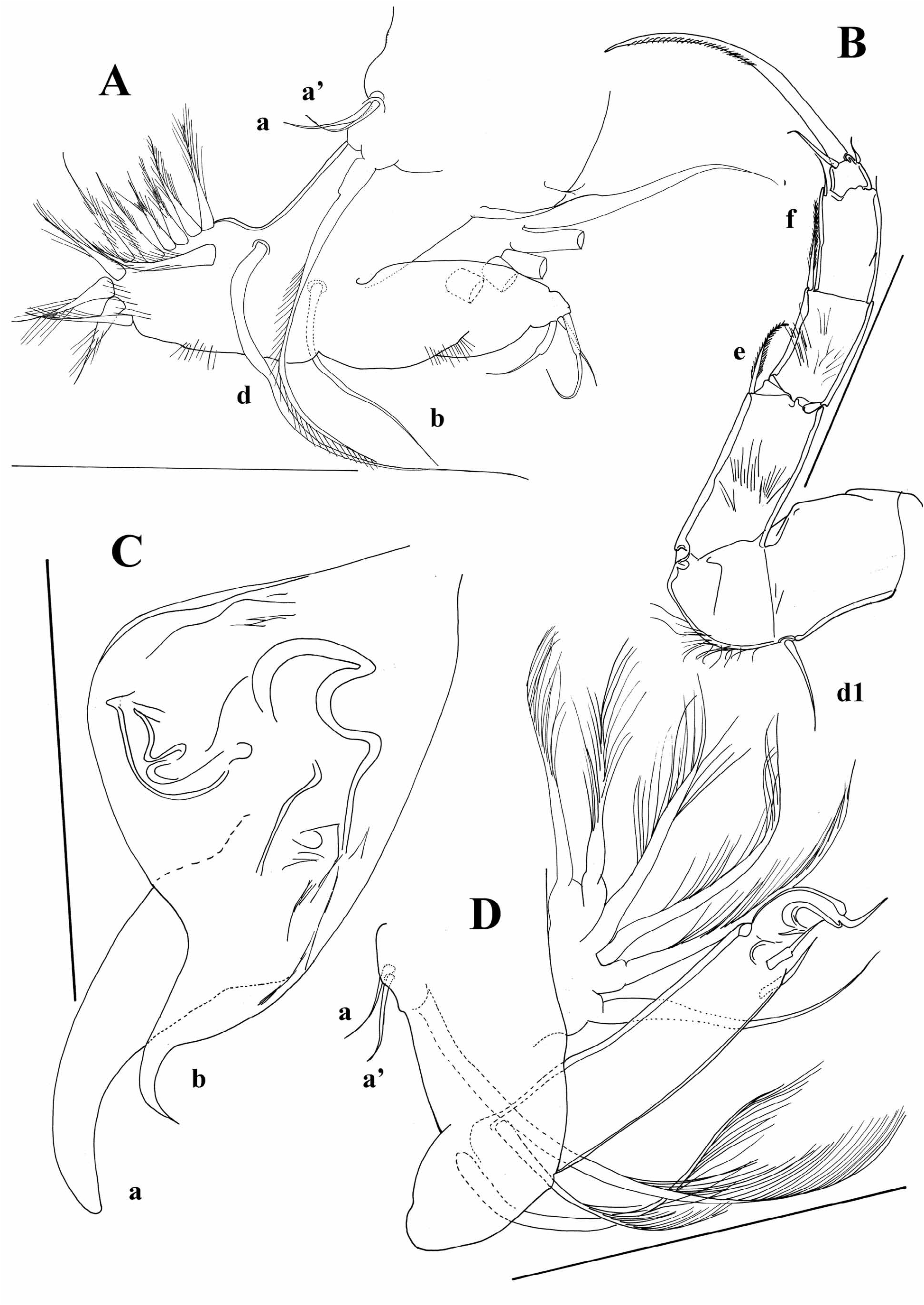
T1 ( Figure 28A View FIGURE 28 ): Two short “a” setae and one long (plumose) seta on protopod. Exopod with 5 or 6 rays. Terminal setae on endopod very short and subequally long.
T2 ( Figure 28B View FIGURE 28 ): Basal seta present. Seta “e” reaching middle of following segment, seta “f” reaching distal end of penultimate segment, while seta “g” very short. Seta h3 on terminal segment short. Terminal claw as long as three distal segments combined. T2 covered with long and dense pseudochaetae, setae “e” and “f” plumose.
T3 ( Fig 27D View FIGURE 27 ): Basal segment with d2 seta. Setae “e” and “f” well developed, while “g” seta tiny. Terminal segment with setae h1 and h2 equally long ( Figure 25C View FIGURE 25 ).
UR ( Figure 29C View FIGURE 29 ): L ratios between anterior margin, anterior, and posterior claw equaling 2.7: 1.3: 1. Posterior seta very long and inserted quite low on posterior margin, posterior margin with short setae.
Male: Carapace same as female ( Figures. 25A, B View FIGURE 25 , 26C, D View FIGURE 26 ) except for the variability of some specimens not having such a well developed flange on RV ( Figure 27A View FIGURE 27 ).
A2 ( Figure 27B View FIGURE 27 ): Setae z1 and z2 transformed into claws, former one being shorter than latter, seta z3 normally developed. G2 long, while G1 and G3 reduced. Setae t2 and t3 transformed into sexual bristles and penultimate segment subdivided.
Prehensile palps ( Figures 25D View FIGURE 25 , 27C View FIGURE 27 , 28D View FIGURE 28 ): Asymmetrical, right palp having a stronger finger and one terminal seta ( Figure 27C View FIGURE 27 ). Both palps with very long trunks compared to fingers.
Hemipenis ( Figure 28C View FIGURE 28 ): Two lobes present, both curved towards inner side, lobe “b” especially curved and with very thick point.
UR ( Figure 29A View FIGURE 29 ): Same as in female.
Remarks and affinities. Physocypria bullata is very closely related to P. lenticularis ( Müller, 1898) and P. denticulata ( Daday, 1910) , all three species having a similar carapace appearance, i.e. RV dorsally overlapping LV with a flange. The main difference between the three species is the appearance of the hemipenis. In P. lenticularis both lobes are evenly rounded while in P. denticulata lobe “b” is much wider and lower in comparison with the lobe “a”. All other representatives of the genus have more or less symmetrical carapaces.


No known copyright restrictions apply. See Agosti, D., Egloff, W., 2009. Taxonomic information exchange and copyright: the Plazi approach. BMC Research Notes 2009, 2:53 for further explanation.
|
Kingdom |
|
|
Phylum |
|
|
Class |
|
|
Order |
|
|
Family |
|
|
Genus |