
Polana (Angusana) eliasi, Domahovski & Cavichioli, 2022
|
publication ID |
https://doi.org/ 10.11646/zootaxa.5104.2.6 |
|
publication LSID |
lsid:zoobank.org:pub:DD63079F-39E4-424F-9DDF-4132F1149064 |
|
DOI |
https://doi.org/10.5281/zenodo.6317779 |
|
persistent identifier |
https://treatment.plazi.org/id/42543D2C-10E3-4D7D-BB7B-7AEFC5D4F44F |
|
taxon LSID |
lsid:zoobank.org:act:42543D2C-10E3-4D7D-BB7B-7AEFC5D4F44F |
|
treatment provided by |
Plazi |
|
scientific name |
Polana (Angusana) eliasi |
| status |
sp. nov. |
Polana (Angusana) eliasi View in CoL sp. nov.
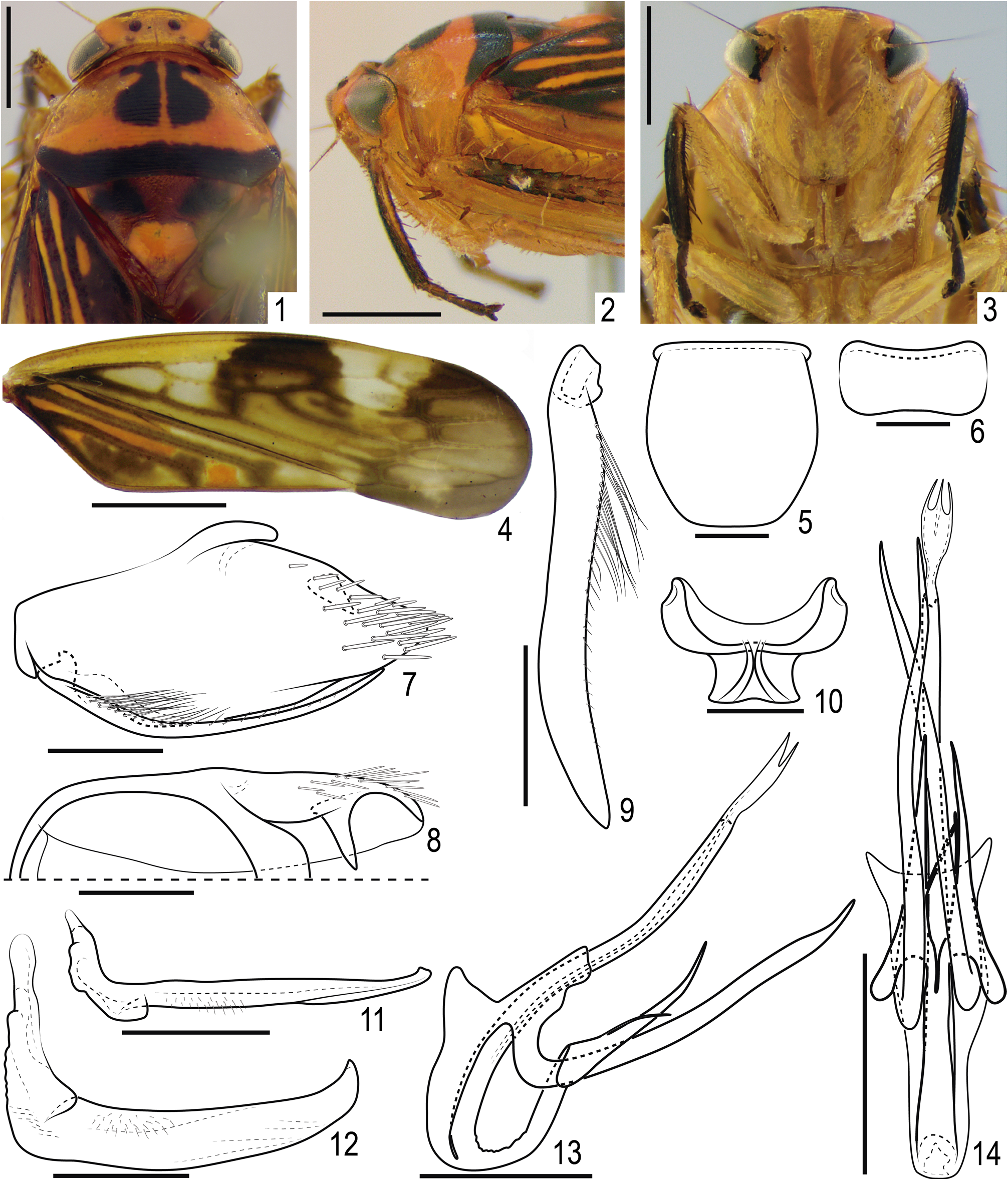
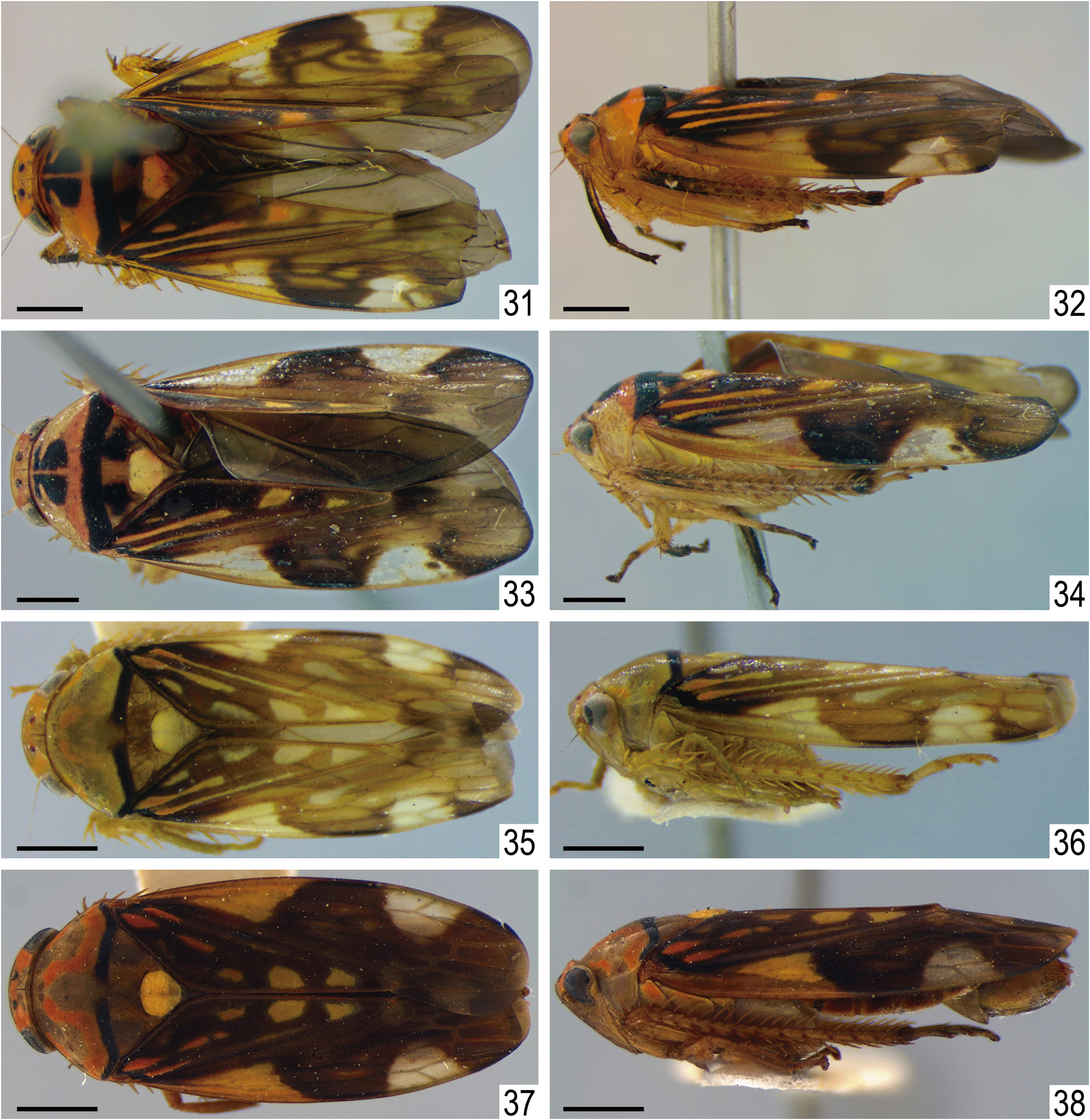
( Figs 1–22 View FIGURES 1–14 View FIGURES 15–22 , 31–34 View FIGURES 31–38 )
Diagnosis. Pronotum ( Fig. 1 View FIGURES 1–14 ) with pair of large and narrowly separate black maculae resembling a goblet; pygofer ( Fig. 8 View FIGURES 1–14 ), in dorsal view, with acute process near mid-length of dorsal margin, directed inward; subgenital plate ( Fig. 9 View FIGURES 1–14 ), very narrow, 9x longer than wide; aedeagus ( Figs 13, 14 View FIGURES 1–14 ) with shaft contiguous to dorsal apodeme and bearing conspicuous ventral process near base, bifid apically; apex of shaft weakly sclerotized bearing three spine-like processes.
Measurements. Holotype male: total length 8.0 mm. Paratype female 8.4 mm.
Description. Head ( Figs 1 View FIGURES 1–14 , 33 View FIGURES 31–38 ), in dorsal view, not produced; median length half as long as interocular width; transocular width seven-tenths of maximum pronotum width; crown with transverse parallel striae, anterior and posterior margins approximately parallel, anterior margin weakly produced over margin of eye, surface slightly concave near ocelli. Ocellus closer to median line than to eye margin and slightly closer to anterior margin of crown. Head ( Figs 2 View FIGURES 1–14 , 34 View FIGURES 31–38 ), in lateral view, with crown-face transition rounded and with several parallel striae; frons and clypeus not inflated. Head ( Fig. 3 View FIGURES 1–14 ), in ventral view, with face almost as wide as long; frontogenal suture distant from eye margins by half width of clypeus and surpassing antennal ledge, extending to anterior margin of crown; antennal ledge carinate, oriented obliquely downwards in relation to frons and extending over frons by short distance; frons approximately as long as wide, surface with texture shagreen, not excavated below anterior margin of crown, lateral margins slightly rounded; epistomal suture indistinct medially; maxillary plate produced ventrally as far as clypeus apex; gena with ventrolateral margin rounded, not excavated below eye margin; clypeus 1.1x longer than wide, lateral margins weakly divergent towards apex, apex carinate and emarginated. Pronotum ( Fig. 1 View FIGURES 1–14 , 33 View FIGURES 31–38 ), in dorsal view, with transverse striae on disc and posterior third, lateral margins longer than eye length; in lateral view ( Figs 2 View FIGURES 1–14 , 34 View FIGURES 31–38 ), slightly declivous and convex. Forewing ( Fig. 4 View FIGURES 1–14 ) 3.4x longer than wide; venation distinct, without extra veins; M vein with segment after the divergence between R+M and before the cross vein m-cu1 as long as the length of m-cu1; inner discal cell shorter than half length of outer discal cell; 3 subapical cells and 5 apical cells (R1 vein present); appendix well developed, wider than maximum width of first apical cell and bordering first and second apical cells; apex rounded. Profemur elongated, approximately 4x longer than wide; AD, AM, and PD rows reduced and poorly defined, with exception of apical setae AD 1, AM 1 and PD 1 respectively; AV and PV rows formed by 6–7 setae. Protibia, in cross-section, more or less cylindrical, with longitudinal carina adjacent to PD row; AV row formed by long setae, gradually increasing in thickness and length towards apex; AD formed by many small undifferentiated setae; PD row with 3 long setae and intercalary undifferentiated setae; dorsal surface with apical setae AD 1 and PD 1 developed; PV row with undifferentiated setae and 5 long setae on apical two thirds. Metafemur with setal formula 2:2:1. Metatibia rows PD, AD, and AV with 24–26, 12, and 16–17 macrosetae, respectively; AD row without intercalary setae between macrosetae; PV row with setae of apical half formed by sequence of 1 thicker and 3–4 thinner setae, ending with a thick seta. Metatarsomere I inner row of plantar surface with 5–6 setae, outer row very reduced in size; apex with 3 patellae flanked by 2 tapered lateral setae on inner and 1 on external corner. Metarsomere II pecten with 2 platellae flanked by 2 tapered lateral setae on inner and 1 on external corner.
Coloration. Head and thorax ( Figs 1–3 View FIGURES 1–14 , 31, 33 View FIGURES 31–38 ) background yellow. Crown ( Figs 1 View FIGURES 1–14 , 31, 33 View FIGURES 31–38 ) orange between ocellus and eye. Ocellus red. Pronotum ( Figs 1 View FIGURES 1–14 , 31, 33 View FIGURES 31–38 ) with pair of large and narrowly separate black maculae resembling a goblet, near anterior margin, behind ocelli; posterior margin with wide transverse black stripe; disc orange. Proepimeron ( Figs 1 View FIGURES 1–14 , 32, 34 View FIGURES 31–38 ) without maculae. Mesonotum ( Figs 1 View FIGURES 1–14 , 31, 33 View FIGURES 31–38 ) with pair of large black maculae near lateral angles. Scutellum ( Figs 1 View FIGURES 1–14 , 31, 33 View FIGURES 31–38 ) orange or light yellow. Face ( Fig. 3 View FIGURES 1–14 ) without maculae. Forewing ( Figs 4 View FIGURES 1–14 , 31, 33 View FIGURES 31–38 ) clavus dark-brown with two orange stripes and two orange maculae, one at base and one at middle length of anal margin; female with a third orange macula near apex; corium with costal margin yellow on basal half, pair of large white area at basal and apical third; middle portion of costal margin black; veins of external half dark-brown; apical portion smoky. Legs yellow ( Figs 2, 3 View FIGURES 1–14 , 32, 34 View FIGURES 31–38 ) except black portions of protibia, pro- and mesotarsus, and base and apex of metatibia.
Male terminalia. Sternite VIII ( Fig. 5 View FIGURES 1–14 ) 1.1x longer than wide; lateral margins slightly rounded; posterior margin straight. Valve ( Fig. 6 View FIGURES 1–14 ) 2.2x wider than long; integument thickening present only on dorsal margin; posterior and anterior margins weakly excavated. Pygofer ( Fig. 7 View FIGURES 1–14 ) 2.2x longer than maximum height; dorsal margin oblique and almost straight; ventral margin slightly and broadly rounded; apex tapered and subacute; macrosetae dispersed on apical third; in dorsal view ( Fig. 8 View FIGURES 1–14 ) lateral lobe with acute process near mid-length of dorsal margin, directed inward. Subgenital plate ( Fig. 7 View FIGURES 1–14 ), in lateral view, not surpassing pygofer apex; in ventral view ( Fig. 9 View FIGURES 1–14 ), very narrow and slightly sinuous, 9x longer than wide; lateral margins approximately parallel; external margin with hair-like setae on basal half; apex subacute. Connective ( Fig. 10 View FIGURES 1–14 ) Y-shape, wider than long; dorsal keel small; stalk short and wide. Style ( Fig. 11 View FIGURES 1–14 ), in dorsal view, with outer lobe small and rounded; blade compressed laterally; in lateral view ( Fig. 12 View FIGURES 1–14 ), portion before outer lobe forming angle of 90 degrees in relation to the blade; blade approximately straight and slightly tapered toward apex; ventral margin smooth; apex curved dorsally and subacute. Aedeagus ( Figs 13, 14 View FIGURES 1–14 ) preatrium not developed; dorsal apodeme enfolding basal portion of shaft; pair of long apodemal processes curved dorsally near base, approximately cylindrical, apex tapered and acute; shaft contiguous to dorsal apodeme bearing a conspicuous ventral process near base, bifid apically, forming two rami which enfold basal portion of apodemal processes, each ramus with rounded lobe near base, slender filiform process on dorsal margin and apex strongly tapered and acute; apex of shaft weakly sclerotized and slightly expanded laterally, forming three spine-like processes.
Female terminalia. Sternite VII ( Figs 15, 16 View FIGURES 15–22 ) 2x wider than long; posterior margin produced medially forming subtriangular lobe. Pygofer ( Figs 15, 16 View FIGURES 15–22 ) about 2x longer than maximum height; posterodorsal and ventral margins rounded; macrosetae dispersed on dorsoapical fourth and ventroapical half; apex rounded. Internal sternite VIII membranous. First valvifer ( Fig. 17 View FIGURES 15–22 ) about as long as wide, fused each other by anterior sclerotized expansion of anteroventral angle. First valvula ( Fig. 17 View FIGURES 15–22 ) slightly curved dorsally, 5.7x longer than wide; ventral interlocking device extending over basal half; dorsal sculptured area striated in oblique lines with inconspicuous scale-like processes; apical portion ( Fig. 18 View FIGURES 15–22 ) without lateral carina, apex abruptly tapered and acute. Second valvula, in cross-section, with apical triangular portion reduced, weakly expanded laterally forming lateral carina, in lateral view ( Fig. 19 View FIGURES 15–22 ), 8x longer than wide, not broadened medially; dorsal protuberance absent; dorsal margin with about 20 very small teeth on apical two-fifths; apical portion ( Fig. 20 View FIGURES 15–22 ) gradually narrowed to acute apex; ventral margin without denticles. Second valvifer ( Fig. 21 View FIGURES 15–22 ) about 3x longer than wide. Gonoplac ( Fig. 21 View FIGURES 15–22 ), 4.3x longer than wide; dorsoapical margin straight, long, with four-tenths length of gonoplac; ventral margin and apical potion ( Figs 21, 22 View FIGURES 15–22 ) with dentiform cuticular projections and few short setae; apex rounded.
Etymology. The new species name is in honor to Claudionor Elias who was a technologist at the Departamento de Zoologia, Universidade Federal do Paraná who in the 1960s to 80s worked as an insect collector mainly in the States of Minas Gerais and Espírito Santo, but also collected in other Brazilian States such as Bahia and Rondônia. His collection effort continues to yield new species.
Material examined. Holotype male: “Vilhena, RO\ 15/X/1986 \ C. Elias, leg.\ POLONOROESTE” ( DZUP) . Paratype: 1♀, same data of holotype ( DZUP) .
Remarks. The new species is easily separated from the other two species of Angusana by the diagnostic characters mentioned above, especially the unusual shape of the subgenital plate and aedeagus ( Figs 9, 13, 14 View FIGURES 1–14 ). Interestingly, the female genitalia seem to represent a transitory stage between the Polana - type and other Gyponini genera due to the absence of the lateral carina on the apical portion of the first valvula ( Fig. 18 View FIGURES 15–22 ) and the reduced lateral expansion on the apical portion of the second valvula ( Fig. 20 View FIGURES 15–22 ). Another characteristic common in species of Polana is the dorsal sculptured area formed by scale-like processes arranged in oblique lines that is inconspicuous in this new species.

| DZUP |
Universidade Federal do Parana, Colecao de Entomologia Pe. Jesus Santiago Moure |
No known copyright restrictions apply. See Agosti, D., Egloff, W., 2009. Taxonomic information exchange and copyright: the Plazi approach. BMC Research Notes 2009, 2:53 for further explanation.
|
Kingdom |
|
|
Phylum |
|
|
Class |
|
|
Order |
|
|
Family |
|
|
Genus |