
Prionospio cerastae, Radashevsky, Vasily I., 2015
|
publication ID |
https://doi.org/ 10.11646/zootaxa.4019.1.22 |
|
publication LSID |
lsid:zoobank.org:pub:88F2DB05-58C4-4726-89D5-99302FABB908 |
|
DOI |
https://doi.org/10.5281/zenodo.4658160 |
|
persistent identifier |
https://treatment.plazi.org/id/5E51D737-FFC9-FFB4-FF4A-A33E1B36F820 |
|
treatment provided by |
Plazi |
|
scientific name |
Prionospio cerastae |
| status |
sp. nov. |
Prionospio cerastae View in CoL n. sp.
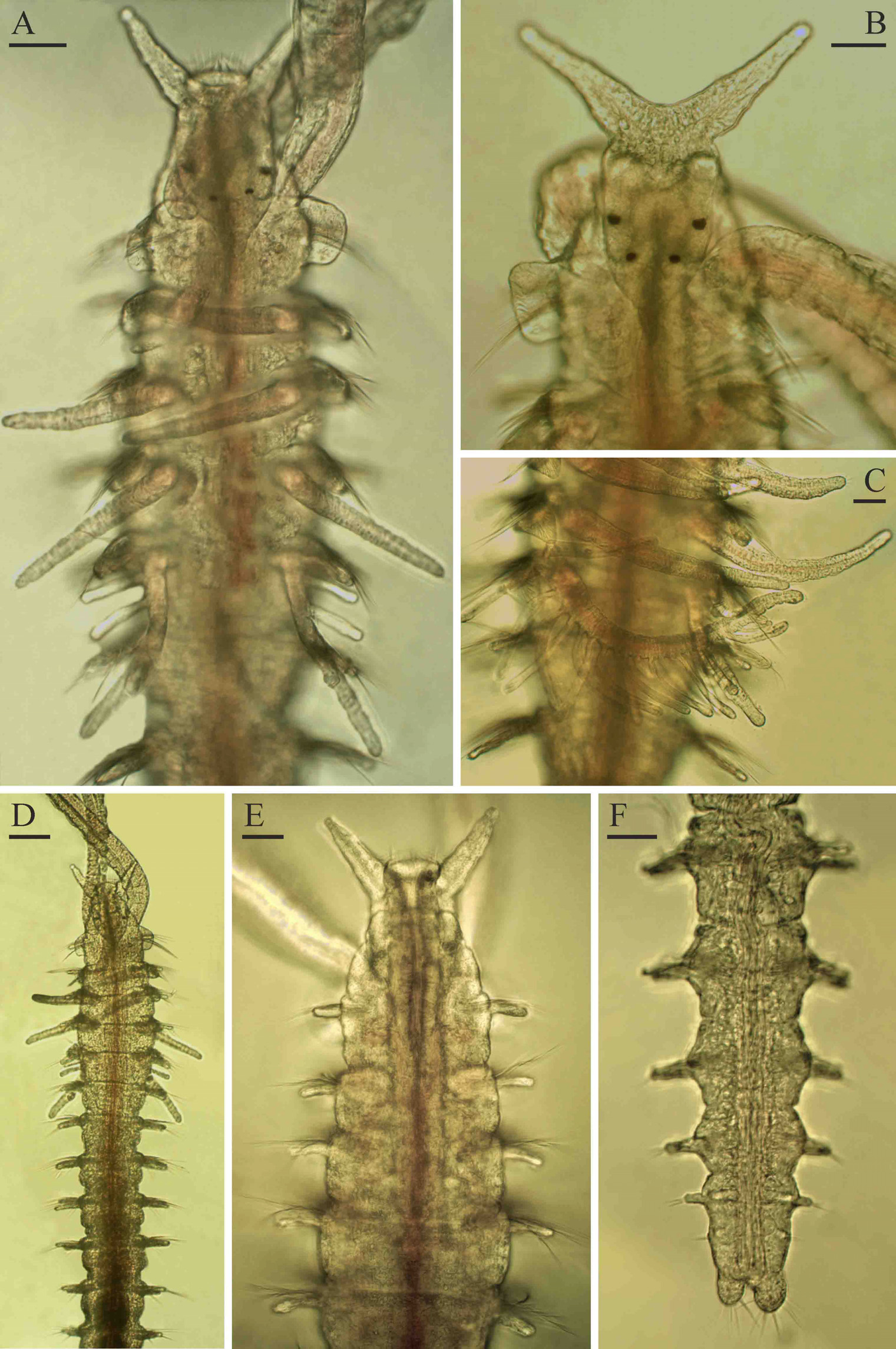
( Figs 12–14 View FIGURE 12 View FIGURE 13 View FIGURE 14 )
Type material. Queensland: Holotype: AM W.47463, MI QLD 2379. Paratypes: AM W.45267, MI QLD 2365 (1); AM W.45268, MI QLD 2373 (4); AM W.45270, MI QLD 2410 (1); AM W.45269, MI QLD 2379 (3); MIMB 28119, MI QLD 2379 (2).
Adult morphology. Up to 7 mm long, 0.28 mm wide for 60 chaetigers. Cuticle and epithelium thin, worms extremely fragile and break easily during handling and fixation. Pigmentation in life absent. Prostomium with one pair of long, pointed fronto-lateral horns ( Figs 12 View FIGURE 12 A, B, D, F, 14A), extending posteriorly to middle of chaetiger 1 as a low caruncle. Occipital antenna absent. Two pairs of small red eyes arranged trapezoidally ( Fig. 12 View FIGURE 12 B). Nuchal organs U-shaped ciliary bands on sides of caruncle, posteriorly extending almost to end of chaetiger 1. Peristomium small, not forming dorso-lateral wings. Palps as long as 15–20 chaetigers, with frontal longitudinal groove lined with fine cilia, short transverse bands of cilia regularly arranged on inner surface, and narrow longitudinal band of cilia running on outer fronto-lateral side along frontal groove on distal half; cilia of inner transverse bands beating towards distal end of palp, while cilia of outer longitudinal band beating towards frontal groove.
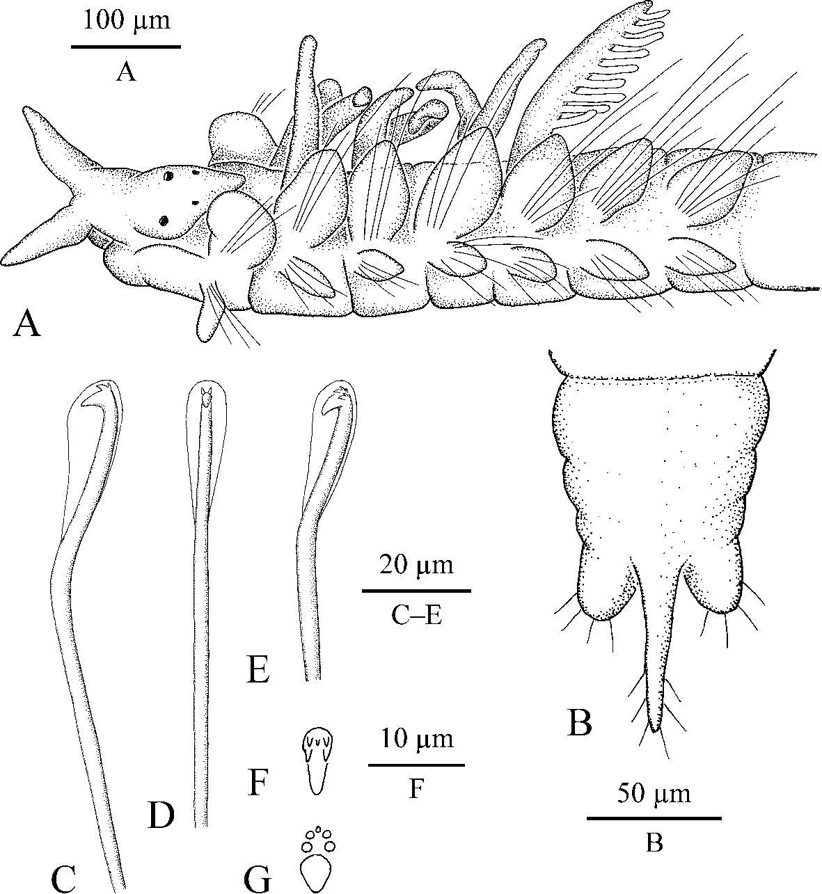
Chaetiger 1 with capillaries and postchaetal lamellae in both rami; notopodial lamellae large, flat, semicircular; neuropodial postchaetal lamellae with lower part rounded and directed ventrally ( Fig. 14 View FIGURE 14 A). Notopodial postchaetal lamellae of chaetiger 2 and succeeding chaetigers leaf-like, large on branchiate chaetigers, gradually diminishing in size on succeeding chaetigers. Neuropodial postchaetal lamellae of chaetiger 2 and succeeding chaetigers semi-oval, elongated posteriorly ( Fig. 14 View FIGURE 14 A). Notopodial lamellae of middle chaetigers with large glandular cells with granular content ( Fig. 13 View FIGURE 13 A). Dorsal crests, folds, lateral pouches and ventral flaps absent.
Hooks in notopodia from chaetigers 12–30, up to three in a series among capillaries. Hooks in neuropodia from chaetigers 10–15, up to five in a series, accompanied by 1–2 inferior capillaries and 2–3 alternating capillaries throughout. Hooks with only outer hood, with main fang surmounted by five apical teeth comprising two pairs of small teeth situated in two vertical rows, and one smaller superior median tooth; upper part of shaft characteristically bent in middle ( Fig. 14 View FIGURE 14 C–G). In lateral and frontal views, hooks appearing tridentate ( Figs 13 View FIGURE 13 B, C, 14C, D). Sabre chaetae absent in neuropodia.
Four pairs of branchiae on chaetigers 2–5 ( Figs 12 View FIGURE 12 A, C, 14A); those on chaetigers 2–4 apinnate, flattened, with surfaces oriented perpendicular to body axis, similar in length, up to two times as long as notopodial lamellae. Branchiae on chaetiger 5 slightly flattened to cylindrical, slightly longer than branchiae on previous chaetigers, each with up to 15 pairs of digitiform pinnules arranged on inner lateral and posterior sides; short apinnate branchiae on this chaetiger in some large individuals probably resulted from regenerating after occasional lost. Each branchia with longitudinal bands of cilia along inner and outer edges. Afferent and efferent branchial blood vessels interconnected by numerous radial capillaries giving branchiae annulate appearance; radial capillaries forming loops inside pinnae in fourth pair of branchiae.
Short nototrochs present on chaetigers 2–5, each composed of one transverse row of 4–6 ciliated cells. Transverse and longitudinal intersegmental ciliation absent.
Pygidium with one thin middorsal cirrus and one pair of short fleshy ventral cirri or lappets, all bearing numerous non-motile sensory cirri up to 35 µm long ( Fig. 14 View FIGURE 14 B). Middorsal cirrus often broken during handling and fixation ( Fig. 12 View FIGURE 12 F).
Oesophagus extending through 6–7 anterior chaetigers. Ventral buccal bulb below oesophagus extending to end of chaetiger 1. Gizzard-like structure in digestive tract absent.
Main dorsal blood vessel transformed into gut sinus in anterior part of midgut. Soft heart body up to 11 µm in diameter extending inside main dorsal vessel from level of chaetigers 3–4 to chaetigers 9–10. Blood red, without globules or other elements.
Nephridia in chaetigers 4–6, greenish in life.
Reproduction. Prionospio cerastae n. sp. is gonochoristic. Both in females and males gametes develop from chaetigers 12–13 to chaetigers 36–40. Oogenesis is intraovarian. Vitellogenic oocytes develop in ovaries attached to segmental blood vessels. Intraovarian oocytes were up to 110 µm in diameter, with germinal vesicle about 50 µm and single nucleolus 17 µm in diameter. Oocyte envelope is about 1 µm thick. Spermatogonia proliferate in testes; spermatogenesis occurs in the coelomic cavity. Spermatids are joined in tetrads. Spermatozoa are ect-aquasperm with small acrosome, spherical nucleus 2–3 µm in diameter, spherical mitochondria probably four in number, and a long flagellum.
Remarks. Adult P. cerastae n. sp. are characterized by the pair of long fronto-lateral horns on the prostomium, two pairs of small eyes, chaetiger 1 with large semicircular notopodial postchaetal lamellae and elongated ventrally directed semi-oval neuropodial postchaetal lamellae, apinnate branchiae on chaetigers 2–4 and pinnate branchiae on chaetiger 5, large glandular cells with granular content in notopodial lamellae of middle chaetigers, hooks with main fang surmounted by five apical teeth, inferior capillaries instead of sabre chaetae in hook-bearing neuropodia, and three pairs of excretory nephridia in chaetigers 4–6. They share short U-shaped nuchal organs, four pairs of pinnate and apinnate branchiae on anterior chaetigers, hooks in both rami in parapodia, and three-cirrate pygidium with members of Prionospio sensu lato and are therefore referred to this group.
Adult P. cerastae View in CoL n. sp. are unique among Prionospio View in CoL in having long fronto-lateral horns on the prostomium. Two rounded fronto-lateral “horns”, actually short extensions, are present on the prostomium in P. cornuta Hylleberg & Nateewathana, 1991 View in CoL from the Andaman Sea. Adults of the latter species differ from P. cerastae View in CoL n. sp. in having pinnate branchiae on chaetigers 2 and 5, and smooth, robust flattened branchiae on chaetigers 3 and 4, dorsal membranous folds on chaetigers 10–37, multidentate hooks, and sabre chaetae in neuropodia from chaetiger 12 onwards.
By the characteristically bent shaft in hooded hooks, and the presence of glandular cells in postchaetal lamellae, adult P. cerastae View in CoL n. sp. appear similar to adults of Aonides View in CoL .
Etymology. The species name refers to one of the characteristic features of adults, the presence of long and pointed fronto-lateral horns on the prostomium. The Latin word cerastes (horned) is derived from the Greek word keras, κέρας (horn).
Habitat. Adult Prionospio cerastae n. sp. were found in fine coral sand at 5–10 m depth.
Distribution. Australia, Queensland, Great Barrier Reef.

| MIMB |
Museum of the Institute of Marine Biology |
No known copyright restrictions apply. See Agosti, D., Egloff, W., 2009. Taxonomic information exchange and copyright: the Plazi approach. BMC Research Notes 2009, 2:53 for further explanation.
|
Kingdom |
|
|
Phylum |
|
|
Class |
|
|
Order |
|
|
Family |
|
|
Genus |
Prionospio cerastae
| Radashevsky, Vasily I. 2015 |
P. cornuta
| Hylleberg & Nateewathana 1991 |