
Procamallanus (Spirocamallanus) tomsici Ramallo & Ailán Choke
|
publication ID |
https://doi.org/ 10.11646/zootaxa.4810.3.4 |
|
publication LSID |
lsid:zoobank.org:pub:9832DCB8-9460-4A8F-93A4-6F552442B7CC |
|
persistent identifier |
https://treatment.plazi.org/id/039C87E7-4305-D52D-E4BF-FB43FB91A4E0 |
|
treatment provided by |
Plazi |
|
scientific name |
Procamallanus (Spirocamallanus) tomsici Ramallo & Ailán Choke |
| status |
|
Procamallanus (Spirocamallanus) tomsici Ramallo & Ailán Choke
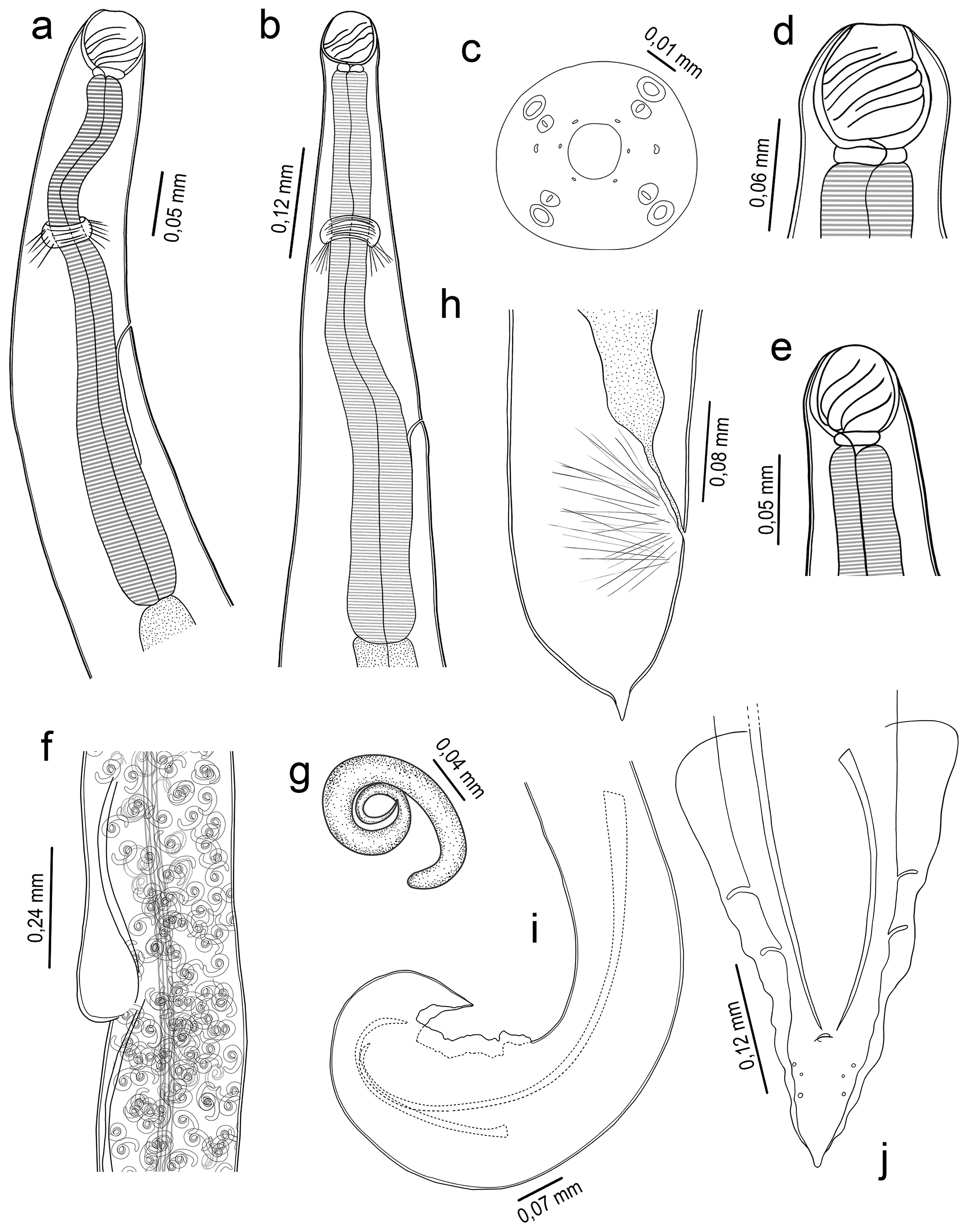
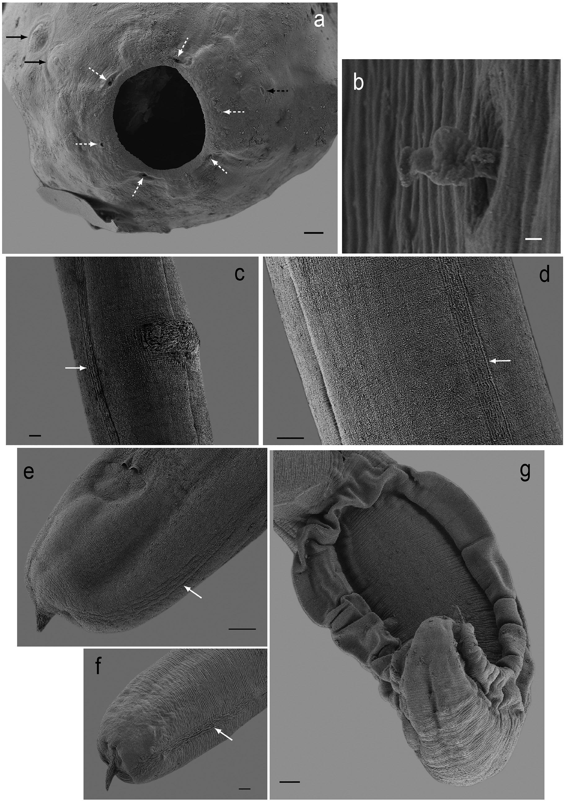
( Figs. 1–2 View FIGURE 1 View FIGURE 2 )
Type material. Holotype: female CH-N-FML #07766; allotype: male CH-N-FML #07767; paratypes (four females, five males) CH-N-FML#07768.
Type host. Pimelodus albicans Valenciennes, 1840 ( Siluriformes , Pimelodidae ), Ichthyology Collection CI- FML #2472, collected 1 December 1997.
Additional hosts: Pygocentrus nattereri Kner, 1858 ( Characiformes , Serrasalmidae ), Ictiology Collection CI- FML #2749, collected 6 October 1998; Hoplias misionera ( Characiformes , Erythrinidae ) Ichthyology Collection CI-FML #2471 (54)).
Type locality. Misión La Paz, Pilcomayo River, Rivadavia Department , Province of Salta (22º22’10”S- 62º32’14”W) GoogleMaps .
Etymology. The new species is named in honor Dr. Zlatko Tomsic (1923-2000), professor of Invertebrates (Fac. of Cs. Naturales e I.M.L, U.N.T) and Director of the Institute of Invertebrates of Fundación Miguel Lillo.
Site of infection. Intestine.
Quantitative descriptors. P. albicans (Prevalence 60%; media intensity 6.5); P. nattereri (Prevalence 67%; mean intensity 4); H. misionera (Prevalence 17%; mean intensity 2)
General description. Medium-sized nematodes with finely, transversely striated cuticle. Females possess a lateral cuticular ornamentation along the body ( Figs.1f View FIGURE 1 ; 2c, d, e, f View FIGURE 2 ). Mouth aperture rounded, surrounded by six visible pores, eight submedian cephalic papillae arranged in two circles, each formed by four papillae, and two medium amphids ( Fig. 1c View FIGURE 1 ; 2a View FIGURE 2 ). Buccal capsule orange-brown, thick walled, barrel-shaped, oral capsule has the same length as width, with simple well developed basal ring; wall of middle part of capsule strengthened by conspicuous thickening appearing in lateral view as drop-shaped, extending anteriorly to anterior margin of capsule. Inner surface of capsule of both sexes provided with 3-4 spiral ridges in lateral view, 1-3 of them being incomplete (not extending from 1 lateral margin of capsule to other) ( Figs. 1a, b, d, e View FIGURE 1 ). Muscular esophagus somewhat shorter than glandular esophagus; both parts of esophagus slightly expanded near their posterior ends ( Figs.1a, b View FIGURE 1 ). Intestine narrow. Deirids small, simple, with globose shape situated at about mid-way between posterior end of buccal capsule and nerve ring ( Fig. 2b View FIGURE 2 ); only observed in females. Excretory pore approximately at the middle region of the muscular esophagus ( Figs. 1a, b View FIGURE 1 ). Tail of both sexes, conical.
Males (based on eleven adult specimens). Length of body 12.03±0.80 (10.55–13.19), maximum width 0.17±0.02 (0.14–0.20). Buccal capsule 0.05±0.003 (0.05–0.06) long and 0.05±0.003 (0.05–0.06) wide with 3–4 inner spiral thickenings, 1-3 of them being incomplete. Basal ring 0.01±0.002 (0.010) long and 0.03±0.005 (0.02-0.04) wide. Muscular esophagus 0.56±0.06 (0.50–0.70) long. Glandular esophagus 0.84±0.07 (0.75–0.90) long. Nerve ring and excretory pore 0.22±0.02 (0.20–0.27), 0.30±0.04 (0.22–0.33), respectively, from anterior extremity. Spicules unequal and very different in length, with pointed distal ends; left spicule 0.25±0.01 (0.25–0.27) long and right spicule 0.72±0.01 (0.71–0.73) long. Posterior end of body ventrally bent, provided with narrow caudal alae, 0.40 long and 0.03 long. This structure is supported by two pairs of pedunculate preanal papillae; and three pairs of sessile postanal papillae. Gubernaculum absent. Tail conical 0.12±0.02 (0.10–0.15) long ( Figs.1i, j View FIGURE 1 ; 2g View FIGURE 2 ).
Females (based on nine gravid specimens). Length of body 22.00±2.1 (19.00–25.00), maximum width 0.34±0.06 (0.24–0.43). Buccal capsule 0.06±0.01 (0.06–0.08) long and 0.06±0.01 (0.05–0.07) wide with 3–4 inner spiral thickenings, 2-3 of them being incomplete. Basal ring 0.01±0.01 (0.01–0.012) long and 0.03±0.004 (0.03–0.04) wide. Muscular esophagus 0.63±0.04 (0.57–0.71) long. Glandular esophagus 1.09±0.13 (0.89–1.30) long. Nerve ring, excretory pore and deirids 0.24±0.02 (0.21–0.28), 0.39±0.07 (0.30–0.48), 0.10, respectively, from anterior extremity. Vulva 14.1±1.2 (12.4–15.53) from posterior end. Vulva pre-equatorial and very conspicuous. ( Figs. 1f View FIGURE 1 ; 2c View FIGURE 2 ) Vagina directed posteriorly. Uterus filled with first-stage larvae (0.2-0.3 long x 0.02 wide) ( Fig. 1g View FIGURE 1 ). Conical tail of females with a terminal cuticular spike, 0.12±0.02 (0.10–0.15) long ( Figs. 1h View FIGURE 1 ; 2e, f View FIGURE 2 ).
| FML |
Fundacion Miguel Lillo |
No known copyright restrictions apply. See Agosti, D., Egloff, W., 2009. Taxonomic information exchange and copyright: the Plazi approach. BMC Research Notes 2009, 2:53 for further explanation.
|
Kingdom |
|
|
Phylum |
|
|
Class |
|
|
Order |
|
|
Family |
|
|
Genus |