
Chilicola (Prosopoides) granulosa Packer
|
publication ID |
https://doi.org/ 10.5281/zenodo.176627 |
|
DOI |
https://doi.org/10.5281/zenodo.6249375 |
|
persistent identifier |
https://treatment.plazi.org/id/59368781-A44B-FFE2-FF7D-FEE4E6DDFAD1 |
|
treatment provided by |
Plazi |
|
scientific name |
Chilicola (Prosopoides) granulosa Packer |
| status |
sp. nov. |
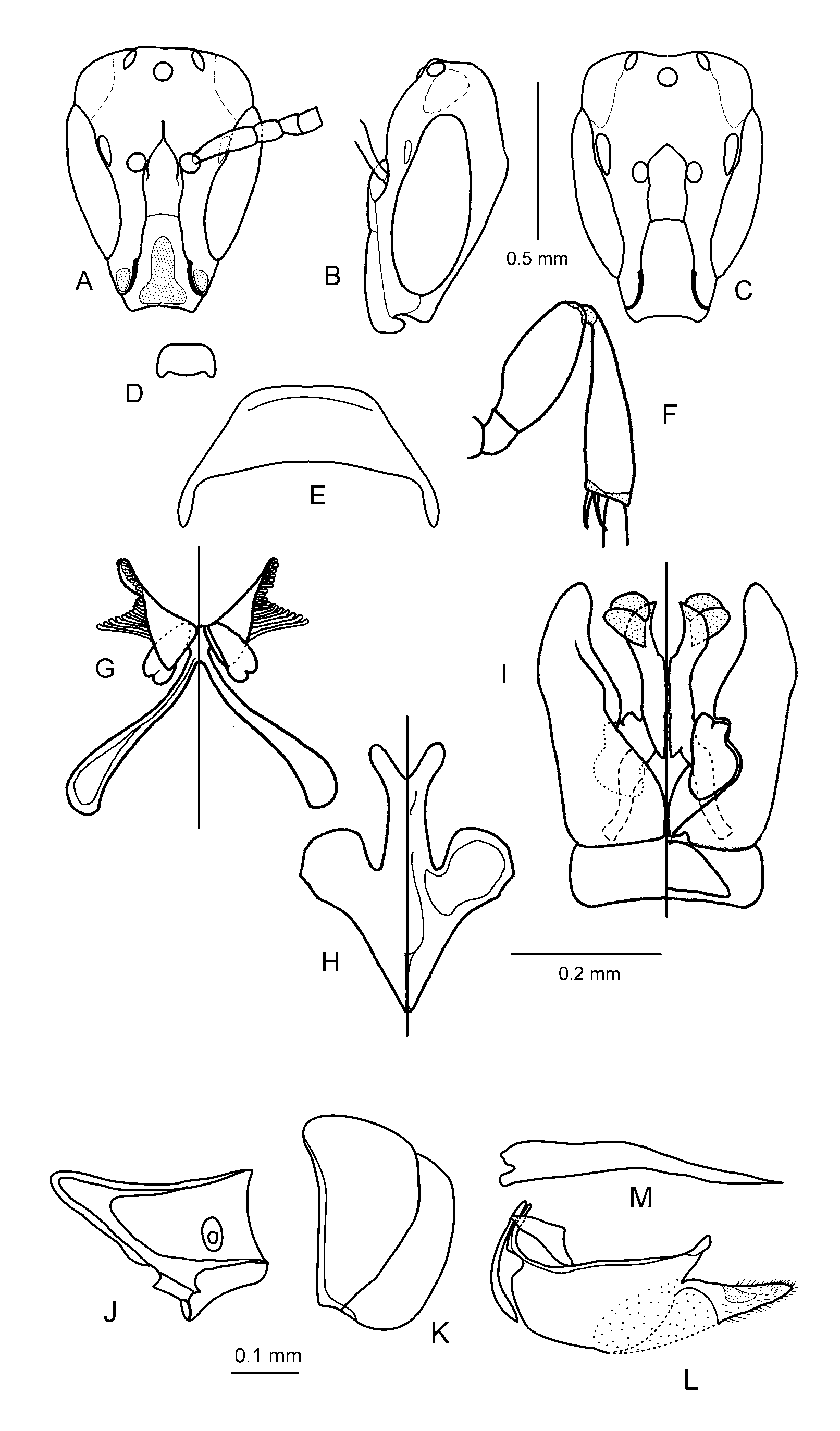
Chilicola (Prosopoides) granulosa Packer View in CoL , n. sp.
( Figs. 8A–M View FIGURES 8 A – M )
Diagnosis: This species is clearly a member of the subgenus Prosopoides as indicated by the combination of an elongate head and pronotum, no supra-antennal depression, an elongate groove below the anterior tentorial pit and well-defined facial fovea ( Figs. 8A–C View FIGURES 8 A – M ). It differs from the two described species in the subgenus by its denser, stronger microsculpture, weaker punctation, stronger swellings on the frons (similar to those of C. obesifrons described above, but not hiding the median ocellus as the more medial swellings are located lower on the face) and the elongate facial fovea, 4X longer than wide ( Figs. 8A and C View FIGURES 8 A – M ), the facial fovea also has a carinate margin that rises above the surrounding frons ( Fig. 8A View FIGURES 8 A – M ), whereas those of the other species are not so margined. There are additional new species to be described in this subgenus, including some from Brazil, which have yet to be seen by the authors: the apparently unique features described above may prove not to be diagnostic once all other taxa have been studied.
Description. Male: body length 3.5mm, wing length 2.4mm, head width 0.7mm.
Colouration: Black-brown with following parts yellow: Labrum medially (yellow-brown laterally), mandible (extreme base brown, apex red), elongate mark on clypeus, oval spot on lower paraocular area circumscribed mesally by elongate tentorial pit ( Fig. 8A View FIGURES 8 A – M ), anterior surface of scape and pedicel, apicodorsal spot on forefemur, dorsal and anterior surfaces of foretibia, apicodorsal spot on midfemur, basal spot on midtibia. Following parts orange: anterior surface of F1–F9 (rest of flagellum brown), foretarsus, spot on pronotal lobe, apical ring to midtibia, basal ring to hind tibia. Mid- and hindtibia brown. Tegula translucent amber. Wing veins dark brown.
Surface Sculpture: Microsculpture dense and strong on head and dorsal surface of mesosoma , appearing granular tending to rugulose on vertex and pronotum, weaker on genal area, mesopleuron and scutellum, which are somewhat shiny. Labrum with scattered punctures on dull microsculptured background. Swollen area between lateral ocellus and compound eye regularly imbricate and impunctate; vertex rugose; elsewhere on head and mesosoma punctures very shallow and difficult to discern especially in areas of stronger microsculpture. Punctures moderately dense on anterior portion of mesoscutum and on scutellum (i~d), sparser and more irregular on mesopleuron (i>d). Dorsal surface of propodeum with few rugae, strong median carina and posteriorly converging lateral carinae, lateral surface shining, dorsolateral portion coarsely rugose. Metasomal terga with very shallow, sparse punctures on shiny but clearly microreticulate background; apical impressed areas smooth.
Pubescence: White; very sparse and very short, fine. Areas of specialized vestiture on gena, metasomal terga and sterna lacking. Transverse row of hairs at base of submarginal zones on S2 to S6, interrupted medially on S2.
Structure: Head: ( Figs. 8A and B View FIGURES 8 A – M ) Longer than broad 67:45. Labrum with apex gently convex with strong lateral protuberance ( Fig. 8D View FIGURES 8 A – M ). Mandible 2X as long as basal depth. Clypeus longer than broad ( Fig. 8A View FIGURES 8 A – M ), 40:32, extending below lower ocular tangent for slightly less than 0.5X its length. Epistomal suture gently sinuate, reflexed at right angle apically; anterior tentorial pit with elongate shining narrow ventral groove. Subantennal sutures weakly sinuate; supraclypeal area ( Fig. 8A View FIGURES 8 A – M ) almost 3X longer than apical width (length:breadth 20:7), strongly protuberant between antennal bases, produced laterad such that small portion of medial margin of antennal socket hidden in frontal view. Frontal line distinctly raised for lower one half of distance between upper margin of antennal socket and median ocellus, otherwise flush with surface of frons forming shiny line. Frons slightly swollen around median ocellus, strongly swollen between lateral ocellus and compound eye, produced immediately mesad of compound eye. Facial fovea deep and distinct, 4X longer than wide and with raised margin. Compound eye weakly emarginate ( Fig. 8A View FIGURES 8 A – M ) such that UOD difficult to define, strongly convergent below UOD:LOD 36:20. Head produced above compound eyes ( Fig. 8A View FIGURES 8 A – M ), upper ocular tangent below lower margin of median ocellus by a distance subequal to ocellar diameter. OOC:IOC 16:12, area between ocelli expanded such that median ocellus is more anteriorly oriented and lateral ocelli more laterally oriented than normal, with eyelid-like cuticular extension over dorsal portion of median ocellus and medial portion of lateral ocellus. Scape 3X as long as greatest width, length slightly less than pedicel plus F1 plus F2; pedicel and all flagellomeres longer than broad on ventral surface, F1 only slightly so; F11 relatively longest, length to breadth 10:7; flagellum slightly broadened towards apex; flagellomeres lacking unusual patterns of setation or structural modifications. Vertex flat in profile, as long as LOL. Malar space subequal to basal depth of mandible, malar suture distinct ( Fig. 8B View FIGURES 8 A – M ). Ratio of gena to eye 8:18.
Mesosoma: Slightly more than 2X as long as greatest depth, 85:39. Pronotum elongate, ratio of medial length of collar to anterior width 12:34, collar 3MOD in length; with short weak medial depression posteriorly ( Fig. 8E View FIGURES 8 A – M ). Episternal suture strong below scrobal groove, strongly curved anteriorly below; scrobal groove absent anterior to scrobe, weakly developed posteriorly. Propodeum subequal in length to scutellum, ratio of scutellum:metanotum:propodeum 13:7:14; propodeal sulcus imperceptible; posterior surface of propodeum separated from lateral and dorsal surfaces by irregular, but strong, carina. Hind trochanter unmodified ( Fig. 8F View FIGURES 8 A – M ). Hind femur slightly expanded, dorsal and ventral surfaces convex, length to greatest depth 53:18 ( Fig. 8F View FIGURES 8 A – M ). Hind tibia slightly expanded towards apex, length to apical depth 73:20 ( Fig. 8F View FIGURES 8 A – M ). Hind tibial spurs unmodified. Hind basitarsus less than 6X as long as greatest depth, parallel sided. Hind tarsal claws bifid. Basal vein weakly curved; marginal cell short, ratio of length of stigma to free portion of marginal cell 20:25; stigmal margin in marginal cell angularly convex; distal stigmal perpendicular goes through second submarginal cell at or just beyond midlength; 1st recurrent vein basal to first submarginal cross-vein.
Metasoma: T1 with length and apical breadth subequal. Apical impressed areas of terga 0.25X length of entire tergum. Sterna unmodified; gradulus of S2 with long posteriorly directed lateral portion, graduli lacking on S3–S6.
Terminalia : S7 ( Fig. 8G View FIGURES 8 A – M ) with two membranous lobes; dorsal lobe triangular, broadly attached to disk at base, posterolaterally directed, with fan of robust, peg-like capitate setae laterally, these much longer anteriorly than posteriorly; ventral process short, anterolaterally directed, apex narrowly incised. S8 ( Fig. 8H View FIGURES 8 A – M ) with apical process long and narrow, apex deeply concave forming a rounded Y. Gonobase with broadly concave apicoventral process. Volsella V-shaped, angularly excised at base of cuspis, apex of digitus acute. Gonostylus not differentiated from gonocoxite. Penis valve with single large membranous lobe recurved medially ( Fig. 8I View FIGURES 8 A – M ).
Female: Body length 4.0mm, wing length 2.7mm, head width 0.8mm.
Colouration: Black with anterior surface of antennal flagellum pale brown, rest of antenna, legs and metasoma dark brown except anterior and dorsal surfaces of foretibia yellow-brown.
Surface Sculpture: Sculpture less coarse than in male, microsculpture more even; labrum densely and deeply punctate; punctures small, more distinct especially on mesoscutum (i=1–4d) and scutellum (i=1–3d).
Pubescence: As in male except as follows: Hind femur with weak scopa, hairs <1.3MOD, hind tibia with some slightly longer hairs 1.5MOD. Scopa of S2 corbiculate, hairs long <3MOD with short posteriorly directed branches.
Structure: Maxillary palpus elongate, 0.6X as long as prementum; prementum elongate, less than 0.17X as broad as long, premental fovea almost covering ventral surface, strongly carinate laterally. Lacinia triangular, 5X longer than greatest breadth. Lorum weakly sclerotised, 0.167X as long as cardo. Rest of body as in male except for usual secondary sexual characteristics and as follows: Area between lateral ocellus and compound eye less strongly expanded, OOC shorter as a result (OOC:IOC 15:14). Clypeus extending below lower ocular tangent for 0.4X its length (10:25) ( Fig. 8C View FIGURES 8 A – M ). Supraclypeal area expanded more laterally, larger portion of medial margin of antennal socket hidden in frontal view. Raised portion of frontal line shorter, raised for lower one-third of distance between upper margin of antennal socket and median ocellus. Malar space slightly shorter than basal width of mandible. Median depression of pronotum stronger. Apical lunule of S5 almost an equilateral triangle.
Sting apparatus: Hemitergite 7 ( Fig. 8J View FIGURES 8 A – M ) broad posteriorly converging to very narrow and elongate apodemal region, lateral process short and broad complex in structure with short anterior and longer posterior angulations, medial portion of marginal ridge somewhat concave, spiracle much closer to lateral than medial ridge, posterior margin of lamina spiracularis evenly and gently concave. Hemitergite 8 ( Fig. 8K View FIGURES 8 A – M ) with apodeme particularly deep much larger than disk, apodemal margin straight but strongly inflected anteriorly for apical 0.25. First valvifer with dorsal arm very short forming right angle. Second valvifer with strong apical process, apicoventral region mostly membranous, gonostylus short and deep with very short setae ( Fig. 8L View FIGURES 8 A – M ). Sting shaft strongly bent ventrad at mid length ( Fig. 8M View FIGURES 8 A – M ). Furcula with ventral arms very broad.
Material studied. Holotype male, allotype female, two male and ten female paratypes: BOLIVIA, Santa Cruz, 11km N. Boyuibe, 20o23’75”S, 63o22’25” W, 2900 feet, 5.iii.1999, M. Irwin and F.D. Parker, malaise trap; additional paratypes as follows: same data except 6.iii.1999, two males and one female; Santa Cruz, 24km S. Carriri, 20o18’81”S, 63o28’5” W, 3550 feet, 5.iii.1999, M. Irwin and F. D. Parker, malaise trap one male and two female paratypes; Santa Cruz, 20km S. Carriri, 4.iii.1999, 20o10’57”S, 63o28’74”W, M.E. Irwin and F. D. Parker, malaise trap, one female. All specimens are at Logan except for two male and two female paratypes in senior author’s collection at York University and one individual of each sex at MACN.
Etymology. The specific epithet refers to the strongly imbricate sculpture on much of the body of this species, appearing granular in many places, especially on the head.
Comments. The new species expands the range of the subgenus from Argentina, Paraguay and Brazil into Bolivia.
| MACN |
Museo Argentino de Ciencias Naturales Bernardino Rivadavia |
No known copyright restrictions apply. See Agosti, D., Egloff, W., 2009. Taxonomic information exchange and copyright: the Plazi approach. BMC Research Notes 2009, 2:53 for further explanation.