
Pseudobiceros murinus Newman & Cannon, 1997
|
publication ID |
https://doi.org/ 10.11646/zootaxa.4438.2.2 |
|
publication LSID |
lsid:zoobank.org:pub:40AA328A-C8EB-4A35-8434-064190D73040 |
|
DOI |
https://doi.org/10.5281/zenodo.5959238 |
|
persistent identifier |
https://treatment.plazi.org/id/03A02F4D-FFE6-FF86-2BE4-F99CD657FE9B |
|
treatment provided by |
Plazi |
|
scientific name |
Pseudobiceros murinus Newman & Cannon, 1997 |
| status |
|
Pseudobiceros murinus Newman & Cannon, 1997 View in CoL
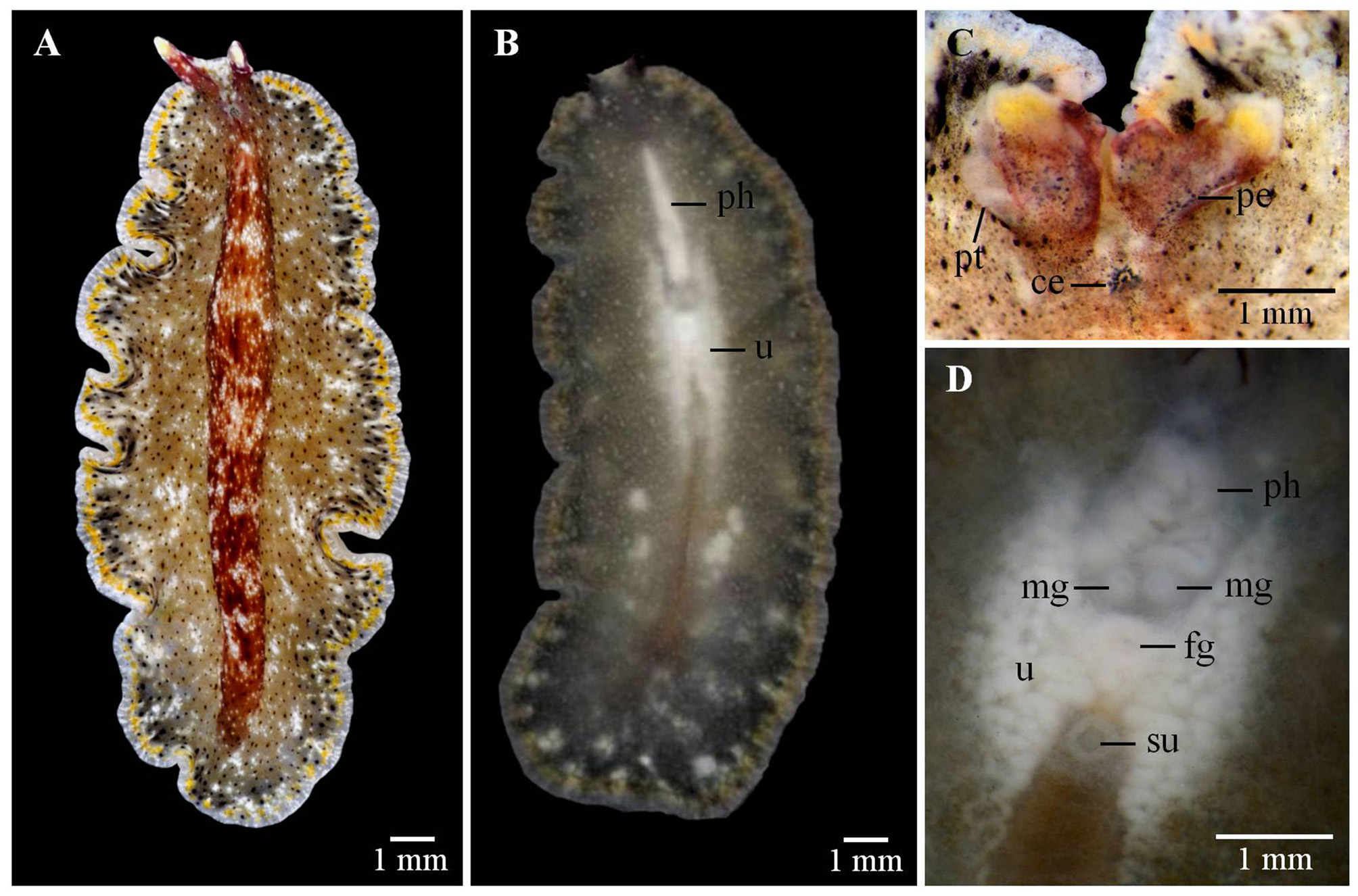
( Fig. 5 View FIGURE 5 )
Material examined and locality: Two mature specimens collected at the Inter University Institute for Marine Sciences ( IUI), Eilat, Israel, northern Gulf of Aqaba, Red Sea (29° 30.211’ N, 34° 55.068’ E) GoogleMaps . a) One specimen (15x 7 mm, live, ZMTAU-VR25134), as sagittal sections of the reproductive structures (8 slides). The remaining portion of the specimen preserved in Ethanol 70%. Collecting date on 1 November 2013. b) One specimen (17x 8 mm, live, ZMTAU-VR25135), preserved in ethanol 70%. Collecting date on 1 November 2013.
Habitat: Specimens found on the rocky shore during low tide (1 m depth), under rocks dominated principally by ascidians and sponges.
Distribution: Madang, Papua New Guinea (Type locality) and Lizard Island, Great Barrier Reef, Australia ( Newman & Cannon 1997); Guam and Yap Islands, Micronesia, and the Red Sea (no information of the exact location) ( Newman & Cannon 2003; 2005); and Kavaratti Island, India ( Apte & Pitale 2011). In this study, the specimens were found in Eilat, northern Gulf of Aqaba, Red Sea.
External anatomy. Oval and elongated body with some deep folds of the margin ( Fig. 5A View FIGURE 5 ). Dorsal background grey to olive green or brownish-green with small black spots evenly distributed over the entire dorsal surface. Few blotches of white scattered around the body. Conspicuous raised dorsal midline with a bright reddishbrown coloration extending to the pseudotentacles. Presence of a fine yellow submarginal band with a dark hue in its inner area surrounding the entire body. Clear rim marked with white spots ( Fig. 5A View FIGURE 5 ). Ventral side translucent grey with opaque white dots and visible yellow line ( Fig. 5B View FIGURE 5 ). Pointed, long, erected ear-like pseudotentacles with yellowish-white tips and red tinge at the base. A triangle of white dots between the pseudotentacles ( Fig. 5A, C View FIGURE 5 ). Small clusters of dorsal and ventral pseudotentacular eyes. Cerebral eyespot horseshoe-shaped located in a clear area ( Fig. 5C View FIGURE 5 ). Ruffled pharynx with simple folds located anteriorly ( Figs. 5B, D View FIGURE 5 ). Two separate male gonopores, one close to the other located posterior to the pharynx. Female gonopore located in the midline, posterior to the male gonopores. Conspicuous sucker behind the female gonopore ( Fig. 5D View FIGURE 5 ).
Taxonomic remarks. Pseudobiceros murinus is included into the color group 4 which is characterized by a mottled pattern ( Newman & Cannon 1994; 1997). This group includes six species of which P. fulvogriseus and Pseudobiceros gardineri ( LAIDLAW, 1902) also show a mottled grayish background with pointed pseudotentacles. Although P. gardineri also exhibits black dots, it lacks the yellow submarginal band. Similarly, P. fulvogriseus lacks the yellow band and the black spots present in P. murinus . As mentioned above, P. damawan closely resembles P. murinus . However, P. damawan has a wide orange submarginal band interrupted with white dots and a conspicuous black rim instead of a fine yellow submarginal band with a clear rim as P. murinus . The pseudotentacles of P. murinus also differ from the three species mentioned before. However, Pseudobiceros mikros NEWMAN & CANNON, 1997 exhibits the same pseudotentacle shape and color pattern as P. murinus but in contrast, P. mikros has a yellow rim and white submarginal band.
Newman & Cannon (2005) mentioned that the color of this species is variable depending on food in the gut and presented three different photographic records for this species. Likewise, Apte & Pitale (2011) reported a morphotype with a lighter dorsal background, more white blotches, a thicker orange-yellowish submarginal band, and purplish-pink pseudotentacles instead of red. Also, the dark coloration associated with the inner area of the yellow band was absent. According to Newman & Cannon (2005), P. murinus has been seen in the Red Sea but there is no additional information, a formal description or specific location. Therefore, this species represents the first record for Eilat, northern Gulf of Aqaba, Red Sea, Israel.
| IUI |
Inha University |
No known copyright restrictions apply. See Agosti, D., Egloff, W., 2009. Taxonomic information exchange and copyright: the Plazi approach. BMC Research Notes 2009, 2:53 for further explanation.
|
Kingdom |
|
|
Phylum |
|
|
Class |
|
|
Order |
|
|
Family |
|
|
Genus |