
Pseudogobius poicilosoma (Bleeker, 1849)
|
publication ID |
https://doi.org/ 10.11646/zootaxa.4961.1.1 |
|
publication LSID |
lsid:zoobank.org:pub:F4C78D3B-590D-4610-9DD1-93310B23D85E |
|
persistent identifier |
https://treatment.plazi.org/id/03EA6777-FFD8-4879-FF53-F3C9FC8AFBF2 |
|
treatment provided by |
Plazi |
|
scientific name |
Pseudogobius poicilosoma (Bleeker, 1849) |
| status |
|
Pseudogobius poicilosoma (Bleeker, 1849) View in CoL
Spotted snubnose goby
Figs 1–3 View FIGURE 1 View FIGURE 2 View FIGURE 3 , 24–25 View FIGURE 24 View FIGURE 25 , Tables 1–5, 18
Gobius poicilosoma Bleeker, 1849a: 31 View in CoL (Pasuruan, Java).— Bleeker, 1849b: 10 (Pasuruan).
Gobius gastrospilos Bleeker, 1853: 477 (Batavia [Jakarta]).
Gobius javanicus Bleeker, 1856: 88 View in CoL (Patjitan, central Java).
Vaimosa piapensis Herre, 1927: 147 View in CoL , pl. 10, fig. 3 (Piapi Creek, Dumaguete, Oriental Negros, Philippines).— Herre 1953: 768 ( Philippines).
Stigmatogobius gastrospilos — Koumans 1932: 8–9 (Pasuruan).
Stigmatogobius poicilosoma View in CoL — Koumans 1932: 9–10 (Pasuruan).
Stigmatogobius javanicus View in CoL — Herre 1953: 764–765 (Malabon, Rizal Province); Rema Devi 1993: 181 (Ennore estuary and surrounding area); Monkolprasit et al. 1997: 251 (Klong Wan (Prachuap Khiri Kan).
Pseudogobius javanicus View in CoL — Akihito & Meguro 1975: 46 (Ishigakijima, Okinawa Pref.); Bleeker 1983: pl. 438, fig. 14 (“colour markings erroneous”); Akihito in Masuda et al. 1984: 268–269 (Amamioshima to Iriomotejima); Kawanabe & Mizuno 1989: 577 ( Japan); Tan & Tan 1994: 356. 356 (Pulau Bintan, Indonesia); Rainboth 1996: 207 ( Cambodia); Ng et al. 1999: 182 (Pulau Tioman, Malaysia); Larson in Randall & Lim 2000: 639 (South China Sea); Kottelat 2001: 61 (Quang Ninh Province); Larson 2001: 202–203 (Patjitan, central Java; Piapi Creek, Philippines); Larson & Murdy 2001: 3601 (western central Pacific); Sakai et al. 2001: 115 (Ishigaki, Iriomote and Okinawa islands; Nansei Islands); Nakabo 2002: 1210 ( Japan); Tan & Lim 2004: 111 (Anambas; Natuna [ Indonesia]); Senou et al. 2004: 235 ( Japan); Shibukawa in Matsuura & Kimura 2005: 63 (Libong Island, Thailand); Larson & Lim 2005: 141 (Sungei Buloh, Sungei Simpang, Singapore); Larson et al. 2008: 143 ( Singapore); Yokoo et al. 2008: 78–79 (Sikao Creek, Trang Province, Thailand); Shibukawa in Kimura et al. 2009: 285 (Andaman Sea); Chen et al. 2014a: 132 ( Taiwan; Hong Kong; Philippines; Singapore; Phuket); Huang et al. 2015: 1 (Taiwan); Tran et al. 2013: 144 (Mekong Delta, Vietnam); Kottelat 2013: 422 ( Indonesia; Philippines); Chen et al. 2014b: 317 (Kinmen Island, Taiwan); Dahruddin et al. 2017: supp. table 2 (Java, Indonesia); Hammer et al. 2021: 2 ( Indonesia, Singapore, Vietnam, Taiwan [distinct mitochondrial DNA lineage]).
Material Examined. INDONESIA: Syntype of Gobius poicilosoma, RMNH 4488, 31 mm SL female, brackish water, Pasuruan , Java, 1847 . Holotype of Gobius javanicus, RMNH 4549, 34 mm SL female, in river, Patjitan , Java, 1879 . Holotype of Gobius gastrospilos, RMNH 4676, 30 mm SL specimen (sex undetermined), Batavia [Jakarta] . NTM S.11125-016, 111(18–33), mangroves at S end Sanur beach, Bali, H. Larson , T. Gloerfeldt-Tarp and P. Kailola, 10 June 1983 ; NTM S.11125-029, 2(30-31), Sanur Beach, South End , Bali, HL 83-19 , T. Gloerfelt-Tarp, P. Kailola and H.K. Larson, 10 June 1983 ; ZSM/CMK 6083 View Materials , 4 View Materials (26–29.5), S of Tanjung Pasir, Tanjerang District, West Java, M. Kottelat and F. Yuwono, 29 May 1988 ; WAM P.30806-003, 11(20.5–34), brackish lake [Danau Haji Buang] in centre of Maratua Island , east Kalimantan, G . R. Allen, 21 May 1994 . SINGAPORE: CMK 6012, 24(12–31), canal into sea, W coast, M. Kottelat and P. Ng, 21 May 1988 ; USNM 366851 View Materials , 27 View Materials (14.5–23), Sungei Buloh, just outside Nature Reserve, small stream entering Straits of Johor , L. Parenti, H. Larson, K. Lim and N. Sivasothi, 5 September 1997 . THAILAND: S.17893-002, 14(24.5–34), Chantaburi Province, Mahidolia Project, T. Wongratana . VIETNAM: NTM S.17895-002, 3(19.5–22), Bac Lieu, K. Shibukawa and party, 19 May 2012 . TAIWAN: NMMB P.3901, 8(18–30.5), Da-pon-wan, Pingtung, I-S. Chen, 12 November 1995 . PHILIPPINES: USNM 241843 View Materials , 42 View Materials (13.5–31), Canauay River about 75 m upstream from mouth, in tidal mangrove pool, Negros Oriental, L. Knapp and party, 9 May 1979 . JAPAN: YCM P-8183, 13(25–31), Kabira Bay , Ishigaki-jima, M. Hayashi, 10 March 1976 ; NSMT P.15776, 8, river on Ishigaki Island, Ryukyu Islands , 29 March 1973 .
Additional material (no data taken). THAILAND: USNM 316047 View Materials , 2 View Materials , Cheh Bilang ; USNM 316048 View Materials , 4 View Materials , Cholburi ; USNM 316044 View Materials , 1 View Materials , roadside ditch near Tah Chalap , T. Roberts and party, 5 May 1970 . HONG KONG: NTM S.15340-001, 2, Tai Tan, Hong Kong area , T. Lam , 6 December 2000 ; CAS / SU 32837 , 1 View Materials , Lan Tau Island , A.W. Herre, 7 January 1936 . INDONESIA: CMK 7264, 8, Padang Island , Sumatra; CMK ex 4546, 1, Padang, Sumatra ; NTM S.14156-003, 11, Benete River , South Sumbawa, AQ 32, K. Martin, 2 April 1995 ; NTM S.16227-001, 3, Buhias, Mantehage Island , North Sulawesi ; NTM S.16226-001, 1, Likupang , North Sulawesi, G. Polgar, 16 September 2005 [last 2 lots genotyped]. SINGAPORE: CAS-SU 30373 View Materials , 33 View Materials , Pulau Ubin ; CAS-SU 33164 View Materials , 20 View Materials , Singapore ; NTM S.13968-002, 23, Sungei Pandan, D. Hoese, K. Lim and P. Ng, 22 December 1993 ; NTM S.13960-011, 22, concrete drain, Marshlands Reserve , H.K. Larson and party, 6 January 1994 ; NTM S.17471-007, 2, Lim Chu Kang , HL 12-7, H.K. Larson and party, 27 October 2012 ; NTM S.17453-006, 4(31-33), creek off Observatory , Pulau Ubin, HL 12-1, H.K. Larson and party, 16 October 2012 . BRUNEI: NTM S.14800-004, 8, Sungai Penabai , Tutong District, HL 97-75, H.K. Larson and party, 23 August 1997 ; PHILIPPINES: CAS-SU 38642 View Materials , 12 View Materials , Cagayan River, Mindanao ; CAS-SU 38641 View Materials , 37 View Materials , Capiz, Panay ; CAS-SU 29845 View Materials , 2 View Materials , Mactan Island ; USNM 316199 View Materials , 45 View Materials , E of Iloilo City, Panay; CMK 9908, 9, Ambacan River at Baybay, M. Kottelat, 2 July 1993 . JAPAN: NTM S.12135-010, 5, Nakama River , Iriomote Island, HL 85-54, H.K. Larson, 19 August 1985 ; NTM S.12731-023, 1, Yonada River, Iriomote Island , H. Senou and Aonuma, 19 August 1985 .
Diagnosis. A moderate-sized Pseudogobius with second dorsal rays I,7–8; anal rays I,6–8; pectoral rays 14–17; 15–16 segmented caudal rays usually in 9/7 pattern; lateral scales 23–28; TRB 7–10; predorsal scales 6–8; opercle with at least two rows of cycloid scales, may be 1–2 cycloid scales on cheek right behind eye; shoulder girdle smooth or with narrow irregular flange; tongue blunt; upper jaw teeth usually widely spaced, conical to slightly flattened, fairly straight with slightly pointed tips (tips may be honey-coloured); colour pattern includes diagnostic dark brown diagonal band from first dorsal fin, a well-spaced pair of dots on the caudal peduncle and many rows of fine dark spots on caudal fin; widespread in estuarine to freshwater habitats of East Asia, South Asia and South-east Asia.
Description. Based on 70 specimens, 16–34.5 mm SL. An asterisk indicates the counts of the 31 mm SL female syntype of Gobius poicilosoma ( Fig. 24 View FIGURE 24 ).
First dorsal VI *; second dorsal I,7–8 (usually I,7, I, 6 in syntype); anal I,6–8 (usually 7*); pectoral rays 13–17 (usually 15, 16 in syntype); segmented caudal rays 8/7–9/7; branched caudal rays 6/6–8/7, modally 8/7; unsegment- ed (procurrent) caudal rays 7/7 (in 1), 8/6 (1); lateral scale count 23–28 (usually 25*); TRB 7*–10 (8); predorsal scale count 6–8 (7*). Gill rakers on outer face of first arch very short, 2+6 (1), 2+7 (1), 2+8 (1), 2+9 (1), 3+7 (2), 3+8 (2) .
Body stout, compressed. Head wider than deep, especially in males (may have inflated cheeks), HL 23.0–29.4% SL (mean 26.0%). Depth at posterior preopercular margin 61.7–73.7 HL (mean 67.6%). Width at posterior preopercular margin 66.2–85.7% HL (mean 73.9%). Mouth small, subterminal, very slightly oblique, upper jaw slightly in advance of lower; jaws reaching to vertical through mid-eye or just posterior to mid-eye in adult males (females with smaller jaws); upper jaw 22.2–45.3% HL (mean 35.6%); lips relatively thin, upper lip fleshier than lower lip; lower broadly fused to chin anteriorly. Eyes lateral, high on head, top usually forming part of dorsal profile, 26.5– 36.2% HL (mean 30.6%). Snout rounded and somewhat inflated, 21.4–34.7% HL (mean 28.2%). Interorbital moderate, 11.3–24.3% HL (mean 17.8%). Body depth at anal origin 17.0–26.2% SL (mean 21.4%). Caudal peduncle compressed, length 23.5–34.3% SL (mean 28.9%). Caudal peduncle depth 11.9–17.6% SL (mean 14.4%).
First dorsal fin low, rounded to roughly triangular, second to third spines longest (usually second). Appressed first dorsal fin just reaching to first second dorsal fin element in adult males, fin falling short of second dorsal in females. Second dorsal spine length 13.3–27.3% SL (mean 17.1%). Third dorsal spine length 12.9–27.3% SL (mean 15.4%). Second dorsal and anal fin heights moderate, fins pointed posteriorly with posteriormost rays longest, rays falling short of caudal fin base when appressed. Pectoral fin oval to rounded, central rays longest, extending back to vertical just above anus, 15.9–26.5 % SL (mean 23.1%). Pelvic fins rounded, just reaching to anus, 18.6–26.2% SL (mean 21.5%). Caudal fin broad, rounded posteriorly, 26.8–37.1% SL (mean 31.4%).
Anterior nostril in short tube, oriented down over upper lip. Posterior nostril oval, with very low rim, placed at mid-level of eye. Gill opening usually extending forward to under mid-opercle. Gill rakers on outer face of first arch very short pointed stubs. Shoulder girdle smooth or with narrow irregular flange. Tongue tip blunt. Upper jaw teeth in 3–4 rows, teeth in outer row always largest and usually widely spaced, conical to slightly flattened, fairly straight with slightly pointed tips (tips may be honey-coloured); teeth in the 2–3 inner rows very small, close-set and sharp; some females have outer row teeth very small and almost blunt-tipped. Lower jaw teeth in 2–3 rows, small, conical, close-set, with rather blunt to sharp tips; 2–3 curved symphyseal canines behind anterior tooth rows in males.
Body scales ctenoid to pectoral fin base and sometimes extending further forward to above opercle, cycloid scales on predorsal and pectoral fin base; opercle with at least 2 rows of cycloid scales, 1–2 cycloid scales occasionally present on cheek right behind eye; belly scales cycloid on posterior half at least, may be ctenoid scales anteriorly. Lateral canals, pores and sensory papillae pattern as in Fig. 2 View FIGURE 2 .
Coloration of preserved material. Colour photographs of freshly dead P. poicilosoma are in Kottelat et al. (1993), Kimura et al. (2009) and Tran et al. (2013) (and see Fig. 25 View FIGURE 25 ). Head and body whitish to pale yellowish brown, with darker brown markings. Head with plain grey to light brown snout, nape with irregular brown mottling and spotting, posteriormost to all nape scales with dark brown spot at rear of each scale. Side of head with one broad brown bar from front of eye to end on jaw about halfway between anterior nostril and corner of mouth; second brown bar from ventral edge of eye running ventrally just past end of jaws and ending just before ventral margin of preopercle; always a paler area on each side of bars. Cheek and opercle with variable scattered greyish to dark brown mottling and irregular small spots; often a third narrow dark brown bar from lower rear edge of eye to rear edge of preopercle. Lips narrowly outlined with dark brown, in both sexes. Underside of head plain whitish to dusky grey.
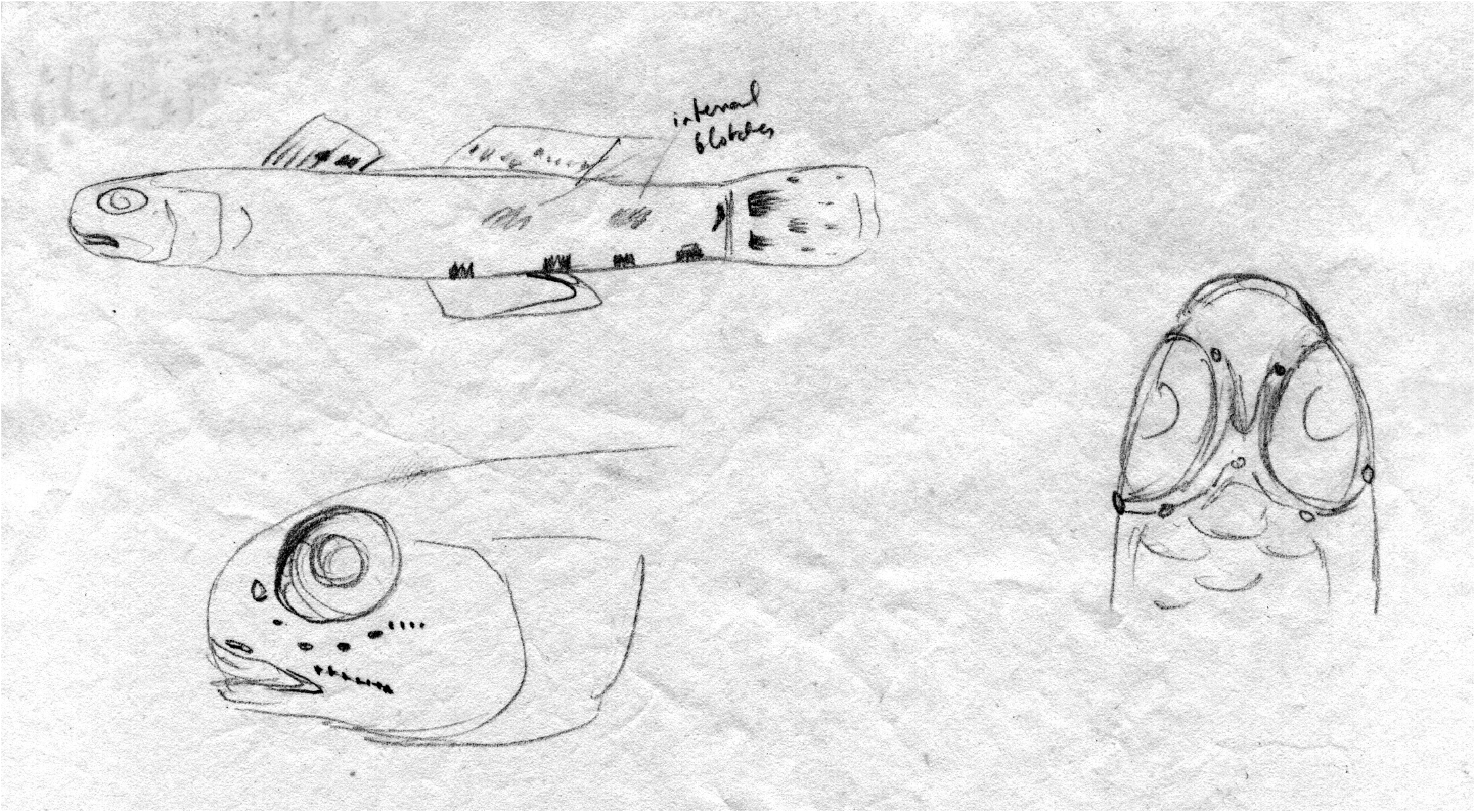
Side of body with mid-lateral series of four pairs of dark brown round to elongate spots, which often fuse to form four elongate dark blotches; anteriormost spots usually subsumed by characteristic anteriorly diagonal dark brown band from below rear of first dorsal fin and ending above abdomen; diagonal dark band may be very dis- tinct and almost black, broken-up or indistinct, but remnants always visible; may be a pair of dark brown spots just behind base of diagonal dark bar on lower side. Remainder of body with scattered small dark brown spots and blotches, which may form irregular rows or pairs of markings. Dorsum with 6–7 indistinct dark brown blotches or small saddles; these often indistinct; scales on dorsal half of body may have thin dark margins. Breast and belly plain whitish to grey to brown, usually darkest in adult males. Peritoneum dark brown to almost black dorsally, fading on sides to pale grey or pale ventrally. Four internal (usually only visible in recently preserved material) dusky grey blotches along ventral midline, two above anal fin and two on caudal peduncle. Posteriormost mid-lateral pair of dark spots, at mid-base of caudal fin, usually coalesced into single spot or blotch. On scaly base of caudal fin itself, two dark brown to blackish often rounded spots, placed above and below last mid-lateral spot, often forming a recumbent Y; lowermost caudal base spot usually smaller than upper and always placed slightly further away from mid-base spot.
First dorsal fin transparent to translucent dusky brownish, with broad brown to dark brown submarginal band and second central brown band ending in black spot on membrane between fourth to sixth spines; the two brown bands may coalesce or become indistinct but black spot always present (may be small); leading edge of first dorsal spine often with 3–4 brown spots. Second dorsal fin transparent to pale grey with 3–4 rows of dark brown small spots and streaks, markings fading or coalescing toward rear of fin.Anal fin plain pale grey to translucent, often with clear margin; fin always darkest in adult males. Caudal fin transparent to greyish with 5–6 curving rows of small dark brown spots, most distinct near the two large basal spots and becoming more scattered and diffuse posteriorly. Pectoral fin base as in body colour with brown to dark brown square blotch or bar on uppermost part. Pectoral fin transparent to slightly greyish, with rays having scattered brown streaks and speckling mostly on upper part of fin (especially in males). Pelvic fins almost translucent white in females; in adult males, fin pale grey to dark brown with sides of disc and most of frenum clear and base of frenum near breast dark brown (may form line or bar).
Coloration of fresh material. The following is based largely on in-situ photographs by Gerry Allen of live Indonesian fish (see also Fig. 25 View FIGURE 25 ). Colour photographs of live or freshly dead P. poicilosoma from other localities are in Senou et al. (2004), Larson & Lim (2005) and Zhou & Gao (2011). When alive, brown to dark brown markings on body and fins are often darker than in preserved material, but the fish look very similar, alive or dead.
Head and body translucent yellowish to whitish to very light brown, with red-brown, dark brown and light brownish markings as per preserved colour (see above), with pearly white to bluish white small spots and speckles (absent in preserved fish); dorsal half of body often with pale yellowish white vertical mark on scales, in staggered to almost random pattern. Peritoneum visible through body wall, yellowish white to yellow in colour. Iris red-gold with narrow bright gold edging to pupil. Four internal blackish blotches visible through body wall along mid-ventral line from middle of anal fin to close by caudal fin base. Black spot in first dorsal fin may have whitish blue to bright blue edging dorsally. Anal fin may show blue iridescence over the grey membrane; fin margin yellowish brown to bright white. Pectoral fins transparent with rays whitish, rays becoming darker in dorsal half of fin. Pelvic fins with creamy white rays. Caudal fin transparent to translucent yellowish, with scattered whitish spots interspersed among brown to black rows of spots.
Comparisons. The dark diagonal band from the first dorsal fin is distinctive (though also present in P. fulvicaudus and P. verticalis n. sp.), in conjunction with the dark bar below the eye, low, usually rounded first dorsal fin, evenly spaced conical teeth with slightly flattened tips (usually) in both sexes, well-spaced dots on the caudal peduncle and the rows of fine dark spots on the caudal fin.
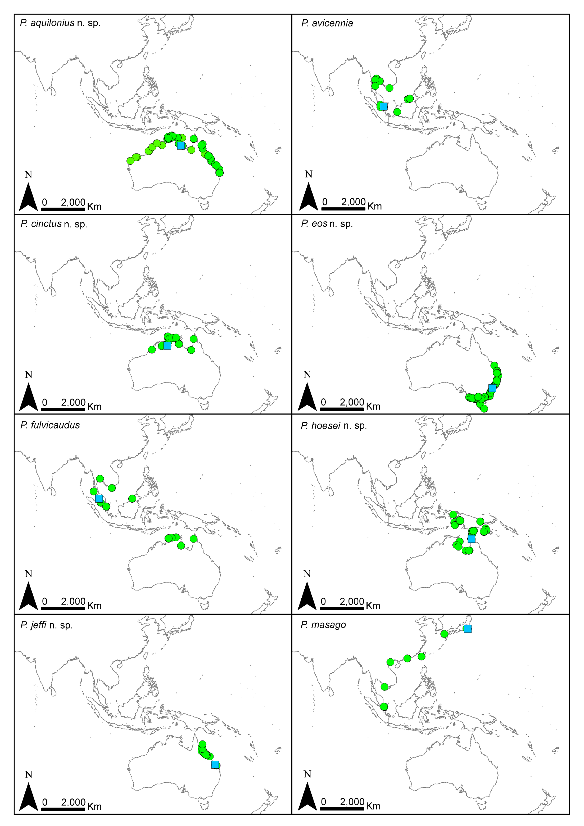
Distribution. Widely distributed throughout western Indonesia, Malaysia, Singapore, Brunei, Thailand, Cambodia, Vietnam, Taiwan, Hong Kong, Philippines and southern Japan; west of Lydekker’s Line.
Ecology. Shallow-water estuarine, found in estuaries of rivers, streams and mangroves, at depths of 0–1 m, especially in mangrove habitats.
Remarks. Bleeker’s seven syntypes of Gobius poicilosoma are not in best condition, with scales mostly missing and one without a head; plus there may be more than one species in the lot (we hope to re-examine them one day and fully resolve this, but have been unable to do so due to RMNH renovation). Two of them are close to Bleeker’s “type” of 39 mm total length. The remaining colour markings on the body and caudal fin (as of 1988 when HKL examined them, see Fig. 24 View FIGURE 24 ), matches the colour patterns observed in recent material (see Fig. 25 View FIGURE 25 ), as do fin ray and scale counts. The colour pattern described in Bleeker’s original description indicates that the side of the body had many irregular dark brown spots and the second dorsal and caudal fins had a mottled or spotted pattern [the name’s etymology: poicilo, mottled, spotted or varicolored; soma, body].
Bleeker (1853) described Gobius gastrospilos from the holotype, RMNH 4676, a 30.5 mm SL specimen in poor condition, with both its jaws missing, fins broken and sensory canals mostly torn open ( Fig. 25D View FIGURE 25 ). It has second dorsal and anal fin ray counts of I,7, 15 pectoral rays, 25 ctenoid lateral scales, 7 TRB scales and 7 predorsal scales that are all cycloid up to close behind the eyes, and the opercle has several rows of cycloid scales. It shares scale and fin ray counts with P. poicilosoma .
Koumans (1932) examined type specimens at RMNH while attempting to revise Stigmatogobius , and considered that Gobius gastrospilos and G. poicilosoma were different species because G. gastrospilos type specimen had scales on the preopercle (but did not say exactly where), while the G. poicilosoma type specimen had no preopercular scales. We have observed that P. poicilosoma occasionally does have one to two scales behind the eye, unlike P. hoesei which always has scales behind the eye but does not occur in the Indonesian archipelago (but does occur in New Guinea and Aru Islands). Koumans described the dark paired blotches at the base of the caudal fin in G. poicilosoma but stated that G. gastrospilos was “uniform brownish” [likely as a result of preservation; Fig. 25D View FIGURE 25 ].
Originally we had considered that Pseudogobius poicilosoma and P. hoesei were conspecific, but as work proceeded we realised that based on biogeography (Sunda/Wallacea versus Sahul regions ( Lohman et al. 2011)), examination of type specimens, their colour patterns and Bleeker’s descriptions, that P. poicilosoma is what has been called P. javanicus . Note that all three type names ( G. poicilosoma , G. gastrospilos and S. javanicus ) were given to specimens collected within close proximity to each other on coastal Java, Indonesia.
The lack of ctenoid nape and opercle scales and colour pattern remnants preclude P. gastrospilos from being a senior synonym of P. melanosticta as proposed by Huang et al. (2014a) (and see under that species).
Vaimosa piapensis Herre, 1927 , is placed in Pseudogobius poicilosoma based on information in Herre’s description and figure as per Larson (2001). Koumans (1940) examined three of Herre’s syntypes at BSM (before they were destroyed in WWII) and stated that they were “Identical with Stigmatogobius javanicus ”.
Nguyen’s 1991 record of Stigmatogobius poicilosoma is illustrated by a drawing of a Pseudogobius , but its species is uncertain.
Huang et al. (2015) presented the mitochondrial genome for P. poicilosoma (as P. javanicus ), based on specimens collected from Kinmen, Taiwan, with additional matching mitochondrial genotypes found by Dahruddin et al. (2017) in Java, Indonesia slightly to the east of Jakarta (topotypic with Gobius gastrospilos ), and by Hammer et al. (2021) in North Sulawesi, Indonesia and Singapore and Vietnam.


| NTM |
Northern Territory Museum of Arts and Sciences |
| T |
Tavera, Department of Geology and Geophysics |
| WAM |
Western Australian Museum |
| R |
Departamento de Geologia, Universidad de Chile |
| YCM |
Yokosuka City Museum |
| NSMT |
National Science Museum (Natural History) |
| CAS |
California Academy of Sciences |
| VI |
Mykotektet, National Veterinary Institute |
No known copyright restrictions apply. See Agosti, D., Egloff, W., 2009. Taxonomic information exchange and copyright: the Plazi approach. BMC Research Notes 2009, 2:53 for further explanation.
|
Kingdom |
|
|
Phylum |
|
|
Class |
|
|
Order |
|
|
Family |
|
|
Genus |
Pseudogobius poicilosoma (Bleeker, 1849)
| Larson, Helen K. & Hammer, Michael P. 2021 |
Pseudogobius javanicus
| Hammer, M. P. & Adams, M. & Unmack, P. J. & Hassell, K. L. & Bertozzi, T. 2021: 2 |
| Huang, S. - P. & Shen, C. - N. & Chen, I-S. 2015: 1 |
| Chen, I-S. & Huang, S. - P. & Huang, K. - Y. 2014: 132 |
| Chen, I. - S. & Weng, C. J. & Chen, Y. - R. & S. - P. Huang & Wen, Z. - H. & Jang-Liaw, N. H. & Tsai, T. H. 2014: 317 |
| Tran, D. D. & Shibukawa, K. & Nguyen, T. P. & Ha, P. H. & Tran, X. L. & Mai, V. H. & Utsugi, K. 2013: 144 |
| Kottelat, M. 2013: 422 |
| Kimura, S. & Satapoomin, U. & Matsuura, K. 2009: 285 |
| Larson, H. K. & Jaafar, Z. & Lim, K. K. P. 2008: 143 |
| Yokoo, T. & Kanou, K. & Moteki, M. & Kohno, H. & Tongnunni, P. & Kurokura, H. 2008: 78 |
| Matsuura, K. & Kimura, S. 2005: 63 |
| Larson, H. K. & Lim, K. P. 2005: 141 |
| Tan, H. H. & Lim, K. K. P. 2004: 111 |
| Senou, H. & Suzuki, T. & Shibukawa, K. & Yano, K. 2004: 235 |
| Nakabo, T. 2002: 1210 |
| Kottelat, M. 2001: 61 |
| Larson, H. K. & Murdy, E. O. 2001: 3601 |
| Sakai, H. & Sato, M. & Nakamura, M. 2001: 115 |
| Randall, J. E. & Lim, K. K. P. 2000: 639 |
| Ng, H. H. & Tan, H. H. & Lim, K. K. P. 1999: 182 |
| Rainboth, W. J. 1996: 207 |
| Tan, S. H. & Tan, H. H. 1994: 356 |
| Kawanabe, H. & Mizuno, N. 1989: 577 |
| Akihito, Prince & Meguro, K. 1975: 46 |
Stigmatogobius javanicus
| Monkolprasit, S. & Sontirat, S. & Vimollohakarn, S. & Songsirikul, T. 1997: 251 |
| Rema Devi, K. 1993: 181 |
| Herre, A. W. 1953: 764 |
Stigmatogobius gastrospilos
| Koumans, F. P. 1932: 8 |
Stigmatogobius poicilosoma
| Koumans, F. P. 1932: 9 |
Vaimosa piapensis
| Herre, A. W. 1953: 768 |
| Herre, A. W. C. T. 1927: 147 |
Gobius javanicus
| Bleeker, P. 1856: 88 |
Gobius gastrospilos
| Bleeker, P. 1853: 477 |
Gobius poicilosoma
| Bleeker, P. 1849: 31 |
| Bleeker, P. 1849: 10 |