
Ptychozoon bannaense, Wang & Liu, 2016
|
publication ID |
https://doi.org/ 10.11646/zootaxa.4084.3.6 |
|
publication LSID |
lsid:zoobank.org:pub:0DB05C07-0166-4B12-BD2F-08DDCD4F0F96 |
|
DOI |
https://doi.org/10.5281/zenodo.6059298 |
|
persistent identifier |
https://treatment.plazi.org/id/923F8E07-5A0A-074D-FF57-A6E618888D5E |
|
treatment provided by |
Plazi |
|
scientific name |
Ptychozoon bannaense |
| status |
sp. nov. |
Ptychozoon bannaense sp. nov.
Figs. 3 View FIGURE 3 , 4 View FIGURE 4 , 5 View FIGURE 5
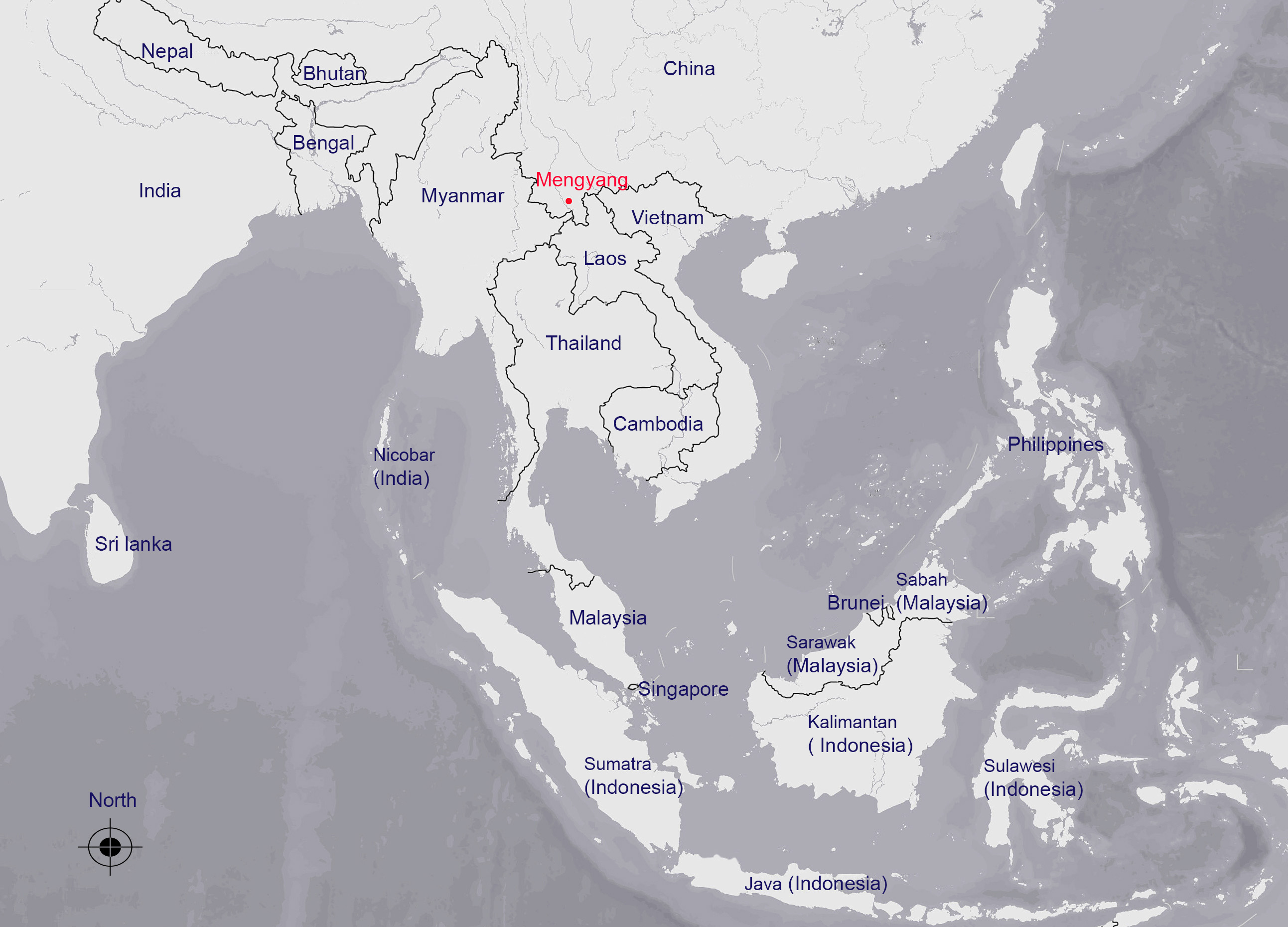
Holotype. SYS ( Sun Yat-sen University Museum of Biology ) r001242, an adult female, from Manheke Village (22° 0819" N, 100°8773" E; 680 m a.s.l., datum = WGS84; see Fig. 1 View FIGURE 1 ), Mengyang Town, Xishuangbanna Dai Autonomous Prefecture, Yunnan Province, China, collected 31 May 2015 by Jian Wang and Hai-Long He.
Paratype. SYS r001237, an adult male, from the same locality as the holotype, collected 30 May 2015 by Jian Wang.
Etymology. The specific epithet “ bannaense ” refers to the type locality, Xishuangbanna, which is usually referred to as “Banna” in Chinese. In the local aboriginal language, the “Xishuang” means “twelve”, the “Banna” means “a small administrative district”. We propose the English vernacular name “Banna Parachute Gecko” and the Chinese vernacular name “Banna San Hu”.
Diagnosis. Ptychozoon bannaense sp. nov. can be diagnosed by the combination of the following characters: (1) body size moderate, SVL 83.2 and 87.5 mm for two specimens; (2) rostral with a short dorsomedian groove; (3) dorsum of head and body covered with granular scales without enlarged tubercles, but male with several enlarged tubercles on the occipital region; (4) supranasals separated from each other by a large internasal; (5) predigital notch in preantebrachial cutaneous flap absent; (6) digits of hands and feet strongly webbed; (7) male possesses 17 preanofemoral pores in a continuous row; (8) the tail with 24 pairs of lateral denticulate cutaneous lobes, ending in a small terminal cutaneous flap; (9) width of tail and caudal lobes progressively decreasing posteriorly; (10) lack of lobe fusion at the terminal caudal flap border; (11) tail tubercle absent; (12) four dark dorsal bands between fore- and hind limbs insertions, the third and fourth dorsal bands fused into an X-shaped mark.
Comparisons. Comparative diagnostic character states, necessary for the recognition of P. bannaense sp. nov., as compared to eight recognized species of Ptychozoon (Table 3) were obtained from the literature ( Boulenger 1890; Taylor 1963; Brown et al. 1997; Brown 1999; Das & Vijayakumar 2009; Sumontha et al. 2012).
Ptychozoon bannaense sp. nov. is the sister taxon of P. lionotum in our phylogenetic tree, and differs from the latter (in parenthesis) by the terminal tail flap short, TFL 5.1 mm (versus 7.8–21.4 mm), not expanded (versus expanded); terminal tail flap edge straight (versus terminal tail flap edge wavy), lacking lobe fusion (versus extensive fusion of 3–9 denticulate lobes); 15 scales across widest portion of tail terminus (versus 24–31); the absence (versus presence) of a predigital notch in the preantebrachial cutaneous flap; the presence of several tubercles on occipital region in male (versus consistently absent enlarged dorsal tubercles); the third and fourth dorsal bands of body fused into a X-shaped mark (versus not fused).
Ptychozoon bannaense is most similar to P. horsfieldii , P. intermedium and P. kaengkrachanense Sumontha, Pauwels, Kunya, Limlikhitaksorn, Ruksue, Taokratok, Ansermet & Chanhome, 2012 in tail morphology. It differs from P. horsfieldii (in parenthesis) by the body size large, SVL 83.2 and 87.5 mm for two individuals (versus SVL 56.8–73.9 mm); the presence of 17 preanofemoral pores in a continuous row (versus 10–11 pore-bearing precloacal and 8–11 pore-bearing femoral scales in a interrupted series); presence of several tubercles on occipital region in male (versus consistently absent enlarged dorsal tubercles); 24 pairs of denticulate tail lobes (versus 15–16); lack 6 6 Ptychozoon bannaense , Ptychozoon F, et al ""2H, """H I = J "H et al H et al G
P. bannaense horsfieldii intermedium kuhli lionotum nicobarense rhacophorus trinotaterra kaengkrachanense F G ’& L’2 % %5 ’L2& " 5’ 5L"’ ’ 5! 5L 2 ’ 2 L"’ 5 (M & %’ ’L5! % 2 %L2 & 2’ L’5
9* 9* L 6 L5 6 9*! 6, 5L, + L 9* *,,
6,
* N + E N M # M E N N E
6
>>>>>
*>>>>> 6
6
* * 4! &!! & F G & &, *
of lobe fusion at the terminal caudal flap border (versus possession of a minimal lobe fusion at its proximal edge). It differs from P. intermedium by the absence of enlarged dorsal tubercles, except occipital region in male (versus consistent presence of dorsal tubercles), the presence of 17 preanofemoral pores in a continuous scale row (versus 8–12 pore-bearing precloacal and 12–19 pore-bearing femoral scales in a interrupted series); lack of lobe fusion at the terminal caudal flap border (versus 2-4 lobes fuse at the anterior border); supranasals separated by an internasal (versus internasal absent, supranasals in contact with each other). It differs from P. kaengkrachanense by supralabials 11–13 (versus 8–10), ventral scales in 37 rows (versus 24–25 rows); supranasals separated by an internasal (versus internasal absent, supranasals in contact with each other); the presence (versus absence) of a short dorsomedian rostral groove; four dark transverse dorsal bands between limbs insertions, the third and fourth fused into a X-shaped mark (versus only three distinct bands).
The new species can be clearly distinguished from the remaining four species in morphology. It differs from P. kuhli by a 5.1 mm long terminal tail flap (versus 21.4–30.3 mm), 15 scales across widest portion of tail terminus (versus 42–51); the terminal tail flap is semicircular in shape, not expanded, and non-denticulate without lobe fusion (versus terminal flap elongated, widely expanded, and with minimal lobe fusion), the absence of enlarged dorsal tubercles, except occipital region in male (versus present 2–6 straight dorsal rows), the absence (presence) of tail tubercles; it differs from P. rhacophorus by the absence of enlarged dorsal tubercles, except occipital region in male (versus present 6–10 dorsal rows), the terminal tail flap present (versus absent), semicircular in shape (versus sharply tapering), SVL 83.2 and 87.5 mm for two individuals (versus 58.8–64.5 mm), supranasals separated by an internasal (versus internasal absent, supranasals in contact with each other), the presence (versus absence) of a short dorsomedian rostral groove; it differs from P. trinotaterra by the terminal tail flap is semicircular in shape, not expanded, and non-denticulate without lobe fusion (versus terminal tail flap widened, at most slightly beyond nearest lobe, and with only a single lobe fusion evident anteriorly), 15 (versus 26) scales across widest portion of tail terminus; the absence of enlarged dorsal tubercles, except occipital region in male (versus the presence of a single midvertebral dorsal tubercle row in a Thai specimen and more numerous differentiated nuchal and temporal region tubercles in a juvenile Vietnamese specimen), SVL 83.2 and 87.5 mm for two individuals (versus 70.5–71.3 mm), the absence (versus presence) of tail tubercles, possession of 17 (versus 19–21) preanofemoral pores, the presence (versus absence) of a short dorsomedian rostral groove; it differs from P. nicobarensis Das & Vijayakumar, 2009 by the absence of enlarged dorsal tubercles, except occipital region in male (versus four irregular dorsal rows), the terminal tail flap is semicircular in shape, not expanded, without lobe fusion (versus terminal tail flap expanded, with “weak” lobe fusion at proximal border), 15 (versus 20–29) scales across widest portion of tail terminus; supranasals separated by an internasal (versus internasal absent, supranasals in contact with each other).
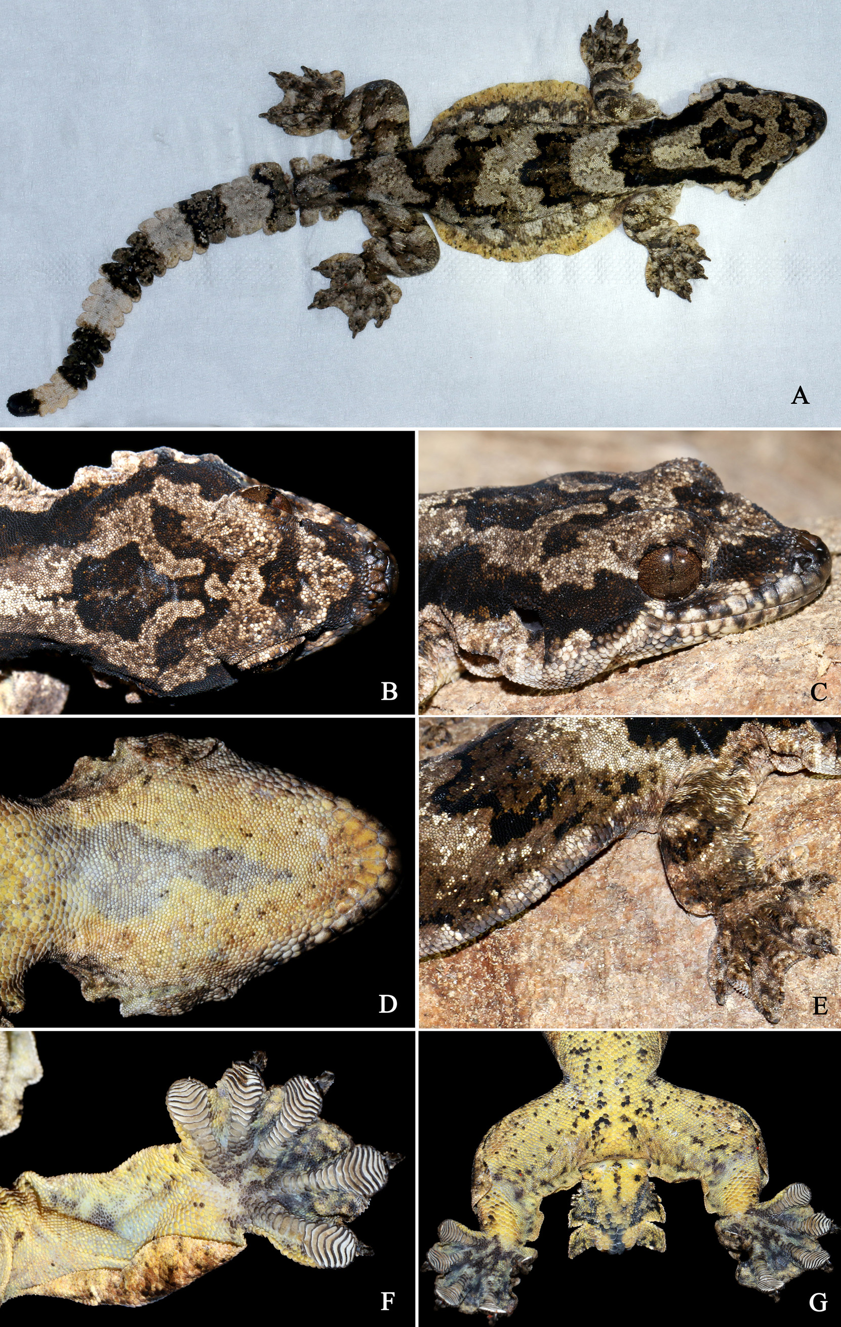
Description of holotype. An adult female individual with an original tail ( Figs. 3 View FIGURE 3 , 4 View FIGURE 4 ); measurements presented in Table 4.
Habitus strongly dorsoventrally depressed; body size moderate, SVL 83.2 mm; AG 39.9 mm, 48% of SVL; head flattened and broad, distinct from the neck, HD 39% of HL, HW 86% of HL; snout long, significantly sloped, acutely tapered, rounded at tip; SL 39 % of HL, and 45% of HW; IND 16% of HW; IOD large, 46% of HW; eye circular, slightly large, ED 70% of HD, 24% of HL, and 63% of SL; pupil vertical, its margin wavy; auricular opening small, circular, AOW 14% of HL and 51% of ED; EAO large, 32% of HL; tympanum deeply sunken; infra-auricular cutaneous flap rounded, broad, measuring 4.0 mm at its widest point, beginning from below angle of jaw at a distance of 1.4 mm, posteriorly extending on side of head below auricular opening, and forming an indistinct second lobe, tapering into the lateral region of the anterior neck; subcutaneous endolymphatic sacs not evident.
Extensive laterally expanded cutaneous parachute with rounded, slightly notched edge, extending on the side of torso from axilla to groin; greatest midbody width 53% of extended parachute width at widest point. Rostral convex and subrectangular, measuring 4.0 mm in width, 1.8 mm in depth; a short dorsomedian groove in upper part of rostral; rostral in contact with two first supralabials laterally, with two supranasals and single internasal posteriorly; external nares rounded, directed laterally, each surrounded by five scales: rostral anteriorly, supranasal dorsally, first supralabial ventrally, two postnasals posteriorly; two triangular supranasals separated from each other by a large internasal; five slightly enlarged oblong scales transversally arranged in a regular row between two upper postnasals along posterior margins of supranasals and internasal; dorsal surface of head and neck covered with granular scales without enlarged tubercles; scales on rostrum, loreal region, upper eyelid and supraorbital region larger than those on between orbits and occipital region; the outer edge of upper eyelid composed of significantly enlarged rectangular scales; supraciliaries triangular, posterior ones bearing minute conical spines; one patch of large scales on upper margin of auricular opening; upper surface of infra-auricular cutaneous flap composed of basal imbricate, enlarged flabellate scales and distal smaller scales; its lower surface composed of small granules; supralabials rectangular, 11/13 (L/R), posterior ones much reduced in size, and series followed by smaller scales to angle of mouth; sixth and succeeding supralabials located under orbit, separated from orbit by one row of enlarged scales, bordered ventrally by supralabials; infralabials rectangular, 11/12 (L/R), followed by several small scales to angle of mouth; mental pentagonal, slightly broader than deep, and bordered by left and right postmentals posteriorly, which contact medially for approximately two thirds of their length; postmentals pentagonal, twice as long as wide; one row of differentiated, enlarged sublabials bordered by all infralabials; one patch of enlarged, imbricate scales below most posterior infralabials; gular scales granular, juxtaposed, gradually transitioning into enlarged, flattened, smooth, subimbricate pectoral scales, and posteriorly imbricate flabellate ventral scales which gradually decrease in size laterally, becoming minute rounded granular scales; ventral scales in 37 transverse rows across the abdomen between the flanks, below the base of the midbody cutaneous parachute; 20 continuous dimpled scales in the precloacal pore-bearing series, arranged in a slightly bowed, inverted V-shaped, followed by six rows of similarly enlarged but not dimpled scales and five rows of gradually reduced scales reaching the horizontal cloacal groove; a pair of cloacal gland openings each followed by a caudolaterally oriented slanted series of five imbricate enlarged scales, ending in a single protuberant cloacal spur on each side of ventral tail base; postcloacal swellings slightly distinct.
Dorsum of body covered with nearly uniform, juxtaposed granular scales without enlarged tubercles; transverse dorsal body scales in 84 rows (above the midbody parachute); upper surface of cutaneous parachute of torso composed of imbricate, serrated trailing edged, strongly elongated rectangular support scales, arranged in regular transvers rows, counting 11–12 scales per row across midbody parachute; lower surface of cutaneous parachute with subimbricate, small, elongate rectangular scales with rounded trailing edges, arranged in regular transvers rows across midbody parachute, with 20–24 scales per row; the outer most edge of cutaneous parachute composed of differentiated triangular scales mixed with minute conical spines.
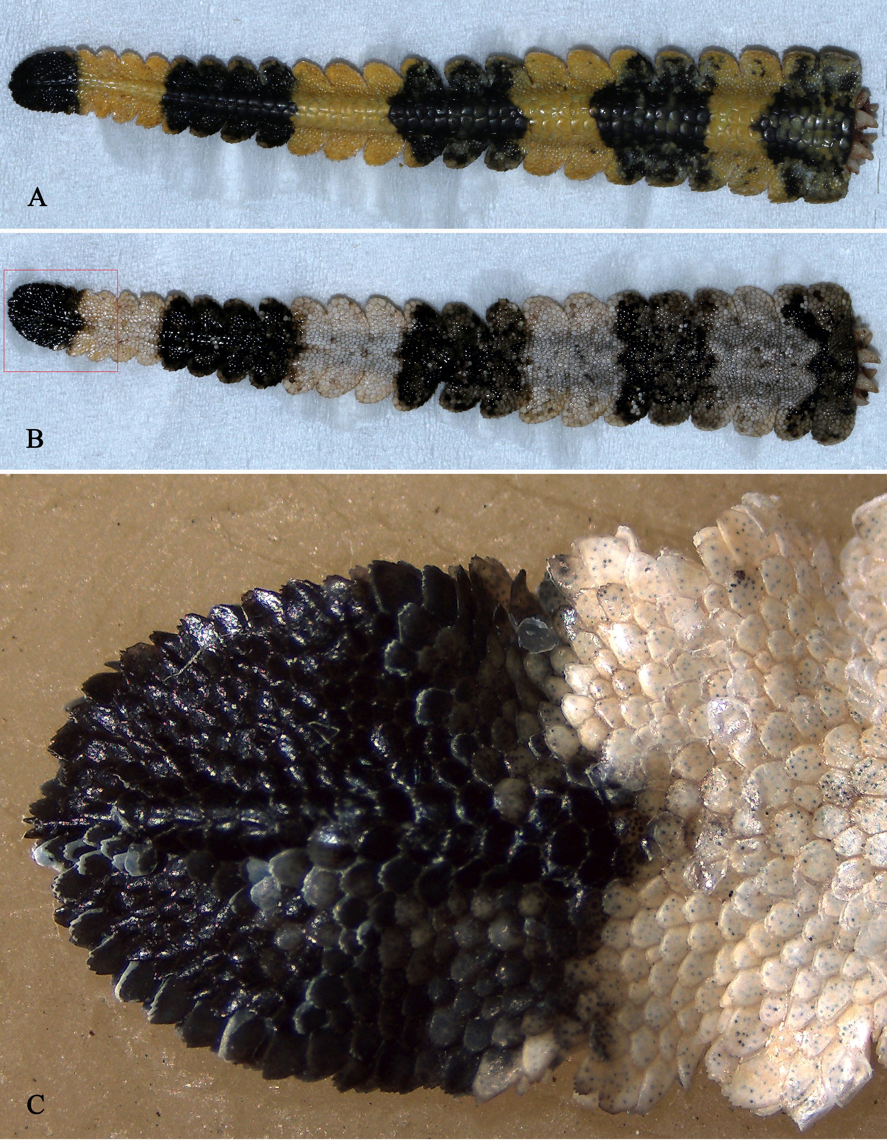
Tail moderate, TaL 79.6 mm, 97% of SVL; tail segmented, strongly depressed dorsoventrally, with 24 pairs of lateral denticulate cutaneous lobes along each side of the tail and a single semicircular terminal cutaneous flap; caudal lobes with posterior angling; width of tail and caudal lobes progressively decreasing posteriorly; basal tail segment bearing first pair of lobes with two smaller sublobes anteriorly, followed caudally by segments each bearing a pair of lobes from second to 24th tail segment; terminal cutaneous flap short, not expanded, TFL 5.1 mm, 6.4% of TaL; terminal tail flap edge straight, lacking lobe fusion; 15 scales across widest portion of tail terminus; tail smooth, lacking tubercles, flattened ventrally; dorsal caudal scales slightly convex, imbricate, round to hexagonal, with weakly serrated trailing edge; four to nine dorsal caudal scale rows to a segment, the most anterior and posterior enlarged; dorsal surface of tail lobes and terminal flap with imbricate scales with serrated trailing edges, their most outer edges composed of large scales with strongly serrated trailing edge, intermixed with conical spines; subcaudal scales enlarged, imbricate, with somewhat serrated trailing edge; median pairs of subcaudal scales largest, usually three rows to a segment; subcaudal series continuing onto tail terminus; lower surface of tail lobes and terminal flap covered with differentiated enlarged, imbricate flat scales.
Limbs brawny, FL 10% of SVL, TBL 15% of SVL; forelimb with wide cutaneous flaps, no predigital notch; cutaneous flap extending along entire anterior margins of forelimb, narrowing and ending in lateral margin of the thumb at level of proximal first lamella, followed by a narrow cutaneous flap along lateral margin of the thumb from the level of the proximal second lamella to widest point of thumb; another extending along entire posterior margins of forelimbs, ending on the wrist joint; a narrow cutaneous flap along posterior margin of the palm from base of finger V to its widest point; hind limbs with wide cutaneous flaps, one of which extends along entire posterior margins of hind limbs, tapering to lateral margin of toe V, ending at its widest point; other beginning at anterior margin of thigh from knee at a distance of 3.8 mm, through the knee, posteriorly extending along anterior margin of tibia, tapering into distal tibia from ankle joint at a distance of 3.1 mm; digits widely dilated; webbing extending to widest point on distal part of each digit; subdigital lamellae undivided, 11–13–15–17 –12 on both right/left fingers I-V respectively, 13–12–15–17 –13/13–13–15–16–15 on right/left toes I–V respectively; two distal phalanges of outer four digits compressed, rising free at distal end, tending to form an angle with remainder of digit, and attached to the widened part; terminal phalanges and claw absent from inner finger and toe; outer four digits bearing recurved claws; dorsal surface of limbs, digits and webbings covered with imbricated, cycloid, scales with serrated trailing edges; the unclawed inner finger and toe with an oval large scale; dorsal support scales of limb cutaneous flaps larger than those on dorsal surface of limbs, digits and webbings; ventral surface of limbs and flaps covered with imbricated, cycloid scales, smaller than dorsal flap surface; ventral surface of webbings with small granular scales.
Coloration in life. Dorsal background color light brown to grey-white; dorsal head decorated with two distinctive black markings: a transverse wavy band in frontal region, another anchor-shaped, extended from parietal region to occipital region; four black bands on dorsum of torso between fore- and hind limb insertions: first band U-shaped, with wavy posterior edge, beginning from tip of snout, covering loreal and temporal region, posteriorly extending along upper margin of auricular opening to the shoulder; second U-shaped with wavy posterior edge, extending from axillae to midbody; third and fourth fused into a X-shaped mark on posterior dorsum of body before hind limbs insertion; another wavy black dorsal band on base of tail, anteriorly in contact with first transvers bands of thighs; dorsal and ventral surfaces of tail with six black transvers bands; fore- and hind limbs with dorsal black transvers bands; ventral surface of head, body, tail and limbs yellow; chin, throat, and breast with little scattered black spots; venter and ventral surface of limbs with denser black spots; subdigital lamellae grey white to yellowish; cloacal opening light colored; iris brown.
Coloration in preservative. In 75% ethanol, coloration and pattern do not significantly differ from coloration in life.
Variation. Measurement, proportions, and scale counts are provided in Table 4. The adult male paratype (SYS r001237) resembles the holotype in overall morphology, but shows secondary male sexual characters (see Fig. 5 View FIGURE 5 ): the presence of 17 preanofemoral pores, and distinct postcloacal swellings. Moreover, the paratype differs from the holotype by the presence of several enlarged occipital region tubercles; the presence of slightly enlarged, flattened paravertebral scales arranged in 2–3 longitudinal rows; possession of a dorsal background color dark brown-red; and by the presence of only indistinct body bands and black markings on top of head.
Distribution, ecological notes and behavior. Currently, Ptychozoon bannaense sp. nov. has only been recorded from Xishuangbanna Dai Autonomous Prefecture, Yunnan Province, China. The paratype (SYS r001237), lacked a tail, was difficult to discern from its surroundings and was captured at 0:25 a.m.on the surface of a road within a moist tropical rainforest. The next night, three adult individuals with original tails, including the holotype (SYS r001242), were simultaneously discovered at about 22:00 on the branches of a tall tree (3 m dbh)at heights between 13– 14 m. The tree was very close (2 m) to the collection site of the paratype and about 10 m from a nearby stream (see Fig. 6 View FIGURE 6 ). The color pattern of these individuals perfectly matched the mottled tree bark, and they could be detected only by noting their occasional slowly crawling movements along the branches. After disturbance from a long bamboo pole, one individual (SYS r001242) parachuted from a height of 13 m to another tree trunk. During this directed areal descent, its cutaneous flaps and digital webbings expanded, limbs were stretched out, tail was held out straight, and mouth was held open, while it travelled in a descending arc for a horizontal distance of 2.5 m, and a vertical distance of about 10 m.


| SYS |
Zhongshan (Sun Yatsen) University |
No known copyright restrictions apply. See Agosti, D., Egloff, W., 2009. Taxonomic information exchange and copyright: the Plazi approach. BMC Research Notes 2009, 2:53 for further explanation.
|
Kingdom |
|
|
Phylum |
|
|
Class |
|
|
Order |
|
|
Family |
|
|
Genus |
Ptychozoon bannaense
| Wang, Ying-Yong Wang Jian & Liu, Zu-Yao 2016 |
Ptychozoon bannaense
| Wang & Liu 2016 |
Ptychozoon bannaense
| Wang & Liu 2016 |
P. kaengkrachanense
| Sumontha, Pauwels, Kunya, Limlikhitaksorn, Ruksue, Taokratok, Ansermet & Chanhome 2012 |
kaengkrachanense
| Sumontha, Pauwels, Kunya, Limlikhitaksorn, Ruksue, Taokratok, Ansermet & Chanhome 2012 |
trinotaterra
| Brown 1999 |
P. intermedium
| Taylor 1915 |
intermedium
| Taylor 1915 |
lionotum
| Annandale 1905 |
Ptychozoon
| Kuhl & van Hasselt 1822 |