
Pugettia intermedia Sakai, 1938
|
publication ID |
https://doi.org/ 10.11646/zootaxa.4672.1.1 |
|
publication LSID |
lsid:zoobank.org:pub:01B7FFA9-1EC8-41A7-8DDE-418D9F4882B8 |
|
persistent identifier |
https://treatment.plazi.org/id/FF0187EC-FF80-CE6A-EAE7-F9CEFDB0F1C3 |
|
treatment provided by |
Plazi |
|
scientific name |
Pugettia intermedia Sakai, 1938 |
| status |
|
Pugettia intermedia Sakai, 1938 View in CoL
[Japanese name: Yotsuha-modoki]
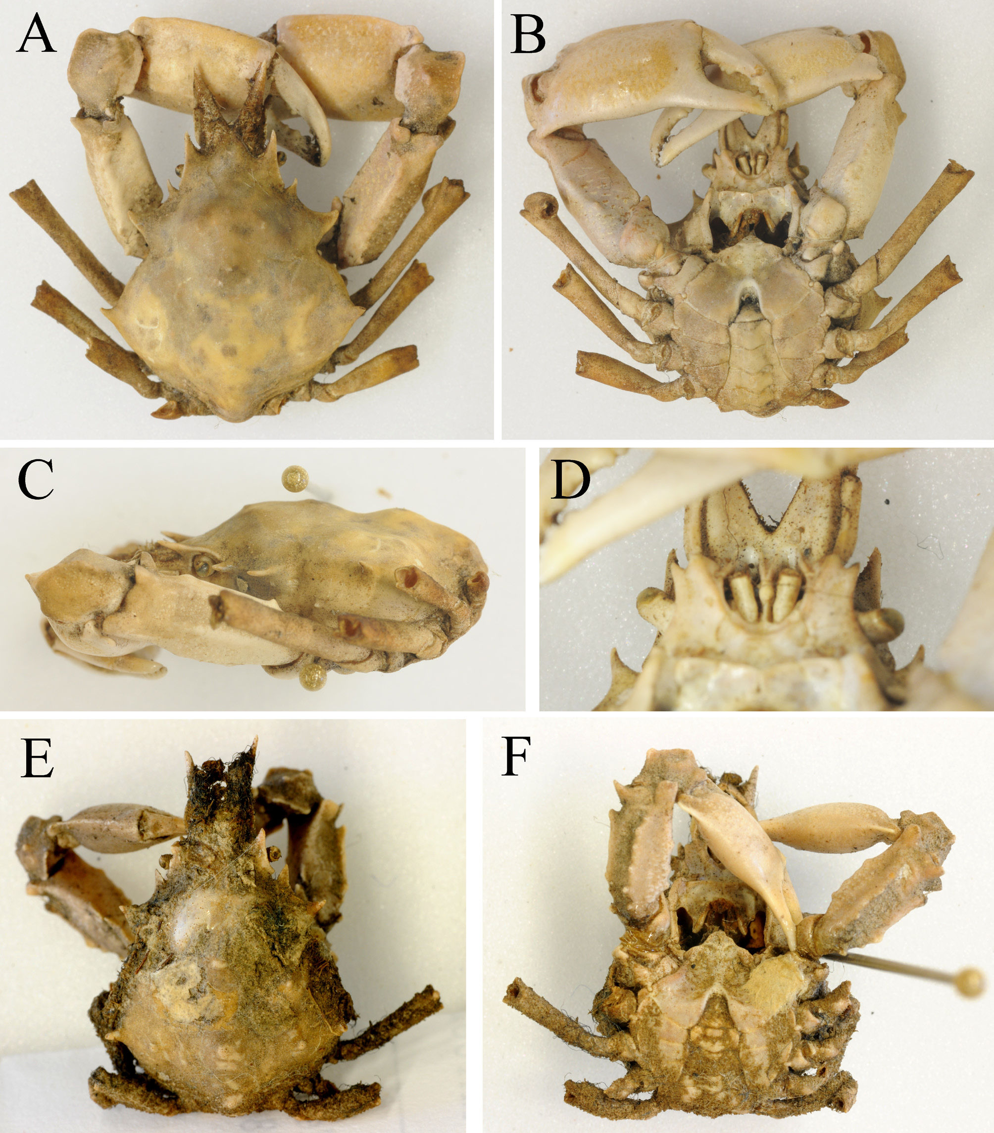
( Figs. 2E, F View FIGURE 2 , 14–24 View FIGURE 14 View FIGURE 15 View FIGURE 16 View FIGURE 17 View FIGURE 18 View FIGURE 19 View FIGURE 20 View FIGURE 21 View FIGURE 22 View FIGURE 23 View FIGURE 24 )
Pisa (Menoethius) [sic.] quadridens . —De Haan, 1839: 97–98 (in part, as paralectotype).— Yamaguchi & Baba 1993: 353, fig. 113; 2003: 38 (in part, as paralectotype). [not Pisa (Halimus) quadridens ]
Pugettia minor View in CoL .— Shen 1937: 287–289, text-fig. 5a, d, f, g. [not Pugettia minor Ortmann, 1893 View in CoL ]
Pugettia quadridens intermedia Sakai, 1938: 258 View in CoL , pl. 36, fig. 2; 1965, 72, pl. 32, fig. 3; 1976: 197, text-fig. 103b [type locality: Simoda (= Shimoda, Izu Peninsula)].— Miyake 1983: 206 (list); 1998: 206 (list).— Ariyama 1995: fig. 2, 3a–c, 4.
Pugettia similis View in CoL .— Sakai 1976: 200, text-fig. 107b [not Pugettia similis Rathbun, 1932 View in CoL ]
Pugettia intermedia View in CoL .— Griffin & Tranter 1986: 93–95, fig. 28a, b.— Yamaguchi et al. 1987: 13, pl. 4, fig. 2.— Muraoka 1998: 24 (in part), tbl. 1 (not holotype).— Marumura & Kosaka 2003: 32 (in part).— Ng et al. 2008: 101 (list).— Yamaguchi & Henmi 2008: 80, figs. 1b.— Wicksten & Stachowicz 2013: 359 (list).
Pugettia quadridens View in CoL .— Yamaguchi & Baba 1993: 353, fig. 113; 2003: 38 (in part).— Muraoka 1998: 24 (in part).— Nabeshima 2011: 124, 1 unnumbered figure; 2013: 124, 1 unnumbered figure [not Pisa (Halimus) quadridens De Haan, 1837 View in CoL ]
Pugettia quadridens pellucens View in CoL . — Marumura & Kosaka 2003: 32 (in part) [not Pugettia quadridens pellucens Rathbun, 1932 View in CoL ] Pugettia vulgaris View in CoL . — Yang et al. 2015: 203–206, figs. 1E, F, 3. [not Pugettia vulgaris Ohtsuchi, Kawamura & Takeda, 2014 View in CoL ]
? Pugettia quadridens intermedia View in CoL .— Ikeda 1981: 15 (in part).— Kim & Kim 1986: 325.
? Pugettia intermedia View in CoL .— Watanabe 2014: 41, unnumbered figure.
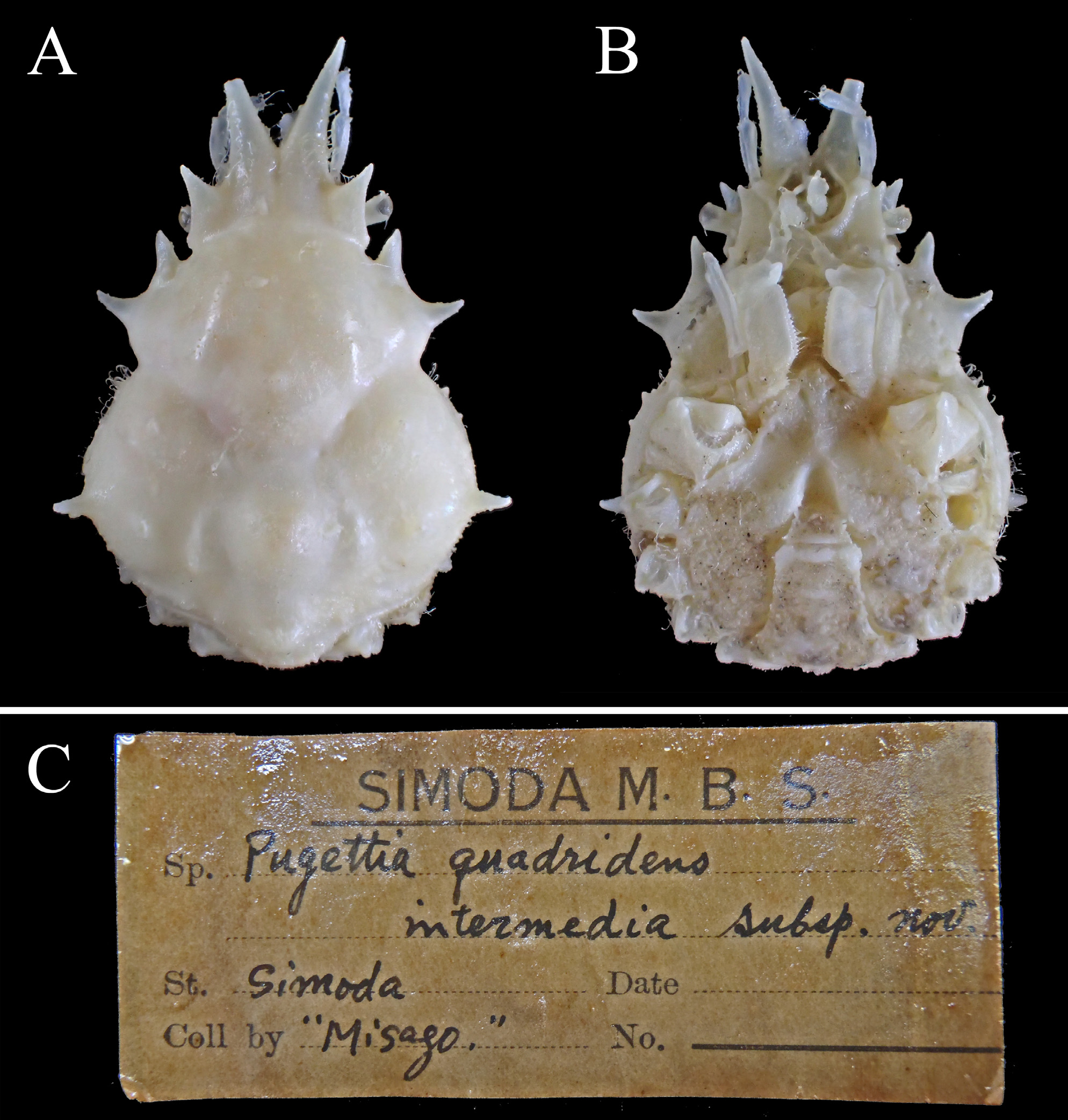
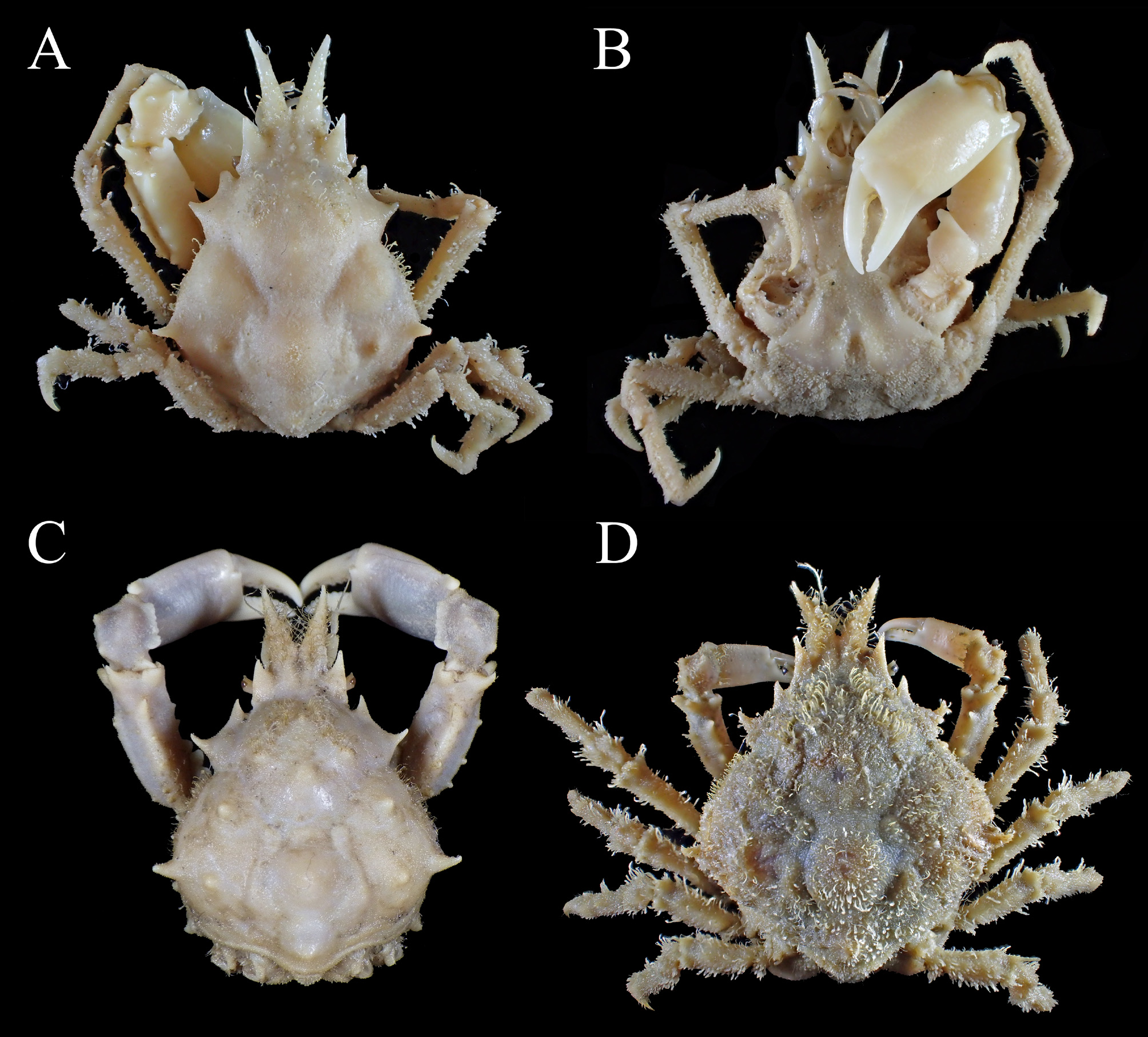
Material examined. Paratype: 1 male (13.9 × 12.5 mm) ( KPM-NH 124160 ), Shimoda, Izu Peninsula, coll. Misago. [not holotype though listed in Muraoka (1998: 24, table 1)]
Non-types: Japan. One male ( RMNH D 42298), Japan, coll. H. Bürger, 1825–1834 (as one of paralectotypes of Pisa (Menaehius) quadridens De Haan, 1839 ); 1 male (15.9 × 12.0 mm) ( CBM-ZC 2574 ), 50–60 m, off Takeoka, Uchibo, Boso Peninsula , gill-net, coll. T. Komai, 1 May 1996 ; 1 male (26.5 × 20.9 mm) ( CBM-ZC 5128 ), 30–40 m, same locality as previous, gill-net, coll. T. Komai, 1 Feb. 1998 ; 1 ovigerous female (14.1 × 10.9 mm) ( KPM-NH 110375), 1 ovigerous female (12.9× 10.1 mm) ( KPM-NH 110376), Sagami Bay ; 2 males (28.0 × 23.1, 28.4 × 23.8 mm) (WMNH-Na-Cr 0314-2), Himaga I., Aichi, coll. S. Nagai ; 1 male (25.0 × 19.7 mm) ( KPM-NH 104634 ), same locality as previous ; 1 male (20.2 × 16.9 mm) ( KPM-NH 104520 ), Shima, Mie ; 1 male (9.5 × 6.4 mm), 3 females (6.8 × 4.8–9.5 × 6.8 mm) (WMNH-Na-Cr 313-1), 30 m, Shionomisaki, Kii Peninsula , Wakayama, coll. S. Nagai ; 3 males (3.9 × 2.7–12.1 × 8.9 mm) (OMNH-Ar 9928), Nishihiro, Hirogawa-cho, Arida-gun , Wakayama, coll. T. Yamahsita, 19 Aug. 2000 ; 1 male (18.4 × 13.8 mm) ( KPM-NH 104049 ), Minabe, Hidaka-gun , Wakayama (examined in Sakai 1976) ; 1 male (13.4 × 9.8 mm) ( KPM-NH 110363), Sakai , Minabe, Hidaka-gun, Wakayama ; 1 male (23.4 × 19.6 mm) (OMNH-Ar 10700), 1 male (13.6 × 9.7 mm) (OMNH-Ar 10701), 1 ovigerous female (20.0 × 15.6 mm) (OMNH-Ar 10702), 1 ovigerous female (15.7 × 12.3 mm) (OMNH-Ar 10703) Yura Fishery Port, Yura, Sumoto , Hyogo, 2 m, trap, coll. T. Watanabe, 3 Jun. 2017 ; 1 ovigerous female (19.5 × 14.9 mm) (OMNH-Ar 6188), 34°21´N– 135°06´E, near large fishing bank, 35–55 m, off Misaki-cho, Sennan-gun , Osaka, coll. H. Ariyama, 26 Mar. 1996 GoogleMaps ; 4 males (18.4 × 14.0–26.7 × 21.1 mm), 3 ovigerous females (16.6 × 13.1–22.9 × 19.3 mm) (OMNH-Ar 6217), Nisikiminami-machi, Hannan roku-ku, Kaizuka-shi , Osaka, coll. H. Ariyama, 28 Mar. 1995 ; 1 male (22.9 × 18.9 mm) (OMNH-Ar 6117), off Sakai-shi , Osaka, stone dredge net, coll. H. Ariyama, 29 May 2000 ; 2 males (26.3 × 21.8, 28.1 × 24.2 mm) (OMNH-Ar 6199), from Kobe-shi, Hyogo, to Sakai-shi, Osaka, Osaka Bay , stone dredge net, coll. H. Ariyama, 15 Feb. 1995 (examined in Ariyama 1995) ; 1 female (28.5 × 23.6 mm) (OMNH-Ar 6208), 2 km off the coast of Kobe-shi to Ashiya-shi, Hyogo, Osaka Bay , coll. H. Ariyama, 8 Feb. 1994 (examined in Ariyama 1995) ; 1 ovigerous female (23.7 × 19.4 mm) (OMNH-Ar 6198), inner part of Osaka Bay , coll. H. Ariyama, 16 Mar. 1998 ; 1 male (19.1 × 15.3 mm), 1 ovigerous female (26.4 × 20.9 mm) (OMNH-Ar 6207), same locality as previous, coll. H. Ariyama, 22 Feb. 1996 ; 1 male, (21.6 × 16.1 mm) (OMNH-Ar 6037), off Nankou, Suminoe-ku, Osaka-shi , Osaka, coll. H. Ariyama, 4 Mar. 1993 (examined in Ariyama, 1995) ; 3 males (5.1 × 3.8–7.9 × 5.7 mm), 2 females (7.3 × 5.1, 8.8 × 6.3 mm) (OMNH-Ar 6025), near Osaka Aquarium Kaiyukan , Osaka, coll. H. Ariyama, Jun. 1998 ; 1 ovigerous female (11.2 × 8.7 mm) (OMNH-Ar 3805), subtidal zone, Maiko, Tarumi-ku, Kobe-shi , Hyogo, coll. R. Yamaguchi, 15–17 Jun. 1993 ; 1 female (15.3 × 11.6 mm) (OMNH-Ar 9922), Shizuki, Awaji-shi , Hyogo, coll. Y. Nakajima & Y. Fukui, 30 Jun. 1974 ; 1 ovigerous female (14.7 × 11.5 mm) (OMNH-Ar 10704), off Karakoto, Kurashiki , Okayama, 0.3 m, muddy sand bottom, coll. T. Watanabe, Apr. 2017 ; 1 male (19.5 × 15.3 mm) ( KPM-NH 104142 ), Tosa Bay ; 1 female (21.3 × 17.0 mm) ( TOYA Cr 10297), 20 m, off Mizuhashi, Toyama-shi , Toyama Bay , coll. N. Miyamoto, 1 Jul. 1990 ; 2 males (16.8 × 13.0, 21.1 × 16.6 mm), 3 ovigerous females (19.3 × 15.8–26.4 × 21.8 mm) (NSMT-Cr 26068), Maizuru Fisheries Research Station, Kyoto University, Maizuru , Tango Sea , coll. K. Sakemi, 13–17 Mar. 2013 ; 2 males (11.3 × 8.3, 18.1 × 14.3 mm) (TRPM-AAr-0000499), Makiya, Iwami , Tottori, coll. Y. Hirata, 20 Sep. 2009 ; 1 female (with rhizocephalan parasite, 16.7 × 12.8 mm) ( KMNH IvR 100011), Nomozaki, Nagasaki, Nagasaki Peninsula, Amakusa Sea , setnet around the rocky reef, coll. K. Matsubayashi, Apr. 1961 ; 1 male (10.8 × 8.1 mm) ( KMNH IvR 100008), Futae, Amakusa Archipelago , trawl net, coll. K. Baba, 26 Feb. 1935 ; 1 ovigerous female (15.4 × 11.4 mm) (OMNH-Ar 4271), Koujiro, Kunimi-cho , Nagasaki, 16 May 1999 .
East China. One female (21.3 × 18.7 mm) ( MBM 160490), around the Qingdao No. 2 high school, coll. Baoling Wu, 23 Dec. 1963 ; 1 male, (22.9 × 18.2 mm) ( MBM 160493), No. 1 Bathing Beach, Qindao, coll. Yang & Zhang, 5 Dec. 1956 ; 1 male (26.2 × 20.7 mm), 1 ovigerous female (22.0 × 17.5 mm) (ZMUC-CRU-20234), Formosa Strait, 23°08'N 117°30'E, 43 m, coll. Kapt. Svenson, 23 Jan. 1912 (figured by Griffin & Tranter 1986); 2 females (7.3 × 5.1, 6.1 × 4.2 mm) (ZMUC-CRU-20235), Formosa Strait, 23°57'N 118°33'E, 50 m, April 1897 (examined in Griffin & Tranter 1986).
Redescription. Male. Full-grown males (18.4–28.6 mm PCL). Carapace ( Fig. 15A View FIGURE 15 ) pyriform, 1.3 longer than width (PCL/CW = 1.3±0.1, N = 18), surface microscopically tomemtose, sparsely with elongate setae; gastric, cardiac, branchial, intestinal regions moderately separated from each other. Gastric region ( Fig. 15A, C View FIGURE 15 ) moderately elevated, always anteriorly with oblique row of hooked setae on either side of midline, posterior end positioned posterior to hepatic lobe basis ( Figs. 15C View FIGURE 15 , 24C, D View FIGURE 24 ); mesogastric, metagastric, protogastric region on both sides each with distinct protuberance ( Figs. 15A, C View FIGURE 15 , 24A View FIGURE 24 ). Hepatic region not markedly elevated, sparsely with long setae. Cardiac, branchial regions ( Figs. 15A, C View FIGURE 15 , 24C, D View FIGURE 24 ) moderately elevated; mesobranchial region elevated, as high as gastric, cardiac regions, with 2 distinct, obtuse tubercles apically, mesial one larger than lateral one; metabranchial region faintly elevated, with low, obtuse tubercle apically. Intestinal region moderately elevated, separated from cardiac region, with obtuse protuberance apically ( Figs. 15C View FIGURE 15 , 24C, D View FIGURE 24 ). Tufts of a few elongate setae on 2 medial protuberances of gastric region, midpoint of epibranchial region, apex of epibranchial spines, summits of cardiac, intestinal regions ( Figs. 15A, C View FIGURE 15 , 24D View FIGURE 24 ).
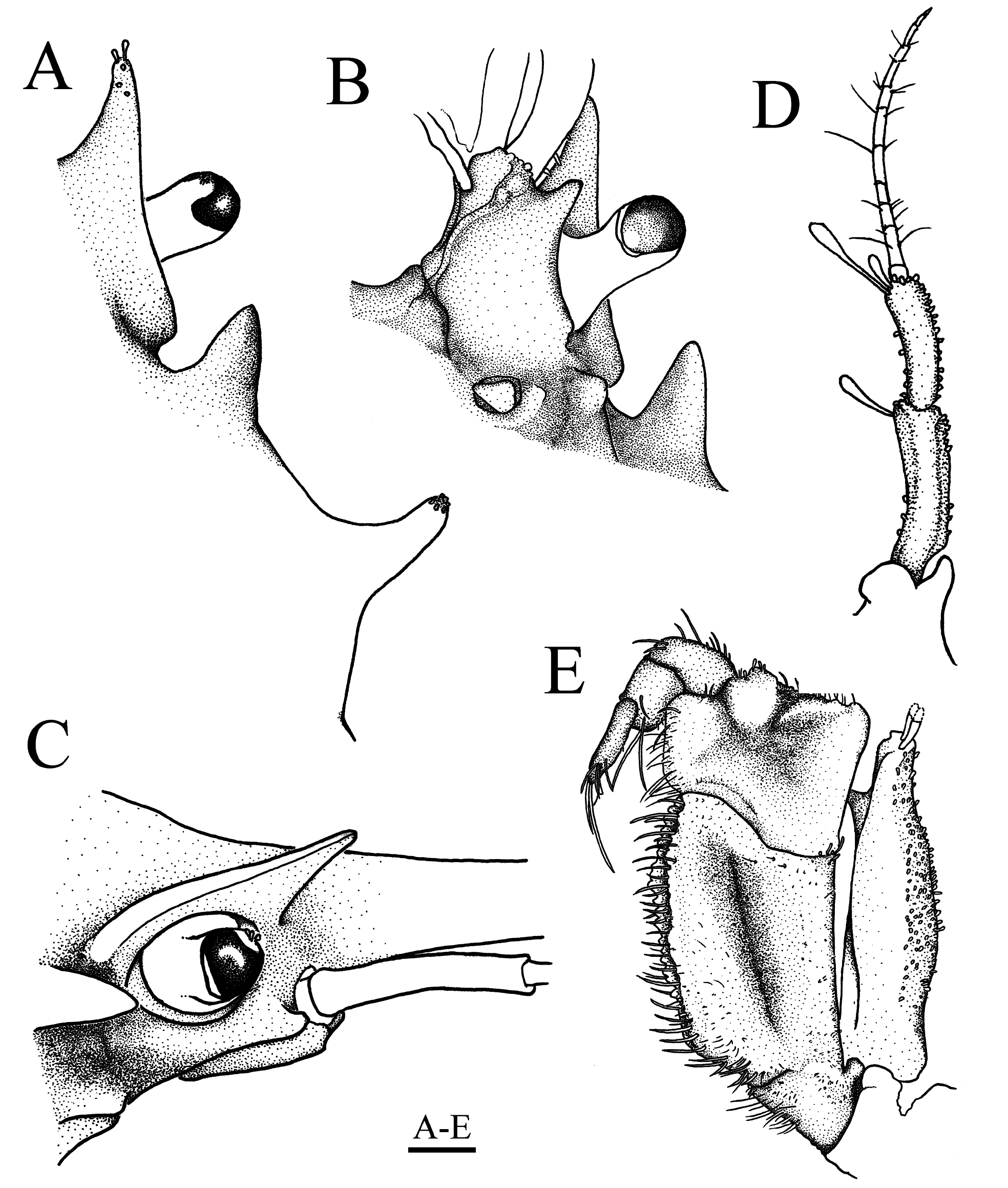
Pseudorostral spines ( Fig. 15A View FIGURE 15 ) relatively long, length 0.2–0.3 of post-pseudorostral carapace length (PRL/ PCL = 0.3±0.0, N = 17), each with two rows of sparse, hooked setae on proximal 0.8 dorsally, nearly connected to rows on protogastric region, single row of simple, long setae on proximal 0.8 mesially; lateral margins divergent anteriorly. Preorbital spine ( Figs. 15A, C View FIGURE 15 , 16A View FIGURE 16 ) triangular, acute at tip, with row of slender setae on mesial margin, compressed dorsoventrally, directed anterolateraly. Supraorbital eave ( Figs. 15A View FIGURE 15 , 16A, B View FIGURE 16 ) moderately extended laterally, slightly concave on lateral margin, distinctly truncated on posterior end ( Fig. 16A View FIGURE 16 ). Orbital hiatus ( Figs. 15A, B View FIGURE 15 , 16A View FIGURE 16 ) large, deep, subrectangular sulcus in dorsal view. Postorbital lobe ( Figs. 15A, B View FIGURE 15 , 16A View FIGURE 16 ) elongate, triangular, subequal to or slightly shorter than supraorbital spine, compressed dorsoventrally, directed anterolateraly, slightly incurved distally. Hepatic lobe ( Figs. 15A View FIGURE 15 , 16A View FIGURE 16 ) faintly demarcated from gastric region, elongate, twice or more than twice longer than postorbital lobe (HpL/PoL = 2.1±0.2, N = 8), directed anterolaterally, acuminate at tip, directed anterolaterally, lateral margin bent outwards in distal half; concavity between postorbital, hepatic lobes extended as low, lamellar plate. Anterolateral carapace margin ( Fig. 15A, C View FIGURE 15 ) with broad patch of sparse, hooked setae; lateral surface inferior to anterolateral margin with 2–5 (3 in general) subacute tubercles. Epibranchial spine ( Fig. 15A View FIGURE 15 ) large, as long as postorbital lobe, distinctly shorter than hepatic lobe, directed laterally, slightly incurved distally, acuminate at tip, positioned at posterior 0.3 of post-pseudorostral carapace length (ESL/PCL = 0.3±0.0, N = 8), distinct from posterolateral carapace margin at base. Posterolateral carapace margin ( Fig. 15A View FIGURE 15 ) faintly convex. Posterior carapace margin ( Fig. 15A View FIGURE 15 ) projected roundly.
Subhepatic region ( Fig. 15B View FIGURE 15 ) narrowly exposed in ventral view, with row of sparse, hooked setae. Pterygostomian region not markedly inflated, with 4–7 (4–5 in general) subacute tubercles along pleural suture. Anterolateral angle of buccal frame ( Figs. 15C View FIGURE 15 ) produced anteriorly, not overlapped by anterolateral angle of merus of third maxiliped when closed, subrectangular in lateral view.
Basal antennal article ( Figs. 15B View FIGURE 15 , 16B View FIGURE 16 ) smooth on surface, bearing low, blunt longitudinal ridge mesial to midline; distolateral angle produced into small, subacute spine directed anterolaterally; lateral margin extended laterally, sinuous, truncate on proximal end, distinct from posterior orbital margin, with strong tubercle basally ( Fig. 16B View FIGURE 16 ). Antennal peduncle ( Fig. 16D View FIGURE 16 ) consisting of two articles; penultimate article ( Fig. 16D View FIGURE 16 ) generally subcylindrical, with lateral, mesial margins carinate over entire length, distal end almost twice broader than proximal end; ultimate article ( Fig. 16D View FIGURE 16 ) two-thirds of penultimate article in length, flattened dorsoventrally, slightly broadened distally; penultimate, ultimate articles with few, noticeably long setae on distomesial angle.
Third maxilliped ( Figs. 15B View FIGURE 15 , 16E View FIGURE 16 ) tomentose. Ischium with broad median groove, lateral margin nearly straight. Merus with dilated, upturned anterolateral angle. Exopod almost half of ischium in maximum width, immediately narrowed in distal one-third, compressed in lateral half, mesial margin with blunt angle on distal one-third ( Fig. 16E View FIGURE 16 ).
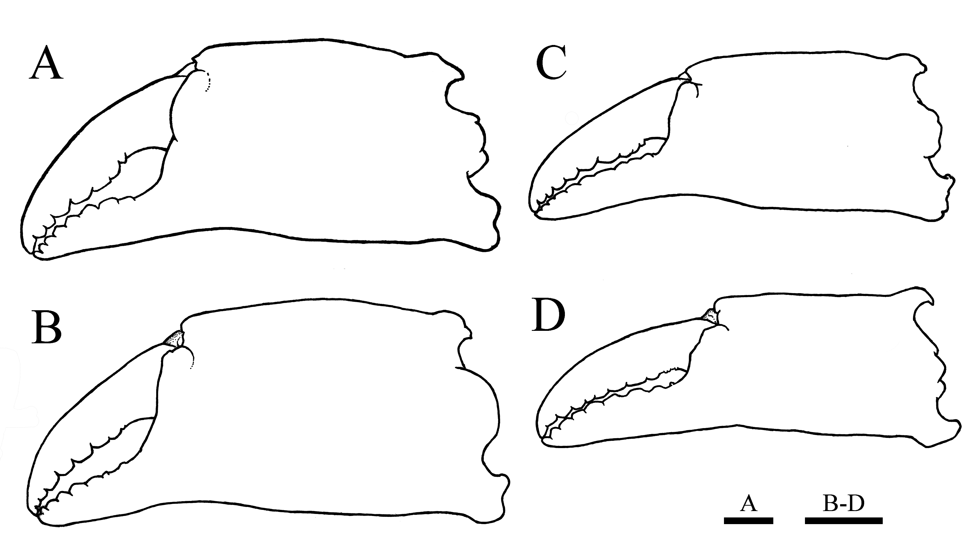
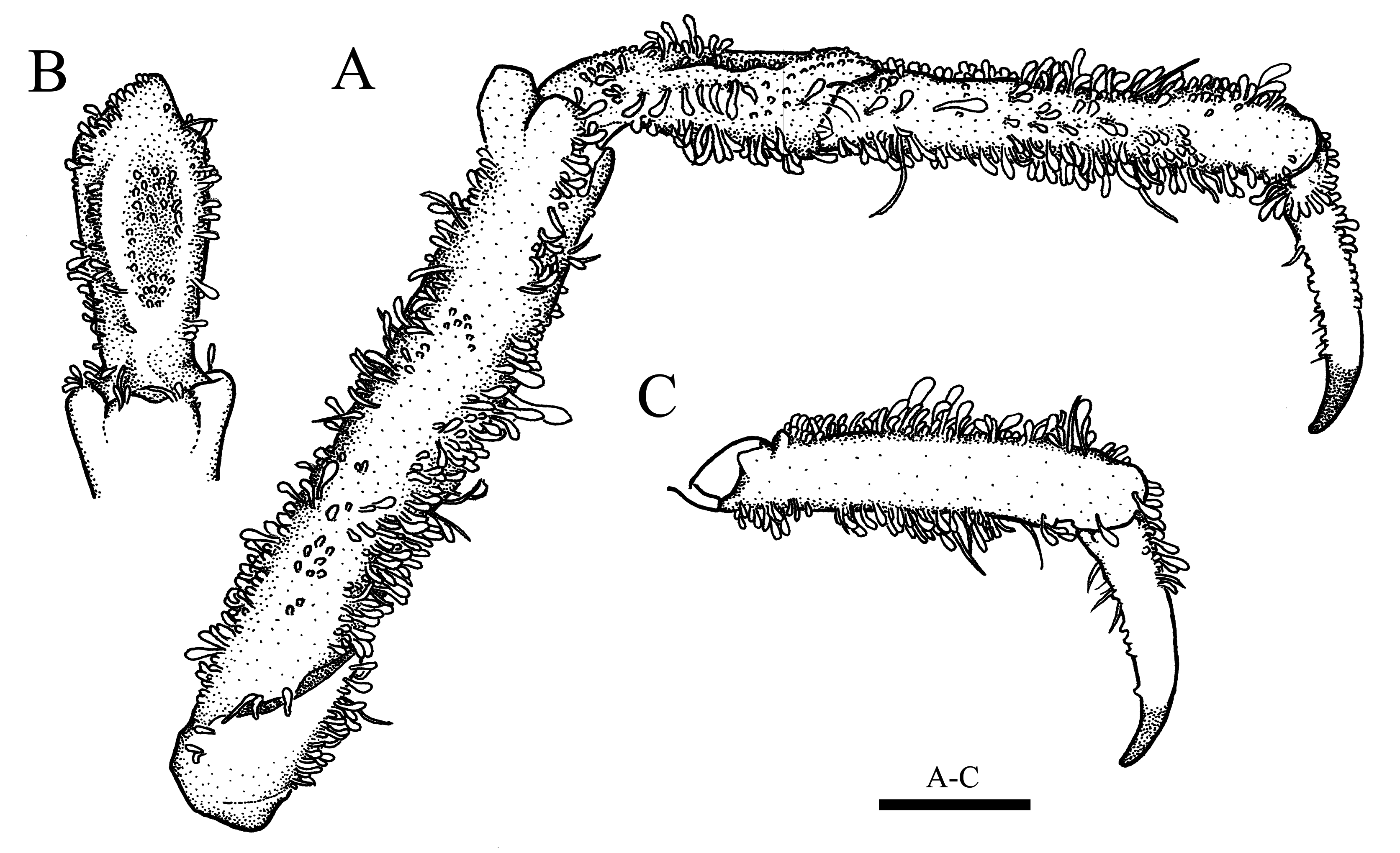
Chelipeds ( Figs. 15A, B View FIGURE 15 , 17 View FIGURE 17 , 18 View FIGURE 18 ) equal in size, similar in shape. Ischium ( Fig. 15B View FIGURE 15 ) weakly swollen ventrally in distal half; mesial margin acutely ridged, cut into 4–6 teeth by faint sinus, distalmost one distinct; distolateral lobe distinct, compressed, rounded apically. Merus ( Fig. 17 View FIGURE 17 D–G) prismatic, length more than twice longer than height (2.3±0.2, N = 4); dorsal surface ( Fig. 17 View FIGURE 17 D–F) with narrow, longitudinal keel with 3–6 (3 in general) lamellar teeth, distalmost largest, directed anteriorly; outer surface ( Fig. 17 View FIGURE 17 E–G) faintly rugose, with blunt, logitudinal ridge, with 4–7 low tubercles; ventral surface ( Fig. 17D, F, G View FIGURE 17 ) with blunt ridge bearing three low teeth; inner surface ( Fig. 17D, E, G View FIGURE 17 ) irregularly rugose, with blunt, longitudinal ridge teminated in subtriangular, proximal lobe; distal margin ( Fig. 17 View FIGURE 17 E–G) with 2 prominent knobs at articulation with carpus (outer knob as large as inner), prominent, obliquely erect, triangular lobe terminated in acuminate tip on upper side. Carpus ( Fig. 17 View FIGURE 17 A–C) moderately inflated, with blunt ridge bearing 3–4 tubercles on dorsal surface ( Fig. 16A View FIGURE 16 ); outer margin obtusely ridged, devided into 4 lobes, both terminal lobes broader than two median lobes ( Fig. 17A, B View FIGURE 17 ); ventral surface with two tubercles ( Fig. 17B, C View FIGURE 17 ); inner margin thinly crested, irregularly dentate, with round, proximal lobe ( Fig. 17A View FIGURE 17 ). Chelae ( Fig. 18A View FIGURE 18 ) more than twice longer than height (ChL/ChH = 2.2±0.2, N = 15); palm strongly expanded, upper margin obtusely ridged, lower margin poorly defined; both fingers bearing broad, subpentagonal teeth along distal two-thirds (proximal 2 or 3 broad), gaped widely in proximal half when closed ( Fig. 18A View FIGURE 18 ).
Ambulatory legs ( Figs. 15A, B View FIGURE 15 , 19 View FIGURE 19 ) decreasing in length posteriorly, surface generally tomentose. Meri subcylindrical, each with distinct, upper distal tubercule ( Fig. 19A View FIGURE 19 ), almost six times longer than height in P2 (5.8±0.5, N = 5), more than four times in P3 (4.4±0.1, N = 3), with tufts of elongate setae on extensor surface, upper, lower flexor margins. Carpi each with deep, medial depression on extensor surface, with tufts of elongate setae on upper, lower flexor margins ( Fig. 19B View FIGURE 19 ). Propodi weakly flattened, moderately narrowed in flexor half, each with pair of cluster of elongate setae on proximal half of extensor margin ( Fig. 19A, C View FIGURE 19 ). Dactyli each with two rows of low, calcareous spines on flexor surface in P3–5, unarmed in P2 ( Fig. 19A, C View FIGURE 19 ).
Thoracic sternites ( Fig. 20A, B View FIGURE 20 ) tomentose on surface, with deep, narrow, rectangular depression on second to fourth sternites on both sides ( Fig. 20A View FIGURE 20 ); first sternite with broad, median depression; second sternite with pair of small depression anteriorly; third to fourth sternites obutusely ridged medially ( Fig. 20A View FIGURE 20 ); sterno-pleonal cavity sparsely with long setae on anterolateral margin ( Fig. 20A View FIGURE 20 ).
Pleon ( Fig. 20B View FIGURE 20 ) with six pleomeres and telson; third to sixth pleomeres fixed, with distinct suture. Third pleomere broadest, lateral margins oblique; fourth, fifth pleomeres trapezoid; sixth pleomere rectangular, dilated on distolateral angle, 0.7 of third pleomere in proximal width (0.7±0.0, N = 4); telson triangular.
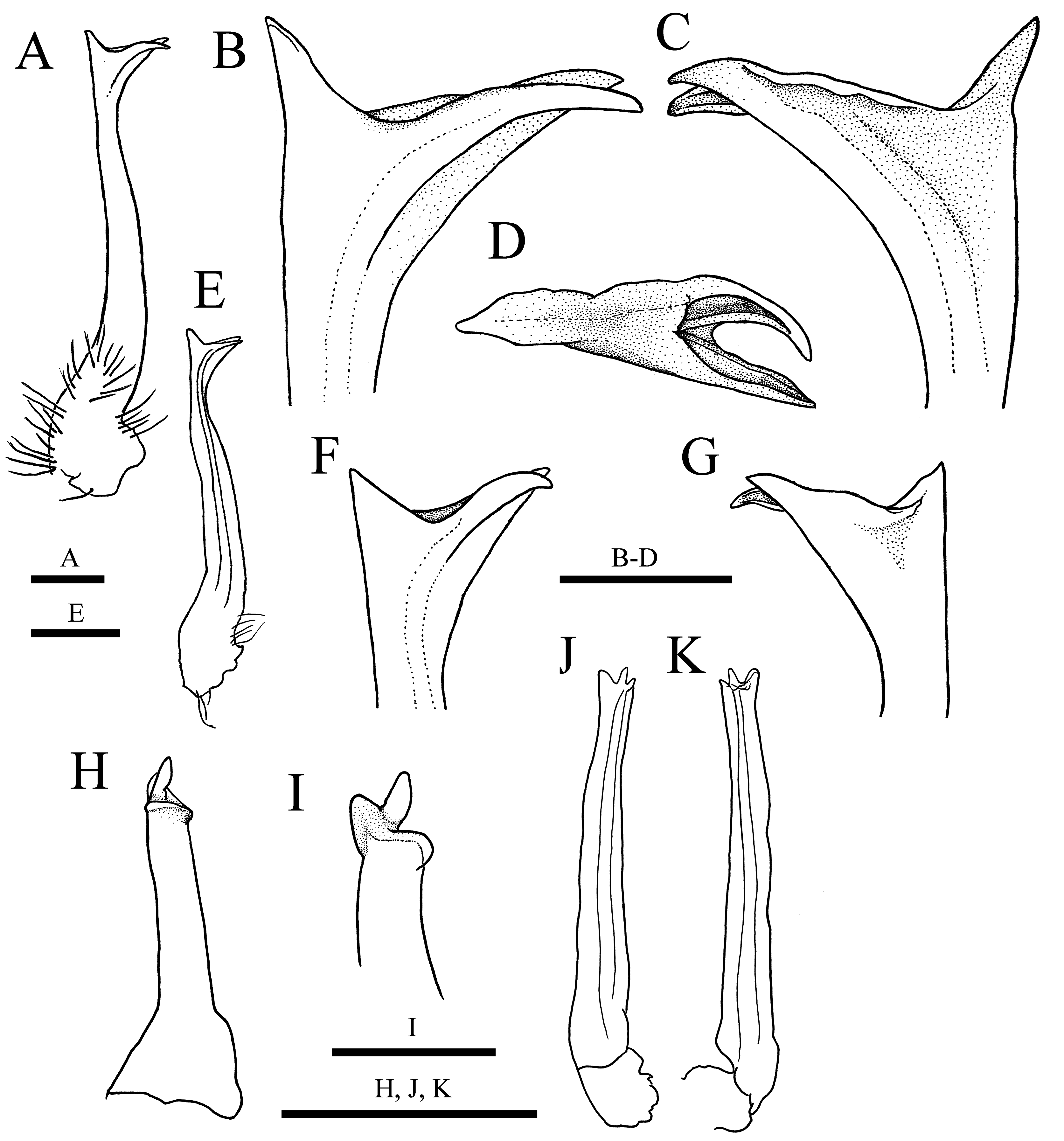
Shaft of G1 ( Fig. 21A, E View FIGURE 21 ) sinuous, trilobate in distal one-fifth; dorsal lobe elongate, triangular, more than twice longer than ventral lobe, weakly curved inwards ( Fig. 21 View FIGURE 21 B–D); ventral lobe triangular, with subacute tip, pointing upwards ( Fig. 21B, C, F, G View FIGURE 21 ); mesial lobe elongate, as long as, or longer than dorsal lobe, directed anteriorly, crossing with dorsal lobe at tip ( Fig. 21 View FIGURE 21 A–C); hiatus between dorsal, mesial lobes narrow ( Fig. 21D View FIGURE 21 ); mesial, lateral margins from dorsal to ventral lobes concave in median part; lateral margin as high as mesial margin, dilated ( Fig. 21 View FIGURE 21 B–D, F, G). Shaft of G2 ( Fig. 21H, I View FIGURE 21 ) stout, narrowed distally, truncated apically; apex with elongate, finger-like projection.
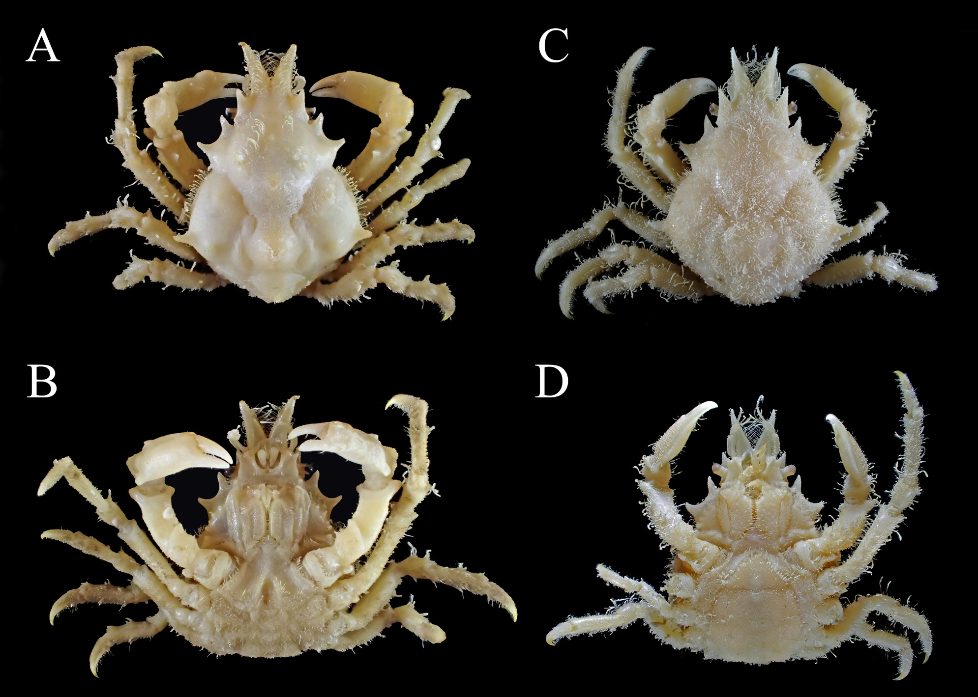
Adolescent males (12.1, 16.8 mm PCL) ( Figs. 14A, B View FIGURE 14 , 22A, B View FIGURE 22 ). Chelae ( Fig. 18C View FIGURE 18 ) proportionally shorter (ChL/ ChH = 2.6±0.1, N = 9) than in full-grown males ( Table 1 View TABLE 1 ), not gaped, both fingers uniformly dentate on cutting margin ( Figs. 18C View FIGURE 18 , 23A, B View FIGURE 23 ). G1 apically trilobate as in full-grown males.
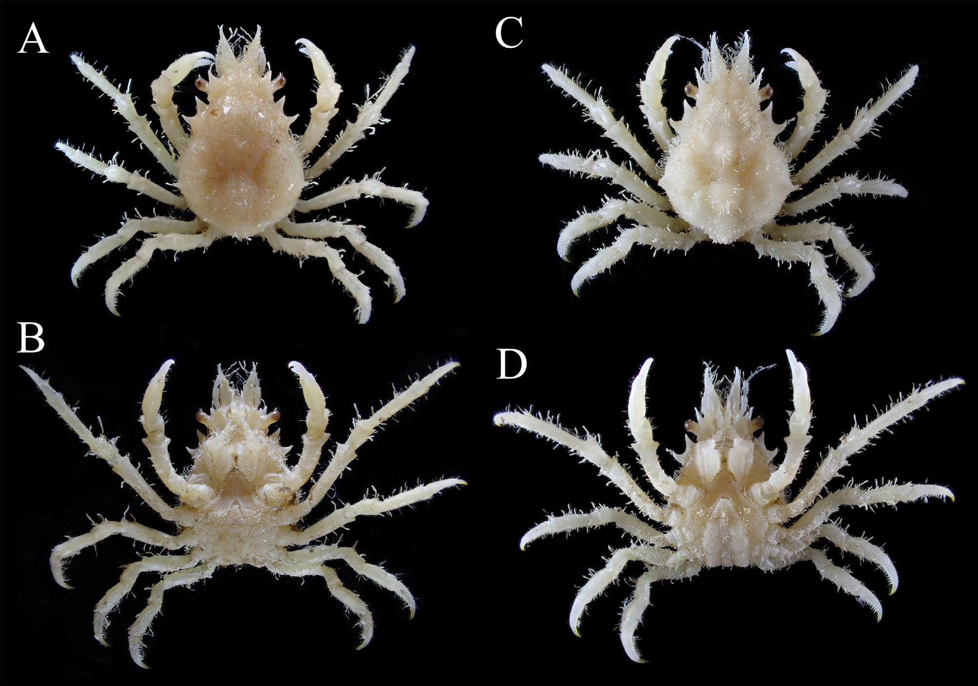
Immature males (<7.5 mm PCL) ( Fig. 23A, B View FIGURE 23 ). Carapace relatively slender (PCL/CW = 1.4±0.1, N = 3). Chela more slender (ChL/ChH = 2.9±0.1, N = 3) than in adolescent specimens ( Table 1 View TABLE 1 ); dentation on both fingers similar to adolescent specimens ( Fig. 23B View FIGURE 23 ). G1 incompletely folded, otherwise folded but provided with short, mesial lobe pointing upwards ( Fig. 21J, K View FIGURE 21 ).
Female. Full-grown females (11.2–28.5 mm PCL). Carapace ( Fig. 15D, F View FIGURE 15 ) similar to males in general proportion (PCL/CW = 1.3±0.1, N = 18; HpL/PoL = 2.2±0.2, N = 7; ESL/PCL = 0.3±0.0, N = 7) (Student t -test, p> 0.05; hepatic, mesobranchial regions more elevated than in males ( Fig. 15D, F View FIGURE 15 ). Pseudorostral spine shorter than in males (PRL/PCL = 0.2±0.0, N = 13) ( Table 1 View TABLE 1 , see also Fig. 15F View FIGURE 15 ) though there was no significant difference (Student t -test, p <0.10). Cheliped merus more slender than in males (2.8±0.2, N = 5; Student t -test, p = 0.01); chelae ( Fig. 18D View FIGURE 18 ) smaller, more slender than in full-grown males (ChL/ChH = 3.0±0.2, N = 15; Student t -test, p <0.01), both fingers uniformely dentated on cutting edges, not gaped when closed. Pleon ( Fig. 15E View FIGURE 15 ) with six pleomeres and telson, expanded (PW6/PW3 = 1.5±0.1, N = 5), fringed with short setae densely. Gonopores ( Fig. 20D View FIGURE 20 ) generally elongate.
Adolescent females (15.3 mm PCL) ( Fig. 22C, D View FIGURE 22 ). Chela slender (ChL/ChH = 2.7, N = 1), both fingers uniformly dentate as in full-grown individuals. Pleon rounded triangular, narrowed toward semicicular telson, lateral margins divergent (PW6/PW3 = 1.3, N = 1).
Immature females (<8.8 mm PCL) ( Fig. 23C, D View FIGURE 23 ). Carapace relatively slender (PCL/CW = 1.4, N = 2). Chela slender (ChL/ChH = 2.9, N = 2). Pleon subtriangular, narrowed toward triangular telson, lateral margins arcuate (PW6/PW3 = 1.1–1.3, N = 2).
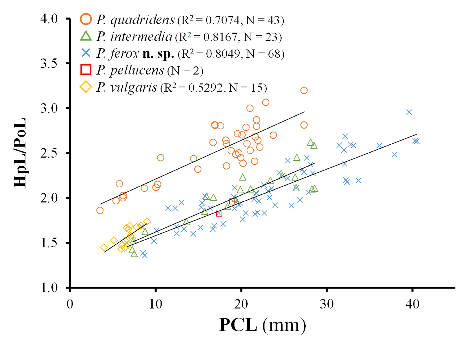
Variations. Carapace tends to be broader (PCL/CW decreases from 1.4 to 1.1) in relation to size growth in both sexes ( Table 1 View TABLE 1 ; see also Figs. 14 View FIGURE 14 , 15 View FIGURE 15 , 22–24 View FIGURE 22 View FIGURE 23 View FIGURE 24 ); the surface is often densely covered with short setae, especially in females ( Figs. 15D, F View FIGURE 15 , 23C View FIGURE 23 , 24D View FIGURE 24 ). Gastric, mesobranchial, and metabranchial regions are less elevated, and the tuberculations on each region are less distinct in small specimens even in the same ontogenetic stage ( Figs. 14 View FIGURE 14 , 22 View FIGURE 22 , 23 View FIGURE 23 , 24A, C View FIGURE 24 ). HpL/PoL increases from 1.4 to 2.6 in relation to size growth ( Fig. 39 View FIGURE 39 ). Both fingers of chela rarely do not contact in distal half in small full-grown individuals ( Fig. 18B View FIGURE 18 ). Spinulation on flexor surface of ambulatory leg dactyli is often reduced or abladed in large specimens. Mesial lobe of G1 is variable in length.
Size. Largest male: 28.4 × 23.8 mm; largest female 22.0 × 17.5 mm; smallest ovigerous female 12.9 × 10.1 mm.
Coloration in life. Based on a few specimens from Sumoto, Osaka Bay. The carapace generally deep red or dark brown, variably with striking patterning by numerous whitish or dark-colored blotches, speckles ( Fig. 25A, C, E View FIGURE 25 ). The thorax, pleon, flexor surface of chelipeds and ambulatory legs white, or whitish light brown ( Fig. 25B, D View FIGURE 25 ). The meri of the cheliped with irregular dark brown band in small specimens ( Fig. 25E View FIGURE 25 ). Chelae generally olive green, both fingers dark brown at basis ( Fig. 25A, D View FIGURE 25 ). Ambulatory legs often with whitish band on meri and propodi. Coloration probably variable, see also Sakai (1965: pl. 32 fig. 3), and Nabeshima (2011, 2013: 124, unnumbered figure).
Distribution. Japan, Pacific coast from Boso Peninsula to Tosa Bay, Osaka Bay, Seto Inland Sea, Sea of Japan coast from off Oga Peninsula to Nagasaki, Goto Islands, Amakusa Archipelago; Gangwon-do, northeast coast of South Korea ( Yang et al. 2015, as P. vulgaris ); both coast of Formosa Strait ( Griffin & Tranter 1986); off Qindao and Jiaozhou Bay, north China ( Shen 1937, as P. minor ; this study).
Habitat. From intertidal to 40 m depth, among algal turfs on the rock reefs, boulder zone, scalop farm ( Sakai 1938, 1976; Yang et al. 2015, as P. vulgaris ). Codium fragile on muddy sand bottom in Okayama, Seto Inland Sea (T. Watanabe pers. comm.). See also Species comparisons.
Decorating materials and epibionts. Branched colonies of hydrozoans, algal pieces, detritus compounds, sometimes encrusted with sponges ( Ariyama 1995; this study).
Ecological notes. Ecology of this species has not been investigated as far as we know.
Remarks. Pugettia quadridens intermedia was described by Sakai (1938) based on three males from Shimoda (Izu Peninsula), one male from Ise Bay, one male and one female from the coast of Gobo, and one male and one female from Wakayama (both Kii Peninsula). He therein designated a full-grown male with 19.5 mm PCL and 15.3 mm CW as the holotype with a photograph of the specimen ( Sakai 1938: 259, pl. 36 fig. 2), but did not indicate its locality. Subsequently, Sakai (1976) noted the type locality as “Shimoda (Sakai)”. This seems to imply that he had intended to designate a full-grown male specimen from Shimoda as the holotype. In the catalogue of major part of extant Sakai’s specimens, one male specimen from Shimoda (KPM-NH 124160: Fig. 14 View FIGURE 14 ) was listed as the holotype ( Muraoka 1998: 24, table 1). However, our re-examination revealed that this specimen is considerablly smaller (13.9 mm PCL and 12.5 mm CW) than Sakai’s measurements ( Fig. 14 View FIGURE 14 vs. Sakai 1938: pl. 36 fig. 2), and despite the lack of both chelipeds, it is likely not a full-grown specimen based on the results of morphometric analyses ( Fig. 39 View FIGURE 39 ). Although there were unfortunately no comments or discussions, Muraoka’s (1998) “ holotype ” is invalid and did not fulfill ICZN 1999, Article 74.6 since there were more than one specimen mentioned in the original description. KPM-NH 124160 is likely one of the three male specimens from “Simoda, the coast of M. B. S.” examined in Sakai (1938) because the specimen is preserved together with the label ( Fig. 14C View FIGURE 14 ), and therefore, this specimen should be regarded as one of paratypes instead (ICZN 1999, Article 72.4.5). In this study, we could locate neither true holotype nor the other paratypes in the extant Sakai’s materials deposited in KPMNH, NSMT, and Shimoda Marine Research Center, University of Tsukuba (SMRC). However, we do not exclude the possibility that the other Sakai’s specimens we could not locate during this study can be found in another institute. Therefore, we here do not designate KPM-NH 124160 as lectotype.


No known copyright restrictions apply. See Agosti, D., Egloff, W., 2009. Taxonomic information exchange and copyright: the Plazi approach. BMC Research Notes 2009, 2:53 for further explanation.
|
Kingdom |
|
|
Phylum |
|
|
Class |
|
|
Order |
|
|
Family |
|
|
Genus |
Pugettia intermedia Sakai, 1938
| Ohtsuchi, Naoya & Kawamura, Tomohiko 2019 |
Pugettia intermedia
| Watanabe, T. 2014: 41 |
Pugettia quadridens pellucens
| Marumura, M. & Kosaka, A. 2003: 32 |
Pugettia quadridens
| Nabeshima, Y. 2011: 124 |
| Muraoka, K. 1998: 24 |
| Yamaguchi, T. & Baba, K. 1993: 353 |
Pugettia intermedia
| Wicksten, M. K. & Stachowicz, J. J. 2013: 359 |
| Ng, P. K. L. & Guinot, D. & Davie, P. J. F. 2008: 101 |
| Yamaguchi, T. & Henmi, Y. 2008: 80 |
| Marumura, M. & Kosaka, A. 2003: 32 |
| Muraoka, K. 1998: 24 |
| Yamaguchi, T. & Harada, K. & Takeda, M. & Kikuchi, T. 1987: 13 |
Pugettia quadridens intermedia
| Kim, H. S. & Kim, I. H. 1986: 325 |
| Ikeda, H. 1981: 15 |
Pugettia similis
| Sakai, T. 1976: 200 |
Pugettia quadridens intermedia
| Miyake, S. 1983: 206 |
| Sakai, T. 1938: 258 |