
Pyura longispina, Rocha & Counts, 2019
|
publication ID |
https://doi.org/ 10.11646/zootaxa.4564.2.9 |
|
publication LSID |
lsid:zoobank.org:pub:FDADF9CA-379C-4980-8AE4-D47F58537E3F |
|
DOI |
https://doi.org/10.5281/zenodo.5930330 |
|
persistent identifier |
https://treatment.plazi.org/id/75898166-70AB-4DDD-9D11-CE57553F385D |
|
taxon LSID |
lsid:zoobank.org:act:75898166-70AB-4DDD-9D11-CE57553F385D |
|
treatment provided by |
Plazi |
|
scientific name |
Pyura longispina |
| status |
sp. nov. |
Pyura longispina sp. nov.
( Figures 7 View FIGURE 7 , 8 View FIGURE 8 )
urn:lsid:zoobank.org:act:75898166-70AB-4DDD-9D11-CE57553F385D
Materials Examined: Holotype: MZUSP555 View Materials Mangrove Island , Bocas del Toro, 9°19'53”N 82°15'00”W, leg. R. M. Rocha, 21.12.2008 GoogleMaps
Paratypes: DZUP PYU-89 , Mangrove Island , Bocas del Toro, 9°17'55”N 82°11'40”W, leg. R. M. Rocha, 15.08.2006 GoogleMaps ; DZUP PYU-128 , 3 individuals, Mangrove Island , Bocas del Toro, 9°19'53”N 82°15'00”W, leg. R. M. Rocha, 21.12.2008 GoogleMaps ; DZUP PYU-129 , 2 individuals, Crawl Key , Bocas del Toro, 9°14'38”N 82°08'25”W, leg. G. Lambert, 25.08.2006 GoogleMaps ; DZUP PYU-134 , Garden Reef , Bocas del Toro, 9°19'28”N 82°13'06”W, leg. R. M. Rocha, 20.08.2006 GoogleMaps ; DZUP PYU-135 , Isla Colon , Bocas del Toro, 9°21'41.9”N 82°16'28.9”W, leg. R. M. Rocha, 22.07.2008 GoogleMaps ; DZUP PYU-140 , Crawl Key , Bocas del Toro, 9°16'01”N 82°07'40”W, leg. R. M. Rocha, 23.09.2008 GoogleMaps .
Other material: USNM 1132564 View Materials pilings in Bocas del Toro town, 9°20'15”N 82°14'26”W, leg. R. M. Rocha, 06.08.2008 GoogleMaps ; USNM 1132853 View Materials pilings in Bocas del Toro town, 9°20'08”N 82°14'32”W, leg. R. M. Rocha, 18.08.2008 GoogleMaps .
Etymology. The name reflects the strong and long spinules in the siphons also visible in living animals.
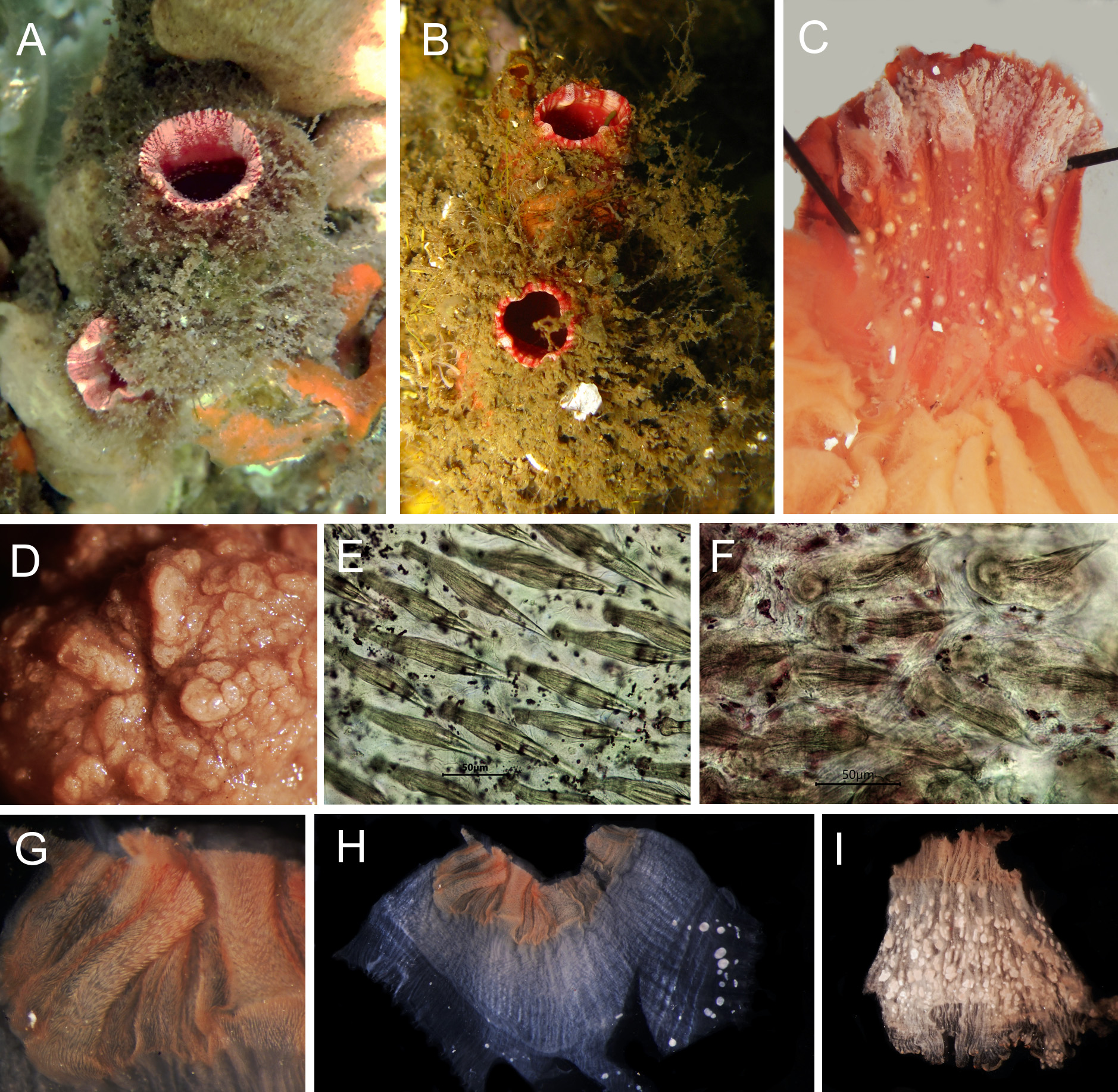
Description. Living specimens are usually heavily covered by epibionts and can be spotted in shallow waters both on mangrove roots and in coral habitats only because of the colorful wide siphons ( Fig. 7A, B View FIGURE 7 ). The holotype is 4.0 cm at the longest length with the tunic, and most other samples are about this size. The tunic has a very irregular and rough surface, and tubercles around the border of the siphons covered by spinules ( Fig. 7D View FIGURE 7 ). It is 1–2 mm thick in the holotype, but can be 5 mm in some regions of contracted animals. After fixation the tunic presents a gradient of yellow brown while the inner layer is a yellow soft membrane.
Siphons lining present a variation of red and pink that can be seen in living animals when siphons are open. A wide zone of dense spinules pointing outwards occurs in both siphons ( Fig. 7C, G, H View FIGURE 7 ). When located close to the margin of the siphon, spinles have an oval plate buried in the tissue from which a curved and pointed projection emerges (~85 µm, Fig. 7F View FIGURE 7 ), but they are more elongated and without the basal plate when inside the siphon (~140 µm, Fig. 7E View FIGURE 7 ). Many individuals had Entoprocta attached to the border of the oral siphon both externally and internally. Posterior to this spiny zone, the internal lining of the siphons show discs of white iridescent material which is not calcareous (Fig. C, H).
Without the tunic the holotype is 2.1 cm at the longest length. The body wall is almost opaque because of the dense musculature. Longitudinal muscle bands radiate from the siphons down the body wall. In some animals those fibers end before the gonadal region making this region transparent through the body wall. There are 24 longitudinal muscle bands on the right side and 25 on the left side in the holotype. Dense circular muscles surround the siphons extending to their base, and a net of thin muscle fibers running in different directions lye over the longitudinal fibers and cover the whole body wall, but are more dense in the anterior region ( Fig. 8A, B, C View FIGURE 8 ). There are minute spicule-like structures on the body wall that cannot be dissolved by hydrochloric acid ( Fig. 8C View FIGURE 8 ). The siphons are 0.5 cm long in the holotype, but can be even longer in other specimens, and maintain the tinge of red even if long preserved.
Some oral tentacles have a variation of brown and orange in preserved specimens. There are three size orders, with one or two exceptionally long tentacle, and the total number of tentacles is 22 in the holotype, 18–30 total variation. The tentacles are rather thick, show little ramifications. First order ramifications are long and form a line along the middle of the lateral side of each tentacle. Second order ramifications are minute or nonexistent in many tentacles ( Fig. 8D, F View FIGURE 8 ). The prepharyngeal groove is made up of two equal size membranes that make a V that surround the dorsal tubercle which is round, protruding, with U-opening with horns slightly or very enrolled ( Fig. 8D View FIGURE 8 ). There is very little space between the tentacle line and the prepharyngeal groove. The dorsal lamina is divided into thin and long languets; 62 in the holotype.
The pharynx has six high folds per side that bends dorsally on themselves or slightly overlap each other in some individuals. The ventral and dorsal folds are smaller ( Fig. 8E View FIGURE 8 ). There are 348–450 total longitudinal vessels. The longitudinal vessel formula in the holotype and two other samples are (from right to left):
E 6 (23) 6 (26) 6 (26) 7 (28) 6 (30) 3 (28) 4 DL 2 (28) 4 (28) 5 (32) 4 (28) 6 (20) 8 (22) 6 E
E 5 (19) 6 (30) 8 (29) 6 (22) 10 (23) 5 (20) 7 DL 6 (18) 6 (27) 6 (27) 8 (26) 8 (23) 5 (23) 0 E
E 10 (20) 14 (26) 14 (27) 9 (36) 9 (25) 9 (16) 9 DL 9 (23) 7 (27) 8 (30) 9 (26) 9 (29) 13 (19) 6 E
There are many parastigmatic vessels and meshes between folds have 4–6 stigmata ( Fig. 8G View FIGURE 8 ). Longitudinal vessels on the right side fray posteriorly forming long, thin languets around the esophagus aperture in both sides.
Endocarps are not present on the body wall on either side of the animal, but they are present along the primary intestinal loop, attached directly to the intestine wall of the ascending portion inside the loop (less numerous and flat), but outside the loop along the descending portion (numerous, larger, flat and irregularly lobed). There are also many irregular endocarps on each gonadal lobe.
The gut is large occupying 2/3 or 3/4 of the left side, with an open primary loop that does not extend up to the prepharyngeal groove ( Fig. 8I View FIGURE 8 ). There is almost no secondary loop and the rectum is either horizontal with a constriction or bent anteriorly before the multilobed anus ( Fig. 8H, I View FIGURE 8 ). The ascending limb of the intestine is also wrinkled in transverse ridges. The bulky digestive gland has one attachment to the stomach wall, and a small portion of tubules can be seen on the right side of the esophagus. It is formed by green flat tubules in preserved animals ( Fig. 8J View FIGURE 8 ).
The gonads are very large: the left one occupies all the space inside the intestinal loop and the right one occupy almost all this side ( Fig. 8I View FIGURE 8 ). They are formed by large and irregularly-shaped hermaphrodite lobes, further divided in other lobes so densely packed that it is not possible to see the gonoducts, unless by their distal ends. Some animals have gonads with more regular undivided oval lobes. There are 12–21 main lobes on the right side and 12– 16 on the left side. The testis form small white patches scattered over the ovary. The gonoducts are tubular, with the sperm duct thinner and with variable length, sometimes slightly shorter than the oviduct, and sometimes slightly longer.
Remarks. This species is similar to Pyura gangelion (Savigny, 1816) known from Martinique ( Monniot C. 1983, Monniot F. 2018) and Brazil ( Skinner et al. 2019) where it has probably been introduced from either the Red Sea, tropical Indian or Pacific ocean ( Monniot 1973, Monniot & Monniot 2001, Kott 2004). Main similarities are the presence of strong spines on the siphons, the white structures inside the siphons lining, and the general shape of the alimentary canal. However, P. longispina sp. nov. has shorter spinules of different shape, larger right gonad, lobed anus and it does not has the internal velum of the atrial siphon divided into four wide lobes, neither the line of endocarps along the heart which is very characteristic of P. gangelion . The heart tube is never close to the right gonad, being more displaced to the left side of the animal close to the ascending limb of the intestine.

| DZUP |
Universidade Federal do Parana, Colecao de Entomologia Pe. Jesus Santiago Moure |
No known copyright restrictions apply. See Agosti, D., Egloff, W., 2009. Taxonomic information exchange and copyright: the Plazi approach. BMC Research Notes 2009, 2:53 for further explanation.
|
Kingdom |
|
|
Phylum |
|
|
Class |
|
|
Order |
|
|
Family |
|
|
Genus |