
Rhyacodrilus quileuticus, Rodriguez, Pilar & Fend, Steven V., 2013
|
publication ID |
https://doi.org/ 10.11646/zootaxa.3664.1.1 |
|
publication LSID |
lsid:zoobank.org:pub:C8136C89-7787-477D-BC64-97AA6C16057B |
|
DOI |
https://doi.org/10.5281/zenodo.6160659 |
|
persistent identifier |
https://treatment.plazi.org/id/154FD01D-4B54-FFD8-FF6E-957EBBD7CE3B |
|
treatment provided by |
Plazi |
|
scientific name |
Rhyacodrilus quileuticus |
| status |
sp. nov. |
Rhyacodrilus quileuticus sp. n.
( Figs 4–5 View FIGURE 4 View FIGURE 5 )
Holotype. USNM 1202053: whole mounted specimen stained in borax carmine and mounted in Canada balsam. Paratypes. USNM 1202054-55: 2 whole mounted specimens, stained in borax carmine, from the type locality (29 April 2004).
Type locality. South Fork Calawah River, Olympic National Forest, Clallam Co., Washington, USA, N47.9627° W124.2941° (29 April 2004).
Other material. 4 mature whole-mounted worms (29 April 2004). 1 mature whole mount, stained in hematoxylin, from West Fork Humptulips River, Washington, Grays Harbor Co., approx. N47.48° W123.68° (2 June 2003).
Etymology. the species has been named after the Quileute People (also called Quilayute) of the Calawah River basin.
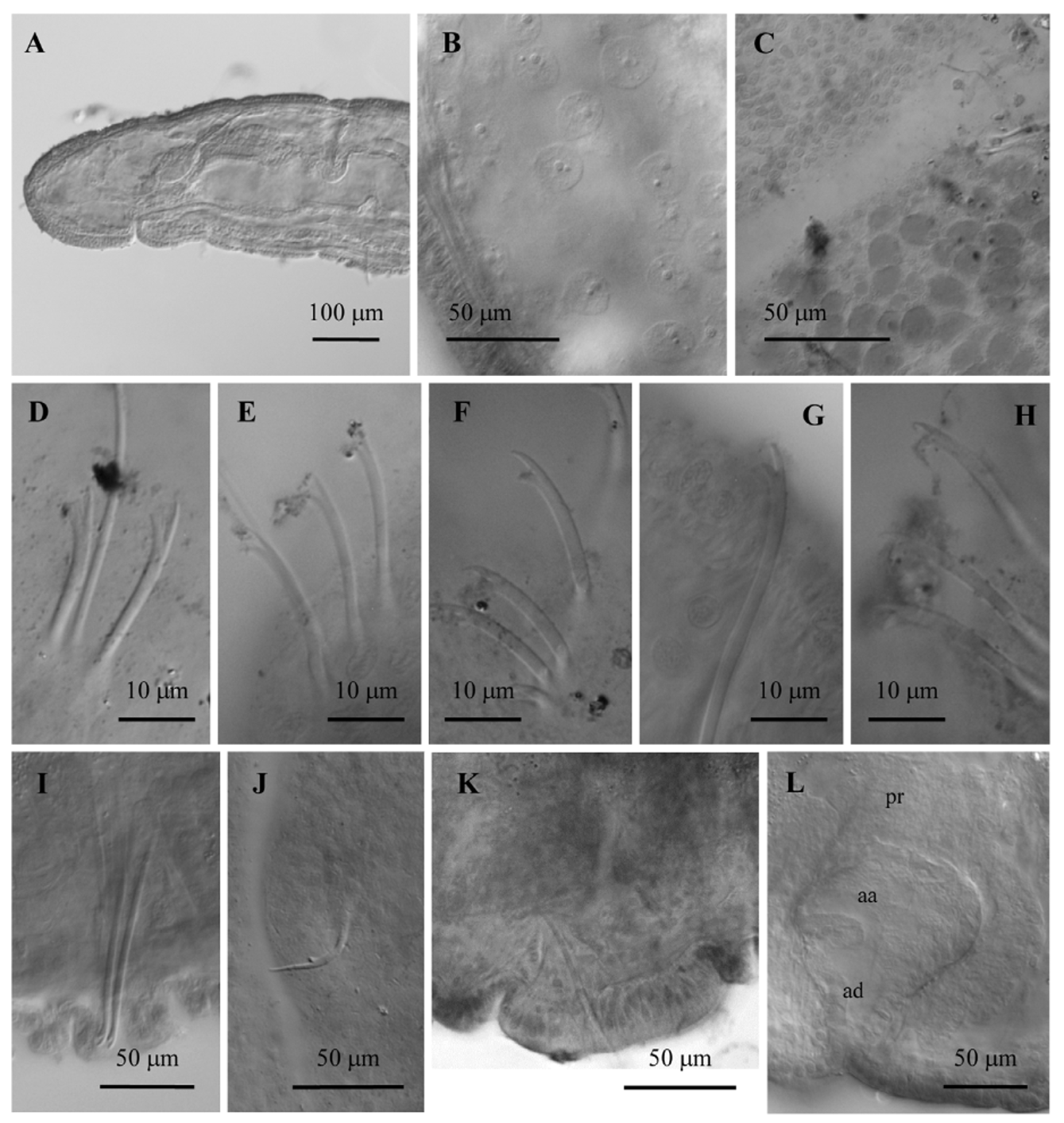
Description. Number of segments: more than 25 (all specimens incomplete). Diameter of the body in segment VIII (whole-mounted specimens) 276–358 µm. Round prostomium (148–217 µm long) ( Fig. 4 View FIGURE 4 A). Epidermis 7–12 µm high, and up to 30 µm high at the clitellum, which extends from the level of chaetae in segment X to the end of XII ( Fig. 5 View FIGURE 5 A). Clitellum formed by large glandular cells ( Fig. 4 View FIGURE 4 C) except for the ventral side around male pores, where there are only small glandular cells. One pair male pores in XI in line of ventral chaetae. One pair spermathecal pores in X, slightly lateral to the line of ventral chaetae, close to intersegment 9/10. One pair female pores opening in intersegment 11/12, about midway between ventral chaetae and the lateral line.
Hair chaetae (0)1–2 per bundle anteriorly, and 0(1) in postclitellar segments, usually absent posterior to segment XII or XV, and shorter than body diameter (length 110–200 µm). Dorsal pectinate chaetae 2–4 per bundle, nodulus 0.3–0.4 from distal end, 64–73 µm long (56–60 µm in II), distal tooth longer or about equal to proximal, intermediate teeth same length or shorter than lateral teeth ( Fig. 4 View FIGURE 4 D, E). Ventral chaetae bifid; anterior ventral chaetae 3–6 per bundle, 75–82 µm long (58–70 µm in II), distal tooth twice length of proximal; 3–5 ventral chaetae in postclitellar segments, 58–76 µm long, with distal tooth thinner, but about the same length as proximal ( Fig. 4 View FIGURE 4 F– H). Nodulus at 0.4–0.5 from distal end in anterior ventral chaetae, 0.3–0.5 in posterior ones.
Modified spermathecal chaetae in segment X, 1–2 per bundle, in line with ventral chaetae, in the posterior third of the segment, well-separated from spermathecal pores (75–97 µm long and ca. 2 µm shaft diameter). The spermathecal chaetae have a very long, sharply-pointed distal tooth (7–17 µm long, most commonly 12 µm), with a short (3 µm), rounded proximal tooth at the base, nodulus at 0.3 from the distal end ( Figs 4 View FIGURE 4 J, 5C). A ventral pad of thickened (to about 30 µm) epidermis surrounds spermathecal chaetae; cells columnar but not distinctly glandular ( Fig. 4 View FIGURE 4 K). Modified penial chaetae (72–84 µm long), 2 per bundle (sometimes an additional, partially developed one growing laterally), arranged in parallel, adjacent to male pores in XI, bifid with long, distinct teeth, and nodulus at 0.2 from distal end ( Figs 4 View FIGURE 4 I, 5D).
Nucleated coelomocytes with granulated cytoplasm (diameter 10–22 µm) ( Fig. 4 View FIGURE 4 B). Pharyngeal glands in IV (ventro-laterally) and V (ventrally), in some specimens also ventrally in VI. Chloragogenous tissue from the back part of segment IV and densely covering the gut from V backwards. No glands observed behind ventral chaetae. Sperm sac may extend anteriorly to segment VIII and back to XV. Egg sac back to XVI. One pair testes in X, one pair ovaries in XI.
Spermathecal duct short and bulbous (60–96 µm long, 55–70 µm maximum diameter), formed by a thick wall of columnar epithelium and a muscular layer ( Fig. 5 View FIGURE 5 B). Oval spermathecal ampullae occupy most of segment X (188–217 µm maximum diameter). Sperm free in lumen of spermathecal ampulla.
One pair sperm funnels on 10/11. Vas deferens 20–26 µm wide and about 150–200 µm long, coiled, and joining the atrial ampulla subapically ( Fig. 5 View FIGURE 5 B). Atrial ampulla globular (84–111 µm long, 57–81 µm wide), completely covered by a diffuse layer of prostate cells up to 40–57 µm high. Atrial duct (39–45 µm long) shorter than the ampulla and associated with a few muscular strands, its diameter widening from the simple male pore towards the ental end, and not clearly separated from the ampulla ( Fig. 5 View FIGURE 5 B). Atrial lumen irregularly filled by epithelial cells ( Fig. 4 View FIGURE 4 L).
Anomalies. In one specimen, genitalia shifted one segment forward.
Distribution and habitat. Known only from two sites in northwestern Washington. The Calawah River (type locality) and tributaries are low gradient, streams; surface flow varies seasonally, with low flows in July-August. Areas of groundwater upwellings result in localized cold spots. Sediment is dominated by cobble-size stones. Dissolved oxygen levels are high (9.7–10.7 mg /L), and the river is rated Class AA (extraordinary water quality) by the Washington Department of Ecology (Hook 2004). There are no domestic water systems or hatcheries located within the South Fork Calawah, and the watershed has substantial populations of several salmonid species (Hook 2004).
Remarks. The species is closely related to R. saelonae based on the presence of both penial and spemathecal chaetae, and the prostate gland covering all of the atrial ampulla. It is, however, clearly distinguished from it and other related species (see Table 1) by the smaller number of genital chaetae, the bifid penial chaetae, the structure of the male duct, and the absence of ventral glands in segment X and adjacent segments. The atrial duct is shorter than the ampulla, gradually narrowing towards the male pore in R. quileuticus sp. n., while it is well-separated and longer than the ampulla in R. saelonae .
Character alcyoneus ardierae carsticus Košel, 1980 gasparoi Martínez- gernikensis Giani & glandulosus (Martínezsp. n. Lafont & Juget, 1993 Ansemil et al. 1997 Rodriguez, 1988 Ansemil et al. 1997)
Dorsal chaetae Hairs and pectinates. No hairs, bifids Bifids, teeth equal Hairs and pectinates. No hairs, bifids with Hairs and pectinates. Hairs absent from variable (modified in XI) equal teeth Hairs absent from XVII XIII–XV backward backward
Penial chaetae 5–7 simple-pointed or 3(4–5), simple-pointed, 2–4 bifid, hook-shaped 2 simple or bifid(?) 2 bifid 1–2 simple- pointed
minutely bifid, fanwise fanwise

No known copyright restrictions apply. See Agosti, D., Egloff, W., 2009. Taxonomic information exchange and copyright: the Plazi approach. BMC Research Notes 2009, 2:53 for further explanation.