
Rhynchelmis (Rhynchelmoides) croatanensis, Fend, Steven V. & Lenat, David R., 2010
|
publication ID |
https://doi.org/ 10.5281/zenodo.196882 |
|
DOI |
https://doi.org/10.5281/zenodo.6206481 |
|
persistent identifier |
https://treatment.plazi.org/id/03EEF043-2A3E-6B65-A1D3-FDB3FDF7A30E |
|
treatment provided by |
Plazi |
|
scientific name |
Rhynchelmis (Rhynchelmoides) croatanensis |
| status |
sp. nov. |
Rhynchelmis (Rhynchelmoides) croatanensis View in CoL n. sp.
( Figures 4–6 View FIGURE 4 View FIGURE 5 View FIGURE 6 )
Holotype: USNM 1142475, a longitudinally dissected worm, stained in hematoxylin and slide mounted.
Type locality: USA, North Carolina, Carteret Co. , seep at Pettiford Creek, 30 Jan 2009, collected by D.R. Lenat. Seep was located on the east side of Pettiford Creek, about 150 meters south of Forest Service Road 128 (Millis Road), Croatan National Forest.
Paratypes: The type locality, 30 Jan 2009: USNM 1142476-1142478, 1 dissected, 1 sagittally sectioned, 1 transversely sectioned.
Other material: The type locality, 24 Jan 2009: 4 longitudinally dissected. 30 Jan 2009: 4 longitudinally dissected, 1 sagittally sectioned, 1 transversely sectioned. All collected by D.R. Lenat.
Etymology. Refers to the type locality, in Croatan National Forest.
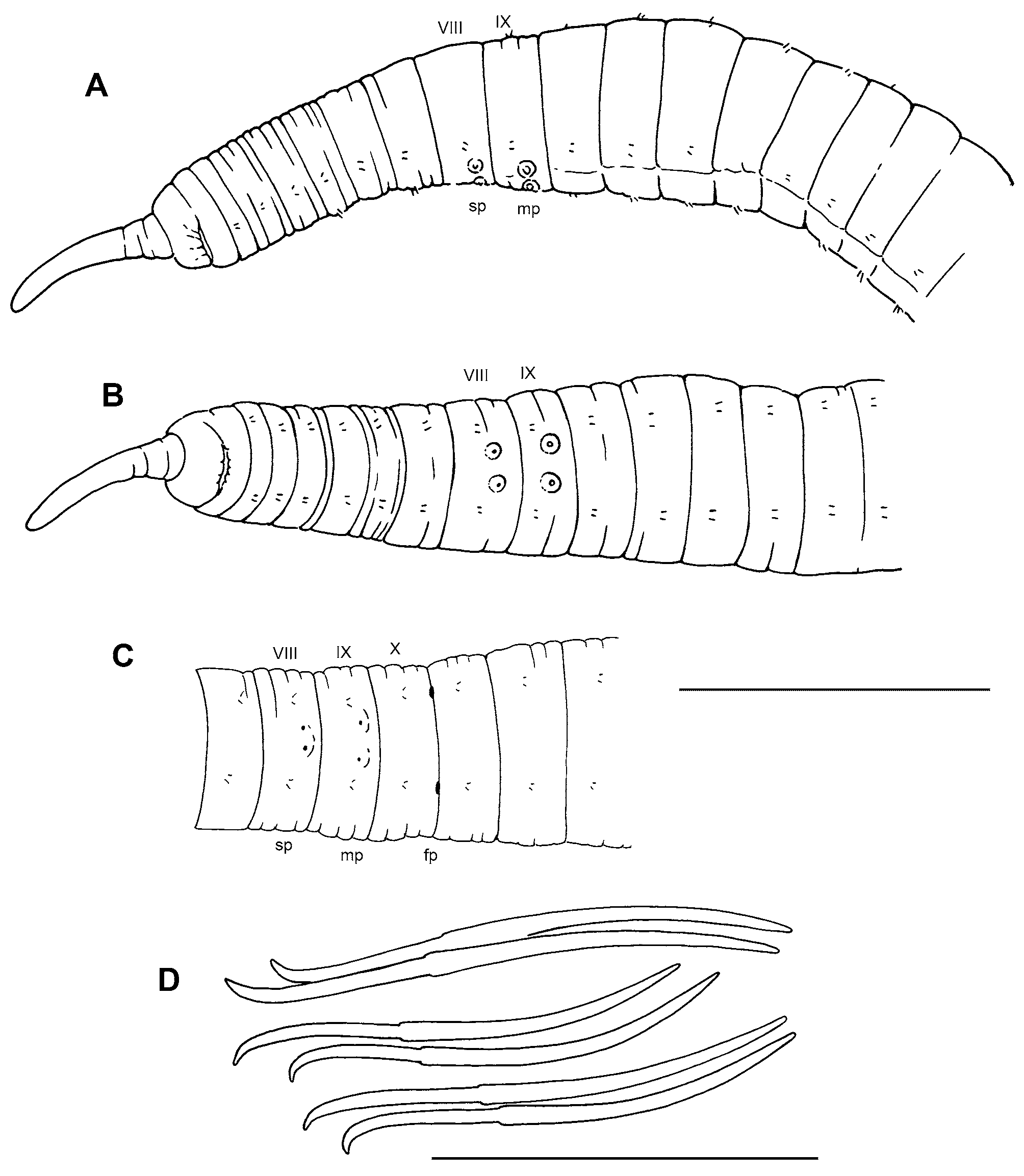
Description. Length of 2 entire, preserved worms 40–42 mm; segments 119–121. Diameter of all worms 0.93 (0.80–1.05) mm in IX (atrial segment), maximum diameter 1.26 (0.92–1.51) mm ( Fig. 4 View FIGURE 4 ). Body round or somewhat concave dorsally in preserved worms. Prostomium with a distinct filiform proboscis 0.9–1.9 mm long, tapering from about 0.1 mm near tip to 0.25 mm at base, faintly ringed, but not distinctly pseudosegmented. Secondary segmentation usually a narrow anterior ring in about IV–VIII, variable and indistinct behind clitellum. Epidermis 15–25 μm thick in preclitellar area, up to 40–70 μm in clitellum, and about 10–15 μm posteriorly. Longitudinal muscle 20–50 μm thick, usually thicker ventrally than dorsally; circular muscle about 5 μm. Clitellum mid-VIII or IX to XVII or XVIII, thickest in X–XIV. Chaetae simplepointed, moderately sigmoid; nodulus approximately 1/3 the distance from tip (0.32–0.43); the inner chaeta in each pair slightly longer than the outer ( Fig. 4 View FIGURE 4 D). In clitellar region, length of ventral chaetae 275 (253–296) μm; dorsals slightly shorter, 214 (194–235); both dorsal and ventral chaetae about 175–245 μm in posterior segments. Pharynx from II–V, without a distinct dorsal pad; about equally developed dorsally and ventrally. Pharyngeal glands usually IV–VI, typically with indistinct dorsal, median and ventral lobes. Brain in the peristomium.
First nephridia usually paired on 11/12; nephridia usually paired in next several segments; paired, single, or absent in posterior segments. Each nephridium with small anteseptal funnel; an ovate postseptal expansion (length about 80–120 μm); and a long, convoluted duct which forms a narrow loop; the conjoined ducts extend dorsally along the anterior lateral blood vessel, then ventrally along the posterior lateral vessel. The ectal end of the duct terminates in an inconspicuous nephropore anterior to the ventral chaetae in the originating segment, without forming a distinct vesicle. Blood vessels as described for R. bolinensis (see above).
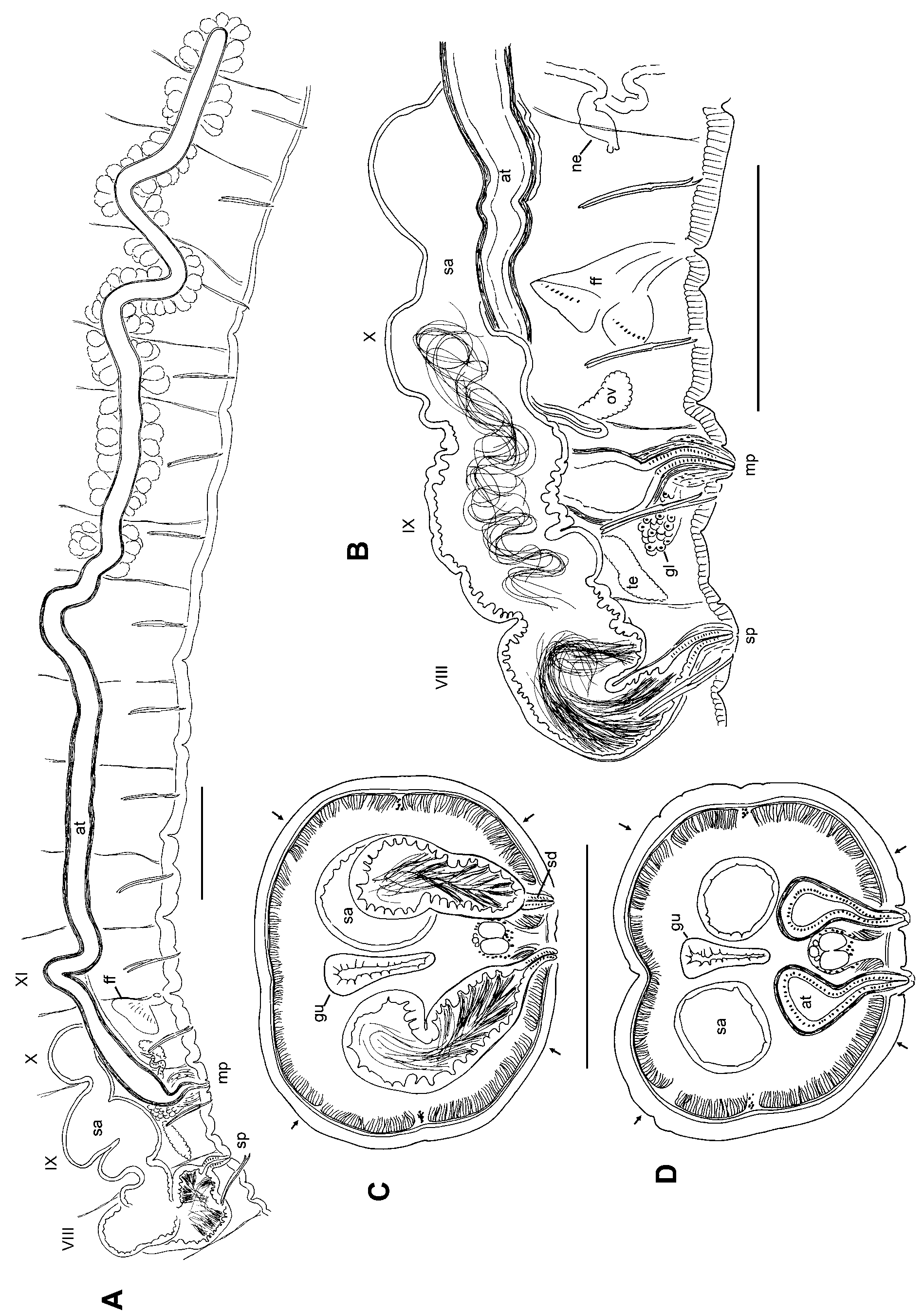
Testes paired in IX, ovaries in X; both testes and ovaries relatively small, not extending beyond midsegment. Sperm sacs extend posteriorly to XXV (XVIII–XXXIII), anterior sperm sacs absent; egg sacs to XXV–XXXVIII. Female pores intersegmental on 10/11, in line with ventral chaetae; female funnels to about 200 μm high. Spermathecae paired in VIII; atria paired in IX; both male and spermathecal pores posterior to, and distinctly median to the ventral chaetae ( Fig. 4 View FIGURE 4 A–D, 5A–D). Male pores within conical papillae, 100–130 μm wide by 20–35 μ high; inconspicuous on some specimens. Spermathecal pores inconspicuous.
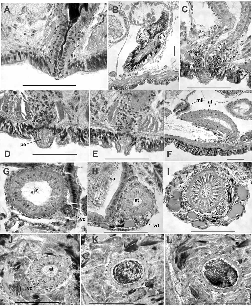
Spermathecae in most cases extending posteriorly to at least IX, but as far as XV within septal sacs ( Fig. 5 View FIGURE 5 A–B). Elongate ampulla sacciform, constricted at septa, 1800 (1050–2700) μm long, by up to 220–350 μm wide. The ectal portion (approximately 1/5 to 1/10) has irregular, deeply incised epithelium 25–30 μm thick, with sperm heads lined up in the incisions ( Fig. 6 View FIGURE 6 B); muscle layer thin (2–3 μm) or indistinct. The main, ental portion has an indistinct muscle layer; epithelial cells irregular, with many small vacuoles near transition, but thinner (10–15 μm) and more regular in the ental half of the ampulla; lumen with sparse, unordered sperm. Ectally, the spermathecal ampulla tapers to a narrow duct, 90–150 μm long; duct is a narrow tube of densely packed epithelium surrounded by a thin (5–10 μm) muscle layer ( Fig. 6 View FIGURE 6 A). Spermathecal pores usually without obvious accessory glands; rarely with a small, petiolate gland.
Male funnels on 9/10 only, about 120–220 μm high, slightly to strongly convoluted; may extend posteriorly through X within the sperm sac. Vasa deferentia enter IX directly, immediately bending up and back into the sperm sacs, without forming a loop in the postatrial segment or penetrating septum 9/10; diameter as much as 35 μm near funnel, 15–25 μm posteriorly. Vas deferens runs adjacent to the prostate-free portion of the atrium within the sperm sac ( Fig. 6 View FIGURE 6 H–I); it penetrates the atrial muscle coat at the beginning of the prostate layer ( Fig. 6 View FIGURE 6 J), and runs within the epithelial layer to join the atrial lumen at the ental end.
Atria elongate ( Fig. 5 View FIGURE 5 A), extending posteriorly within the sperm sacs to XX (XIV–XXIII); total length 4800 (3580–6010) μm. The long (1370–3010 μm) ectal portion of each atrium lacks prostates; this section has a 5–12 μm muscle layer and a relatively thick epithelium (12–26 μm); the epithelium changes over the length of this section, with indistinct cells ectally and somewhat columnar cells entally ( Fig. 6 View FIGURE 6 G–I). The ectal section is initially somewhat expanded in IX (diameter 100–170 μm), with a wide lumen ( Fig. 6 View FIGURE 6 F–G), but then narrows slightly ( Fig. 6 View FIGURE 6 H); posteriorly, the diameter varies (about 80–130 μm), and the lumen remains narrow (20–25μm; Fig. 6 View FIGURE 6 H–I). The approximately equal ental portion is nearly tubular, with a diameter about 75–100 μm. Histology of the ental portion is similar for its entire length: a thin muscle layer about 5–8 μm; a thin epithelial layer about 3–10 μm; and a relatively wide lumen, 55–75 μm wide, filled with sperm cells ( Fig. 6 View FIGURE 6 J– L). Prostates are in pyriform clusters of granular, indistinct cells, 80–160 μm long; cells sometimes with small vacuoles; prostate glands are densely packed over the entire ental part of the atrium.
Ectal end of atrium narrows to a short, cylindrical duct 120–210 μm long, 35–50 μm in diameter ( Fig. 5 View FIGURE 5 B); duct has a columnar epithelium and thin, transverse-circular muscle layer; the duct is surrounded by a thick layer of amorphous tissue that is not distinctly muscular ( Fig. 6 View FIGURE 6 C). The duct terminates in a short papilla or penis (20–36 μm long). Penes are short extensions of the atrial duct, enclosed within a small, conical porophore ( Fig. 6 View FIGURE 6 D–E). Internally, a single, loose cluster of petiolate gland cells, 120–170 μm high, inserts just anterior to the male pore ( Fig. 5 View FIGURE 5 B).
Remarks. Although the location of the male pores in R. croatanensis —in IX —is unique within the genus, the species is otherwise similar in many respects to the only other described eastern Nearctic species, R. bolinensis n. sp. The atria are very long and tubular, with the vas deferens joining the muscle layer at the beginning of the prostate layer, and the ectal end forming a distinct, narrow duct. The spermathecae are sacciform, with a distinct, short-tubular ectal duct; the ampulla has a short ectal portion with a convoluted epithelium, which contains most of the viable sperm; the larger, ental part of the ampulla has a thinner epithelium, and contains sperm tails or degenerate sperm.
Despite general similarities in structure of reproductive organs, R. croatanensis and R. bolinensis differ in several details. Atria of R. croatanensis have a much longer ectal (prostate-free) portion, with a thickened epithelium, the penial structures are smaller, accessory glands are weakly developed at the male pores, and both male and spermathecal pores are inconspicuous. The nearly median position of the male and spermathecal pores of R. croatanensis is unique within R. (Rhynchelmoides), although a median shift is seen in some Rhynchelmis species within the Sutroa group. The unusual pre-clitellar nephridia of R. bolinensis are not present in R. croatanensis ; although nephridia usually begin on 12/ 13 in Rhynchelmis species, the location of the first nephridia on 11/12 can be explained by the apparent anterior shift of male and female pores in R. croatanensis . Spermathecal ampullae tend to be very large in R. croatanensis , commonly extending posterior to the female segment. The length of atria and spermathecae may approach that of the Japanese R. orientalis ( Yamaguchi 1936, Fend & Brinkhurst 2010). Rhynchelmis orientalis atria also have an elongate, prostate-free section, but in that species the ental section becomes markedly distended when filled with sperm.
Like R. bolinensis , R. croatanensis was collected in a small seep with heavy leaf accumulations, which goes dry during summer months. Pettiford Creek is located near heavily developed sections of the North Carolina coast, but its drainage area is entirely within an undeveloped portion of the Croatan National Forest. The headwaters are in the Pocosin Wilderness, producing high quality water, but with pH values often near or below 4.0. It is unclear if seepages to Pettiford Creek also have the same low pH values. The mainstem of Pettiford Creek supports a high diversity of other lumbriculid taxa, some of which are still undescribed. All mature worms were collected late January. Like R. bolinensis , R. croatanensis appears to have a very short, mid-winter period of sexual maturity.
| USNM |
Smithsonian Institution, National Museum of Natural History |
No known copyright restrictions apply. See Agosti, D., Egloff, W., 2009. Taxonomic information exchange and copyright: the Plazi approach. BMC Research Notes 2009, 2:53 for further explanation.