
Saxipendium coronatum Woodwick & Sesenbaugh, 1985
|
publication ID |
https://doi.org/ 10.11646/zootaxa.2408.1.1 |
|
publication LSID |
lsid:zoobank.org:pub:9BBB84BB-239C-41EA-9CFC-682449F96281 |
|
persistent identifier |
https://treatment.plazi.org/id/101A87CA-FF8B-FFA0-DBA6-FF34FF22FEEB |
|
treatment provided by |
Felipe |
|
scientific name |
Saxipendium coronatum Woodwick & Sesenbaugh, 1985 |
| status |
|
Saxipendium coronatum Woodwick & Sesenbaugh, 1985 View in CoL
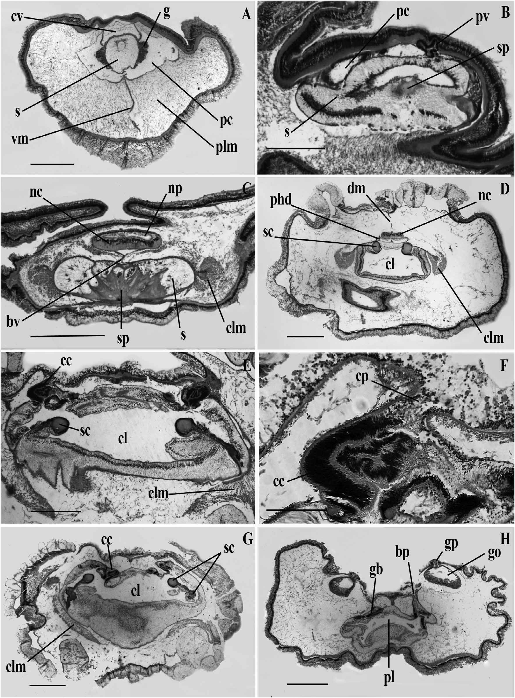
( Fig. 5A–H View FIGURE 5 )
Material examined. Eleven specimens were collected by the deep diving submersible Alvin in 1979 near ''Rose Garden'' geothermal vent, Galapagos Rift (00°47.9' N, 86°13.5' W), 2478 m depth. The holotype is NMNH 97395 View Materials and paratypes are 97396–8. Individuals were loosely attached to the rocks located at the periphery of the vent area GoogleMaps .
External features. Proboscis arrow shaped, longer than broad, with longitudinal dorsal groove. Collar very short with elevated ring at its posterior end. Trunk slightly flattened dorsoventrally, presenting a median longitudinal groove. Dorsolateral genital ridges present on each side of body, the gonopores externally visible. Preserved specimens 25 mm long, about 2 mm thick. Color yellow-white in live material, collar darker than rest of the body and collar ring paler. Holotype measurements: overall length 215 mm, proboscis 11.0 mm, collar 3.0 mm, trunk 201 mm.
Internal features. Nerve-fiber layer of proboscis thickened dorsally ( Fig. 5A View FIGURE 5 ). Proboscis coelom occupying posterior third of proboscis. Ventral septum present in posterior part of organ but dorsal septum formed by the large cardiac vesicle that is in contact with dorsal wall of proboscis ( Fig. 5 A View FIGURE 5 ). Circular-musclefiber layer half thickness of nerve-fiber layer. Longitudinal muscle fibers diffuse. Glomerulus extending over tip of stomochord but poorly developed ( Fig. 5A View FIGURE 5 ). Stomochord with thick walls and large central lumen, expanding ventrolaterally in neck region to form two horns, each with own lumen ( Fig. 5B View FIGURE 5 ). Neck with two coelomic cavities, left vesicle leads to exterior by left proboscis pore ( Fig. 5B View FIGURE 5 ). Skeletal body starts in neck, forming crown-shaped plate with spikes dividing stomochord into subsections ( Fig. 5C View FIGURE 5 ). Skeleton with no keel. Proboscis with dorsal groove that is more conspicuous in preserved specimens.
Only dorsal mesentery of collar present. Anterior neuropore forming at level where dorsal part of collar fuses with neck ( Fig. 5C View FIGURE 5 ). Perihaemal diverticula start at level of cornua, fused in some specimens ( Fig. 5D View FIGURE 5 ). Peribuccal diverticula absent. Skeletal cornua extending to near end of collar ( Fig. 5D,E View FIGURE 5 ), bending ventrally around gut then bending back anteriorly for a short portion ( Fig. 5G View FIGURE 5 ). Collar longitudinal muscles well developed, present as masses on each side of collar lumen ( Fig. 5C–E,G View FIGURE 5 ). Collar canals unique in connecting coelom to outside through pores located dorsomedially ( Fig. 5E,F View FIGURE 5 ). As in other species, collar canals also connect to first gill pouch ( Fig. 5G View FIGURE 5 ).
Branchial portion of pharynx equal in size to ventral portion or slightly smaller ( Fig. 5H View FIGURE 5 ). Dorsal and ventral mesenteries present. Two rows of dorsolateral gonads forming external ridges; gonads connecting to exterior with externally visible gonopores situated on top of genital ridges ( Fig. 5H View FIGURE 5 ). Branchial sacs opening by dorsolateral pores recessed between genital ridges and elevated median line of trunk ( Fig. 5H View FIGURE 5 ). Type specimen had 54 branchial pores and 40 oesophageal pores. Ventrolateral longitudinal muscles of trunk poorly developed.
Remarks. Woodwick & Sesenbaugh (1985) mentioned the presence of an antrum, a special chamber connecting the testis and the gonopore, and thought it to be a distinctive feature of this species. There is now doubt concerning the uniqueness of this character because new observations showed that, in a single specimen, not all the gonads present this feature. Further, so little information is available on the gonads of other species that we cannot certify its absence from other taxa.
The defining characters of Saxipendium coronatum are listed below:
A. Proboscis is arrow shaped with a posterior dorsal groove.
B. Longitudinal musculature of the proboscis is diffuse.
C. Proboscis skeleton is coronate, without a visible keel.
D. Skeletal cornua are recurved.
E. Dorsal and ventral mesenteries are present in the trunk, but only the ventral one is present in the proboscis and the dorsal one in the collar.
F. Cardiac vesicle is in contact with the dorsal wall of the proboscis.
G. Perihaemal diverticula are present and start at the level of the skeletal cornua.
H. Peribuccal diverticula are absent.
I. An anterior neuropore.
J. Collar canals open into the first branchial sac and also to the exterior via collar pores.
K. Gonads form two dorsolateral ridges and the gonopores are externally visible.
No known copyright restrictions apply. See Agosti, D., Egloff, W., 2009. Taxonomic information exchange and copyright: the Plazi approach. BMC Research Notes 2009, 2:53 for further explanation.