
Scotognapha, DALMAS, 1920
|
publication ID |
https://doi.org/ 10.1206/0003-0082(2001)338<0001:AROTGS>2.0.CO;2 |
|
persistent identifier |
https://treatment.plazi.org/id/038A87D4-A559-5C49-FF7E-9ACBFCB2FA51 |
|
treatment provided by |
Carolina |
|
scientific name |
Scotognapha |
| status |
|
SCOTOGNAPHA DALMAS View in CoL
Scotognapha Dalmas, 1920: 119 (type species by original designation Pythonissa convexa Simon ).
DIAGNOSIS: Specimens of Scotognapha can be separated from other gnaphosids by the combined presence of a low, narrow, serrated keel on the cheliceral retromargin and a normal cheliceral tooth situated between the keel and the promarginal tooth row (figs. 2–4). The peculiar shape of the male palpal tegulum, which is extended posteriorly into a distinct prolateral lobe (as in figs. 19, 23, 27, 47) provides an excellent putative synapomorphy for the genus and is here predicted to occur in the as yet unknown males of S. paivai and S. costacalma .
DESCRIPTION: Total length 5.40 –14.05. Carapace oval in dorsal view, widest between coxae II and III, smoothly narrowed opposite palpi, light to dark brown, without darkened lateral margins; cephalic area slightly elevated; thoracic groove long, welldeveloped, longitudinal. From above, anterior eye row procurved, posterior row slightly recurved; from front, both rows slightly procurved. AME circular, dark; PME irregularly rectangular, light; other eyes oval, light; eyes almost subequal; AME separated by slightly less than their diameter, subcontiguous with ALE; PME usually separated by less than their diameter, by more than their diameter from PLE; lateral eyes of each side separated by more than their diameter; MOQ longer than wide, slightly wider in front than in back (fig. 1). Clypeal height more than twice AME diameter. Chelicerae with short, narrow, slightly serrated retromarginal keel, situated terminally, two or three promarginal teeth, and one intermediately situated tooth (figs. 2–4); retromarginal keel often appearing small, toothlike under light microscopy. Mouthparts and sternum dirty yellow to light brown; endites short, 1.5–2.0 times longer than wide, rounded, with oblique depression and weak distal scopula (fig. 5); labium twice as long as wide at middle (fig. 5); sternum oval, broad medially, with long setae at margins, short dark setae covering entire surface, rebordered, with short extensions to and between coxae.
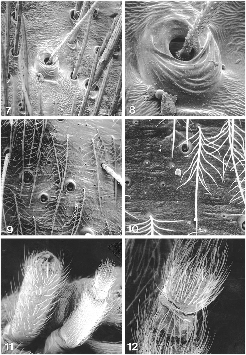
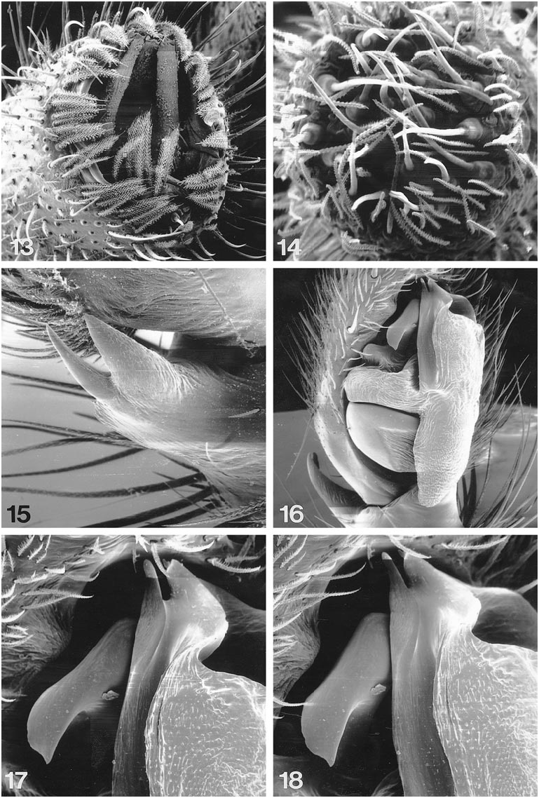
Leg formula 4123. Typical leg spination pattern (only surfaces bearing spines listed): femora: I d110, p001; II d110, p001; III d101, p011, r011; IV d110, p00 1, r001; patellae: III p100, r010; IV r0 10; tibiae: III d201, p211, v222, r21 1; IV d100, p111, v222, r211; metatarsi: I v120; II v222; III d122, p111, v222, r111; IV d122, p111, v222, r1 11. Legs yellowbrown or light brown; tarsi lightly scopulate, with two dentate claws and claw tufts; trochanters not notched; metatarsal preening comb lacking; tibiae I, II of females ventrally without spines, of males with three pairs of spines; tarsi with two rows, metatarsi with single row of trichobothria, trichobothrial bases elevated, bearing 6–8 short, narrow ridges (figs. 7, 8); tarsal organ elevated, circular, smooth, with oval distal opening (fig. 6). Abdomen usually dirty yellow to brown with conspicuous anterior tuft of hairs, dorsum of some species with chevron pattern anteriorly, dark dots and stripes posteriorly; venter same color or lighter, with two closely spaced longitudinal rows of dark spots, few dark spots on sides; males with small anterior scutum. Carapace, abdomen, and legs covered by distally squamose, plumose setae bearing proximally 6–7 pairs of appendages originating from lateral surface (figs. 9, 10). Six spinnerets, anterior laterals large, cylindrical, separated by slightly more than their diameter, with two major ampullate gland spigots and five piriform gland spigots (fig. 13), posterior medians smallest, apically with minor ampullate and aciniform gland spigots, females with two posterior rows of cylindrical gland spigots; posterior laterals narrow, long, longer than ALS, with apical segment bearing aciniform gland spigots (fig. 14); posterior laterals typically strongly retracted into abdomen for 2/3 of their length, hence often appearing very short, even shorter than anterior laterals.
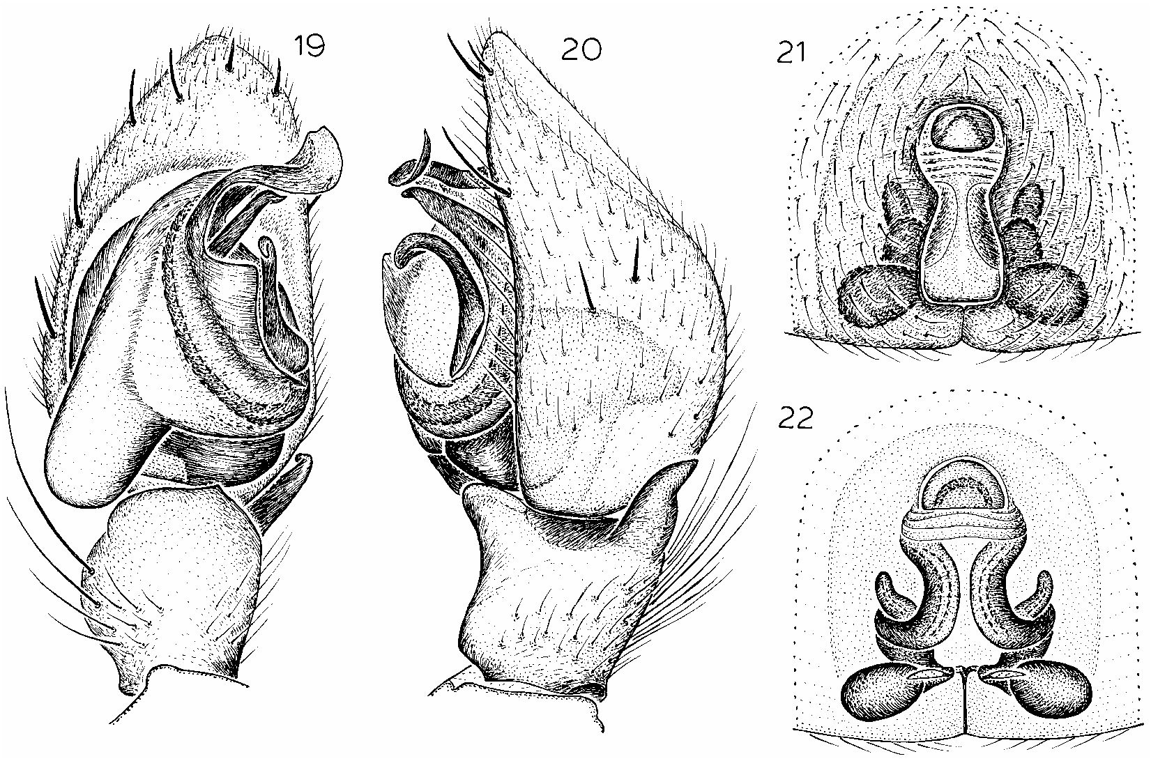
Male palp with prolateral portion of tegulum enormously extended posteriorly (figs. 16, 19, 23, 27, 31, 35, 39, 47); embolus fused with conductor, medially or prolaterally situated, directed anteriorly (figs. 17, 43, 51, 57, 61) or retrolaterally (figs. 19, 23, 27, 31, 35, 39, 47); median apophysis hooked; variously shaped terminal apophysis situated dorsally of embolus (figs. 19, 24, 28, 31, 47). Retrolateral tibial apophysis usually more or less conical, slightly curved at tip (figs. 20, 24, 28) or bifurcate ( S. teideensis only, figs. 15, 52–54). Epigynum with deep, rounded or elongated atrium, wide anteriorly and narrow to very narrow posteriorly, bearing wide anterior hood and raised median septum; copulatory openings situated medially (figs. 21, 25, 29, 33, 37, 41, 45, 49, 55, 59, 63, 65, 67); spermathecal ducts wide, short, with distinctive heads situated anteriorly, spermathecae consisting of posterior pair of large, globular or oval, widely separated receptacles (figs. 22, 26, 30, 34, 38, 42, 46, 50, 56, 60, 64, 66, 68).
Figures 19–22 View Figs
Pythonissa convexa Simon, 1883: 291 (male from Canary Islands, in MNHN, examined; see Note below).
Callilepis convexa: Simon, 1893: 382 .
Scotognapha convexa: Dalmas, 1920: 120 View in CoL .
NOTE: Simon ( 1883) described Pythonissa convexa on the basis of a single female, in poor condition, collected by Verneau in the Canary Islands; no specific locality was mentioned, and there is no definitive indication in the original description as to whether the specimen was adult. In a later listing of specimens collected by Verneau in 1888, Simon ( 1889) recorded the species from named sites on three different islands (Grand Canary, Fuerteventura, and Lanzarote); again, however, no indication of the sex or maturity of any of those specimens was provided. When Dalmas ( 1920) first restudied the material in the Simon collection, he found one adult male and two juvenile females, and indicated that one of the juvenile females was actually the type specimen. He described the adult male, and it has seemed best to follow his decision and accept the choice of this adult male as representing Simon’s species. Unfortunately, there is no indication of an exact island of origin for that male, either. However, the only modern specimens matching that male are from Grand Canary, and we therefore assume that Grand Canary is the source, and type locality, of Simon’s species.
DIAGNOSIS: Males can easily be recognized by the long embolus bearing a wide, strongly curved tip (fig. 19) and the long, narrow terminal and median apophyses (fig. 20), females by the elongated, anteriorly narrowed epigynal atrium (fig. 21), as well as the short, wide heads of the anterolaterally directed spermathecal ducts (fig. 22).
MALE: Described by Dalmas ( 1920).
FEMALE: Total length 7.80. Carapace 3.71 long, 2.67 wide. Femur II 2.02 long. Eye siz
es and interdistances: AME 0.17, ALE 0.17, PME 0.15, PLE 0.17; AMEAME 0.13, AMEALE 0.04, PMEPME 0.06, PMEPLE 0.22, ALEPLE 0.20; MOQ length 0.46, front width 0.43, back width 0.37. Leg spination: femur III d111; tibiae: II v001; III d101, p221, r111; IV r121; metatarsus I v220. Epigynal atrium elongated, narrowed anteriorly, with wide hood (fig. 21); heads of spermathecal ducts short, wide, directed anterolaterally, receptacles situated laterally (fig. 22).
MATERIAL EXAMINED: CANARY IS LANDS: no specific locality (Verneau, MNHN), 13, 2 juveniles . Grand Canary: Llanos de la Gorra, Mar. 23, 1997, under stones, elev. 100 m (J. Murphy, AMNH, JAM), 23, 1♀ .
DISTRIBUTION: Known only from Grand Canary. Records from Graciosa by Dalmas ( 1920), and from Lanzarote and Graciosa by Schmidt ( 1990), were based only on juveniles and are here rejected. The adult female recorded as this species by Schmidt ( 1990) from El Medano on Tenerife presumably be
longs to S. medano instead, whereas the adult female similarly recorded from Gomera presumably belongs to S. canaricola . The male recorded as this species by Schmidt and Krause ( 1996) from Fuerteventura belongs to S. atomaria .
Scotognapha juangrandica , new species
Figures 23–26 View Figs
TYPES: Male holotype and female allotype from a stony area near the sea at Juan Grande , Grand Canary, Canary Islands (Mar. 27, 1997; J. Murphy), deposited in AMNH .
ETYMOLOGY: The specific name refers to the type locality.
DIAGNOSIS: This species seems closest to S. convexa , but can be separated in males by the wide, flat, apically untwisted embolus and wide median and terminal apophyses (figs. 23, 24), in females by the elongated and medially narrowed epigynal atrium (fig. 25), the narrow, anteriorly directed heads of the spermathecal ducts (fig. 26), and the posteriorly situated receptacles.
MALE: Total length 7.54. Carapace 3.90 long, 2.73 wide. Femur II 2.84 long. Eye sizes and interdistances: AME 0.15, ALE 0.14, PME 0.20, PLE 0.15; AMEAME 0.12, AMEALE 0.02, PMEPME 0.07, PMEPLE 0.18, ALEPLE 0.19; MOQ length 0.47, front width 0.35, back width 0.37. Leg spination: femora: I d111, r011; II d111, p011; III d111; IV d111, p011, r01 1; patella IV p010; tibiae: I p112, v222, r101; II p111, v222, r100; III r111; IV d101; metatarsi: I p112, v222, r11 1; II d011, p111, r011; IV d222. Palp with wide, anteriorly flattened embolus, wide, short terminal apophysis, strongly curved, short, distally oval median apophysis (fig. 23); retrolateral tibial apophysis long, narrow, tip slightly bent anteriorly (fig. 24).
FEMALE: Total length 8.71. Carapace 3.91 long, 3.25 wide. Femur II 2.34 long. Eye sizes and interdistances: AME 0.16, ALE 0.18, PME 0.18, PLE 0.19; AMEAME 0.17, AMEALE 0.04, PMEPME 0.08, PMEPLE 0.24, ALEPLE 0.25; MOQ length 0.52, front width 0.49, back width 0.45. Leg spination: femur III d111; tibiae: II v001; IV p211; metatarsi: I v220; III d122. Epigynal atrium elongated, anteriorly deep, posteriorly shallow, narrowed medially, epigynal hood narrow (fig. 25), heads of spermathecal ducts narrowing to tip, directed anteriorly (fig. 26), receptacles situated posteriorly.
OTHER MATERIAL EXAMINED: CANARY ISLANDS: Grand Canary: Juan Grande, Mar. 27, 1997, stony area near sea (J. Murphy, JAM), 1♀ ; Sonnenland, Mar. 16, 1997, dry stony area, elev. 20 m (J. Murphy, JAM), 13 .
DISTRIBUTION: Known only from Grand Canary.
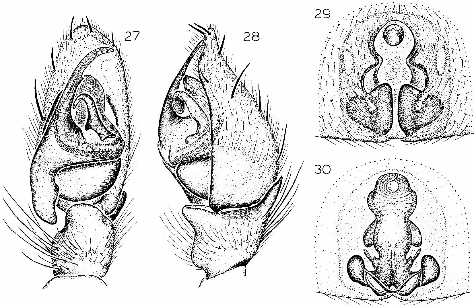
Figures 27–30 View Figs
Scotognapha atomaria Dalmas, 1920: 121 View in CoL (adult male, juvenile male and juvenile female syntypes from Grand Canary , Canary Islands, in MNHN, examined).
Scotognapha gravieri Dalmas, 1920: 121 View in CoL (male holotype from ‘‘Syria’’, probably mislabeled, in MNHN, examined). NEW SYNONYMY.
Scotognapha convexa View in CoL (misidentified): Schmidt and Krause, 1996: 266 (male from National Park El Jable, Fuerteventura).
DIAGNOSIS: Males can easily be recognized by the very long, narrow, apically arched embolus (which bears a small process medially), the large, spoonshaped, and apically oval terminal apophysis and long, narrow median apophysis, and the long, anteriorly narrowed and apically slightly curved retrolateral tibial apophysis (figs. 27, 28), females by the wide, deep anterior portion and extremely narrow, shallow posterior portion of the epigynal atrium (fig. 29), as well as the slender heads of the spermathecal ducts (fig. 30).
NOTE: One of the males collected on Fuerteventura shows some variation in palpal structure: the embolus is slightly wider and more strongly bent anteriorly, and bears a larger medial process. Much larger samples would be needed to corroborate these slight differences as speciesspecific rather than individual variation.
MALE: Described by Dalmas ( 1920).
FEMALE: Total length 10.50. Carapace 4.25 long, 3.00 wide. Femur II 2.31 long. Eye siz
es and interdistances: AME 0.14, ALE 0.17, PME 0.14, PLE 0.12; AMEAME 0.12, AMEALE 0.03, PMEPME 0.06, PMEPLE 0.24, ALEPLE 0.18; MOQ length 0.42, front width 0.36, back width 0.34. Leg spination: femora: III d112; IV d111, p01 1; patellae: III p110, r110; IV p100; tibiae: III d202; IV d110, p212; metatarsi: I v200; II v221. Epigynal atrium anteriorly wide, deep, extremely narrow, shallow posteriorly (fig. 29), heads of spermathecal ducts short, slender, directed anterolaterally (fig. 30).
MATERIAL EXAMINED: CANARY IS LANDS: Fuerteventura: Corralejo, National Park El Jable, Dec. 1992 (G. Schmidt, DMG), 13; Jandia, May 1988 (J. Wunderlich, AMNH), 13, 1♀. Grand Canary: (C. Alluaud, MNHN), 13, 1 juvenile 3, 1 juvenile ♀ (syntypes). ‘‘ Syria ’’ (probably mislabeled): ( Brulerie , MNHN), 13 (holotype) .
DISTRIBUTION: Known only from Fuerteventura and Grand Canary. The female described by Schmidt ( 1976) from Costa Calma, Fuerteventura, belongs to S. costacalma .
SYNONYMY: No genitalic differences were
detected between the male types of Dalmas’ two species.
Scotognapha medano , new species
Figures 31–34 View Figs
Scotognapha convexa (misidentified): Schmidt, 1990: 11 (female from El Medano, Tenerife).
TYPES: Male holotype and female allotype from a stony and sandy area by the sea at El Medano , Tenerife, Canary Islands (Mar. 15, 1996; J. Murphy), deposited in AMNH .
ETYMOLOGY: The specific name is a noun in apposition taken from the type locality.
DIAGNOSIS: This species seems closest to S. atomaria , but can be separated in males by the narrow, anteriorly slightly widened, and apically bent embolus, which lacks a medial process, the small, spoonshaped terminal apophysis, the short, narrow median apophysis, and the short, spiniform retrolateral tibial apophysis (figs. 31, 32), in females by the extremely wide epigynal atrium, slightly narrowed posteriorly, the wide epigynal hood (fig. 33) and the wide, thick heads of the spermathecal ducts (fig. 34).
MALE: Total length 6.85. Carapace 3.25
long, 2.51 wide. Femur II 2.31 long. Eye sizes and interdistances: AME 0.11, ALE 0.14, PME 0.10, PLE 0.12; AMEAME 0.07, AMEALE 0.03, PMEPME 0.03, PMEPLE 0.10, ALEPLE 0.15; MOQ length 0.34, front width 0.24, back width 0.25. Leg spination: femora: I d111, r001; II d111, p011, r001; III d111; IV d111, p01 1, r011; patellae: III p110, r110; IV p0 10; tibiae: I p011, v222; II p111, v22 2; III d202; IV d101, p211; metatarsi: I p010, v222; II p111; III d000, p222, r222; IV d000, p222, r222. Palp with long, narrow, smooth embolus, slightly widened anteriorly, bent apically, without medial process; terminal apophysis small, spoonshaped; median apophysis short, narrow (fig. 31); retrolateral tibial apophysis short, spiniform (fig. 32).
FEMALE: Total length 7.88. Carapace 3.50 long, 2.44 wide. Femur II 1.88 long. Eye sizes and interdistances: AME 0.14, ALE 0.14, PME 0.11, PLE 0.10; AMEAME 0.07, AMEALE 0.01, PMEPME 0.05, PMEPLE 0.18, ALEPLE 0.14; MOQ length 0.32, front width 0.28, back width 0.25. Leg spi nation: femur III d111; patella III p010; tibiae: III d102; IV p211; metatarsi: III d100, p222, r222; IV d100. Epigynal atrium deep, extremely wide, with long, thick lateral margins, narrowed posteriorly; hood wide, bearing transverse ridges anteriorly (fig. 33); heads of spermathecal ducts wide, thick, similar in size to receptacles (fig. 34).
OTHER MATERIAL EXAMINED: CANARY ISLANDS: Hierro: (Cott, MNHN), 1♀ (misidentified by Berland as S. convexa , vial also contains a female belonging to the Drassodes lapidosus group); Las Playas, under stones, Aug. 1985 (J. Wunderlich, AMNH, CJW), 2♀. Lanzarote : between Femes and Uga, Apr. 1996, under stones (J. Wunderlich, AMNH) , 13; Femes , May, elev. 400 m (J. Wunderlich, CJW) , 13. Tenerife: El Medano, Mar. 7, 1996, stony, sandy area by sea, elev. 10 m (J. Murphy, JAM), 1♀; Las Galletas, El Fraile, Mar. 11, 1996, stony waste near sea (J. Murphy, JAM), 1♀ .
DISTRIBUTION: Known from Hierro, Lanzarote, and Tenerife.
Scotognapha canaricola (Strand)
Figures 35–38 View Figs
Gnaphosa canaricola Strand, 1911: 191 (female holotype from Alto Garajonay , Gomera, Canary Islands, in ZMH, examined).
Scotognapha canaricola: Schmidt, 1975a: 221 View in CoL .
DIAGNOSIS: Males can easily be recognized by the wide, curved embolus, which is apically bifurcate with an elongate median portion, and the oval, curved spermathecal duct (figs. 35, 36), females by the narrow, anteriorly slightly widened epigynal atrium (fig. 37) and the wide, elongated, oval heads of the spermathecal ducts (fig. 38).
MALE: Total length 7.28. Carapace 3.64 long, 2.60 wide. Femur II 1.95 long. Eye sizes and interdistances: AME 0.12, ALE 0.13, PME 0.11, PLE 0.14; AMEAME 0.04, AMEALE 0.02, PMEPME 0.04, PMEPLE 0.12, ALEPLE 0.16; MOQ length 0.44, front width 0.32, back width 0.36. Leg spination: femora: I d111; II d111; III d11 1, r001; IV d111; tibiae: I v232; II p0 01, v122; III d221, r111; IV d101, p211; metatarsus II p010. Palp with wide, curved embolus, bifurcate apically, with elongate medial portion; spermathecal duct oval, curved; median apophysis short, strongly bent; terminal apophysis irregularly shaped (fig. 35); retrolateral tibial apophysis long, straight, slightly curved at tip (fig. 36).
FEMALE: Described by Strand ( 1911).
MATERIAL EXAMINED: CANARY IS LANDS: Gomera : Alto Garajonay, Mar. 4, 1908 (W. May, ZMH), 1♀ (holotype) ; Ermita de Nuestra Señnora del Paso , Mar. 17, 1999 (matured by May 4, 1999), stony hillside, elev. 1000 m (J. Murphy, JAM) , 13; Las Hayas , July, laurel forest, under stones (J. Wunderlich, AMNH) , 13; Valle Gran Rey , July, under stones, elev. 20 m (J. Wunderlich, CJW) , 13.
DISTRIBUTION: Known only from Gomera; the male described by Schmidt (1975b) from Lanzarote belongs to S. brunnea .
Scotognapha paivai (Blackwall)
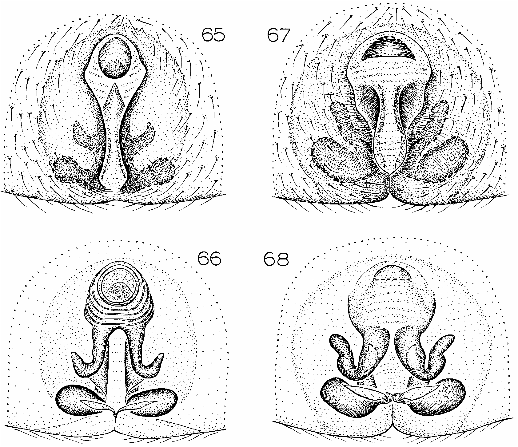
Figures 65, 66 View Figs
Drassus paivani Blackwall, 1864: 175 (female syntype from Great Salvage , Salvage Islands, in HDO, examined; patronym for Paiva).
Drassus bewickii Blackwall, 1864: 176 (female syntype from Great Salvage , Salvage Islands, in HDO, examined; patronym for Bewicke). NEW SYNONYMY.
Drassus paivai: Simon, 1883: 282 .
Drassus bewicki: Simon, 1883: 282 .
Scotognapha paivai: Denis, 1963: 40 , figs. 7–9.
Scotognapha bewickei: Denis, 1963: 41 View in CoL , figs. 10– 12.
DIAGNOSIS: Females resemble those of S. canaricola , but can be recognized by the extremely narrow, anteriorly rhomboidal epigynal atrium (fig. 65) and the large, long heads of the spermathecal ducts (fig. 66).
MALE: Unknown. The male from Tenerife described by Blackwall (1868: 407) could not be found in HDO, but was presumably misidentified.
FEMALE: Described by Blackwall ( 1864).
MATERIAL EXAMINED: SALVAGE IS LANDS: Great Salvage: ( HDO), 2♀ ( S. paivai syntypes) , 2♀ ( S. bewickei syntypes) ; 1983 (J. Murphy, AMNH), 1♀ .
DISTRIBUTION: Known only from the Salvage Islands (see Denis, 1963, for additional records); the juveniles from Tenerife record
ed by Schmidt (1975c: 503, 504) are presumably misidentified.
SYNONYMY: No genitalic differences were detected among the female syntypes. Blackwall and Denis separated the two species only by differences in the size of the posterior lateral spinnerets: S. bewickei has very long spinnerets that are longer than the anterior laterals, with a long basal segment and a small apical segment, whereas S. paivai has a short basal segment on the posterior lateral spinnerets, which are shorter than the anterior laterals. The same character was probably used by Simon ( 1912) when he inferred that S. bewickei belonged to the Agelenidae rather than Gnaphosidae . Our study of the types and additional material indicates that all species of this genus have narrow, very long posterior lateral spinnerets (fig. 11) that are longer than the anterior laterals, but that the posterior laterals are most commonly retracted into the abdomen (at least in preserved specimens) and thus look quite short, even shorter than the anterior laterals.
Scotognapha haria , new species
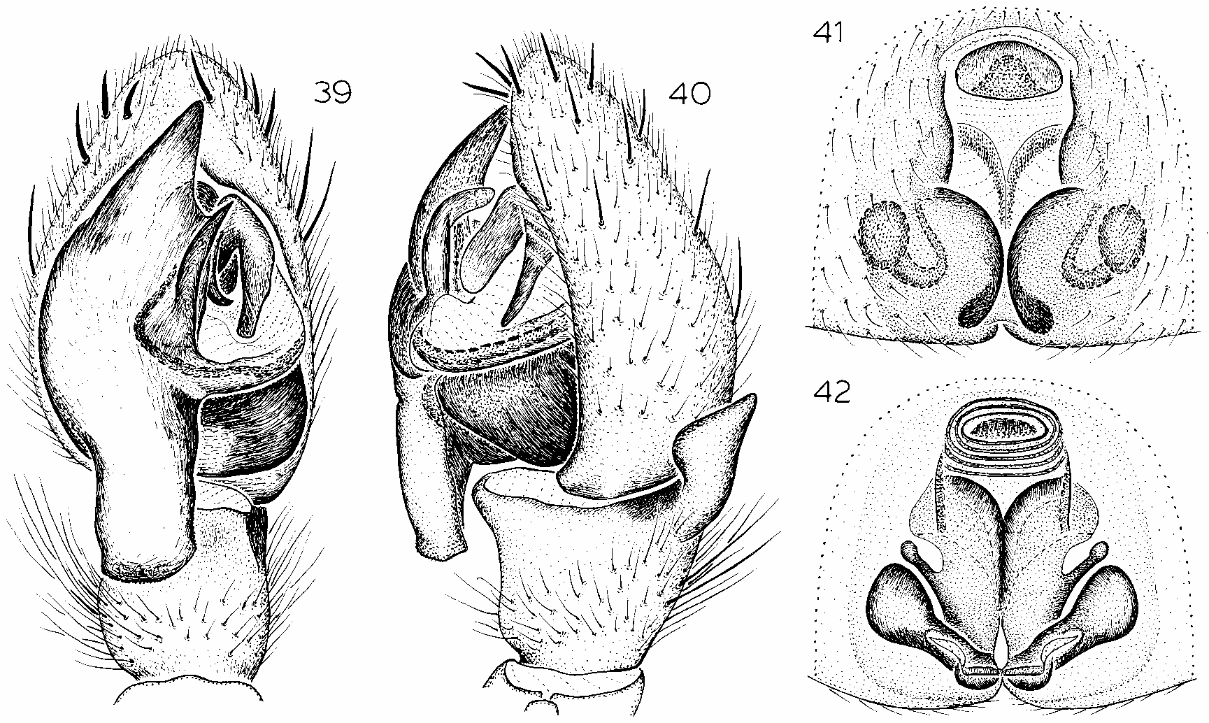
Figures 39–42 View Figs
TYPES: Male holotype and female allotype from a stony area in a montane forest S of Haria , Lanzarote, Canary Islands (end of Apr.–May; J. Wunderlich), deposited in AMNH (courtesy of Mr. Wunderlich) .
ETYMOLOGY: The specific name is a noun in apposition taken from the type locality.
DIAGNOSIS: Males resemble those of S. canaricola but can be recognized by the wide, square distal portion of the tegulum, the straight, apically bifurcate embolus with a greatly elongated lateral branch, and the triangular retrolateral tibial apophysis (figs. 39, 40), females by the wide median portion and strongly narrowed posterior portion of the epigynal atrium (fig. 41) and the narrow, widely spaced heads of the spermathecal ducts (fig. 42).
MALE: Total length 8.43. Carapace 4.21 long, 3.07 wide. Femur II 2.57 long. Eye sizes and interdistances: AME 0.11, ALE 0.15, PME 0.14, PLE 0.12; AMEAME 0.08, AMEALE 0.01, PMEPME 0.03, PMEPLE 0.19, ALEPLE 0.20; MOQ length 0.35, front width 0.31, back width 0.27. Leg spi nation: femora: I left leg d112, r011, right leg d112, r001; II d112, p011, r010; III d112; IV d(left)111, d(right)112, p0 11, r011; patellae: II p010; III p110, r1 10; IV p100, r110; tibiae: I p111, v2 22, r010; II p112, v122, r010; III d2 02; IV d111, p211; metatarsi: I p011, v222; II p012, r101; III p211, r211; IV d222, r211. Palp with wide, straight embolus, bifurcate apically, with elongate lateral portion; distal part of tegulum square; median apophysis short, strongly bent; terminal apophysis small, oval (fig. 39); retrolateral tibial apophysis triangular (fig. 40).
FEMALE: Total length 9.46. Carapace 4.15 long, 2.62 wide. Femur II 2.10 long. Eye sizes and interdistances: AME 0.21, ALE 0.17, PME 0.17, PLE 0.15; AMEAME 0.13, AMEALE 0.04, PMEPME 0.13, PMEPLE 0.23, ALEPLE 0.17; MOQ length 0.59, front width 0.53, back width 0.49. Leg spination: femora: III d111; IV d111; tibiae: III d202, r111; IV d101, p211. Epigynal atrium deep, wide anteriorly, slightly narrowed medially, strongly narrowed posteriorly, copulatory openings situated medially (fig. 41), heads of spermathecal ducts
narrow, widely spaced; receptacles large, directed anterolaterally (fig. 42).
OTHER MATERIAL EXAMINED: None.
DISTRIBUTION: Known only from Lanzarote.
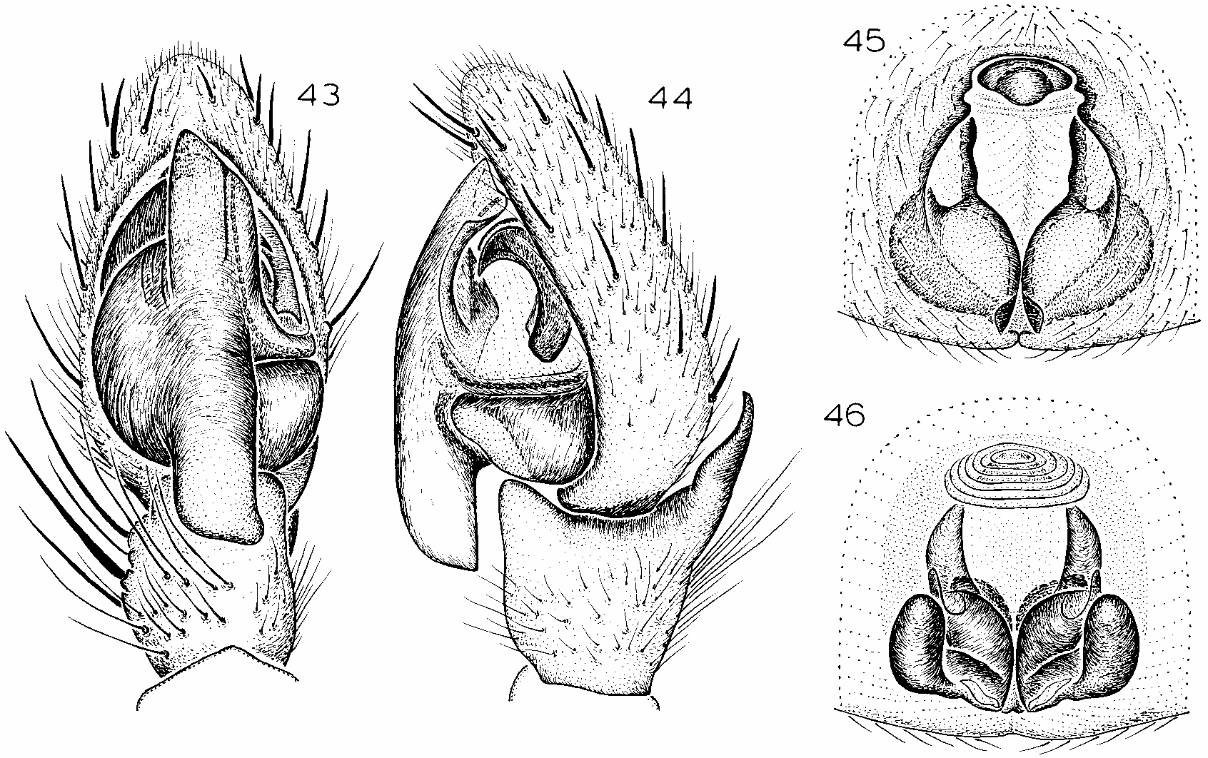
Figures 43–46 View Figs
Scotognapha canaricola (misidentified): Schmidt, 1975b: 242, fig. 1 (male from Bateria de Río, Lanzarote).
Scotognapha brunnea Schmidt, 1980: 331 View in CoL , fig. 1 (female holotype from Playa Famara , Lanzarote, Canary Islands, in NMS, examined).
DIAGNOSIS: Males and females resemble those of S. haria and S. taganana but males can be recognized by the long, wide, and straight embolus, the square posterior portion of the tegulum, the wide, long median apophysis, and the narrow, sharply pointed retrolateral tibial apophysis (figs. 43, 44), females by the deep, yshaped epigynal atrium, the lateral tubercules on the epigynal field (fig. 45), and the slender heads of the spermathecal ducts, which are covered ventrally by the anteriorly directed receptacles (fig. 46).
MALE: Described by Schmidt (1975b). FEMALE: Described by Schmidt ( 1980). Material Examined: CANARY ISLANDS:
Lanzarote: Bateria de Río, Nov. 10, 1972
(G. Schmidt, NMS), 13; Playa Famara, Sept.
12, 1977 (G. Schmidt, NMS), 1♀ (holotype). DISTRIBUTION: Known only from Lanzarote .
Scotognapha wunderlichi , new species
Figures 47–50 View Figs
TYPES: Male holotype taken in pitfall trap at an elevation of 600 m at Barranco de Agaete , Grand Canary , Canary Islands (May–June 1988; J. Wunderlich), and female allotype taken in pitfall trap at an elevation of 400 m at Embalse Parrelillo, Grand Canary, Canary Islands (Aug. 29, 1990; C. G. Campos), deposited in AMNH .
ETYMOLOGY: The specific name is a patronym in honor of the collector of the holotype.
DIAGNOSIS: Males can easily be recognized by the basally wide embolus, which is strongly narrowed anteriorly, and bent and sharply pointed apically (figs. 47, 48), females by the wide, deep, medially narrowed epigynal atrium (fig. 49), the large, round heads of the spermathecal ducts, and the laterally directed receptacles (fig. 50).
MALE: Total length 5.82. Carapace 2.75 long, 2.10 wide. Femur II 1.83 long. Eye sizes and interdistances: AME 0.10, ALE 0.12, PME 0.12, PLE 0.10; AMEAME 0.04, AMEALE 0.01, PMEPME 0.04, PMEPLE 0.11, ALEPLE 0.18; MOQ length 0.31, front width 0.29, back width 0.27. Leg spination: femora: I d111; II d111; III d11 1; IV d111, p011, r011; patellae: III spineless; IV p100; tibiae: I p011, v22 2; II p111, v122; III d101; IV d101, p212; metatarsi: I p010, v222; II p01 1. Palp with embolus basally wide, strongly narrowed anteriorly, bent apically, sharply pointed, converging with long, narrow terminal apophysis (fig. 47); median apophysis short, slightly curved; retrolateral tibial apophysis straight, long (fig. 48).
FEMALE: Total length 6.90. Carapace 3.60 long, 2.70 wide. Femur II 2.70 long. Eye sizes and interdistances: AME 0.21, ALE 0.17, PME 0.17, PLE 0.15; AMEAME 0.13, AMEALE 0.04, PMEPME 0.13, PMEPLE 0.23, ALEPLE 0.17; MOQ length 0.59, front width 0.53, back width 0.49. Leg spination: femora: III d111; IV d111; tibia IV p211. Epigynal atrium wide, deep, equal in width anteriorly and posteriorly, narrowed medially, with wide hood (fig. 49); spermathecal ducts with large, round heads, receptacles oval, directed laterally (fig. 50).
OTHER MATERIAL EXAMINED: None.
DISTRIBUTION: Known only from Grand Canary.
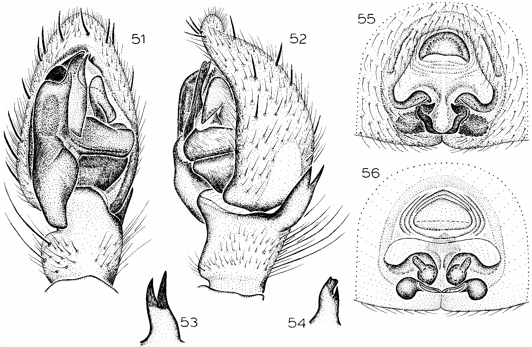
Scotognapha teideensis (Wunderlich) , new combination Figures 1–18 View Figs View Figs View Figs , 51–56 View Figs
Nomisia teideensis Wunderlich, 1991: 486 , figs. 761–764 (male holotype from Las Canadas , Tenerife, Canary Islands, in NMS, examined).
DIAGNOSIS: Males can easily be recognized by the straight, narrow, bifurcate embolus with almost equally sized apical portions, and the bifurcate tip of the retrolateral tibial apophysis (figs. 51–54), females by the extremely wide medial portion of the epigynal atrium (fig. 55) and the large, elongated, anterolaterally directed heads of the spermathecal ducts (fig. 56).
MALE: Described by Wunderlich ( 1991).
FEMALE: Total length 14.05. Carapace 7.54 long, 5.85 wide. Femur II 4.46 long. Eye sizes and interdistances: AME 0.17, ALE 0.20, PME 0.15, PLE 0.17; AMEAME 0.19, AMEALE 0.09, PMEPME 0.12, PMEPLE 0.31, ALEPLE 0.36; MOQ length 0.52, front width 0.45, back width 0.42. Leg spination: femur III d113; patellae: III p010; IV r000; tibiae: III d122, r212; IV d2 10, p211, r132; metatarsi: I, II spineless; III d222; IV d222. Epigynal atrium wide, deep, widened medially, narrowing posteriorly, with wide hood (fig. 55), spermathecal ducts with large, elongate, anterolaterally directed heads, receptacles oval, laterally directed (fig. 56).
MATERIAL EXAMINED: CANARY IS LANDS: Tenerife : Las Canadas, road 821, 48 km, Feb.–May, pitfall, elev. 2000 m (M. Knösel, C. G. Campos, J. Wunderlich, NMS) , 13 (holotype), June 11–19, 1984, pit falls, elev. 2100–2250 m (C. G. Campos, CPO, AMNH) , 33, 1♀, Las Canadas , June 3–12, 1995 (N. Zurita, CPO, AMNH) , 73, July 6–11, 1995 (M. Arechavaleta, CPO, AMNH) , 23, June 3–29, 1995, Oct. 7, 1995, May 18, 1996 (N. Zurita, CPO, AMNH) , 63, 1♀, June 11–12, 1995 (A. Camacho, P. Oromı´, CPO, AMNH) , 23; Tabaiba , Apr. 3– May 30, 1985, pitfall ( AMNH) , 13.
DISTRIBUTION: Known only from Tenerife.
Scotognapha galletas , new species
Figures 57–60 View Figs
TYPES: Male holotype and female allotype from Las Galletas , Tenerife, Canary Islands (Apr.; J. Wunderlich), deposited in AMNH (courtesy of Mr. Wunderlich) .
ETYMOLOGY: The specific name is a noun in apposition taken from the type locality.
DIAGNOSIS: Males resemble those of S. teideensis but can be recognized by the wide, bifurcate embolus with a wide, curved pro lateral tip and a narrow retrolateral tip (fig. 57) and the thick, distally narrowed retrolateral tibial apophysis (fig. 58), females by the wide epigynal atrium with large lateral tubercules (fig. 59) and the short heads of the anteriorly directed spermathecal ducts (fig. 60).
MALE: Total length 7.87. Carapace 3.88 long, 3.00 wide. Femur II 2.44 long. Eye sizes and interdistances: AME 0.16, ALE 0.17, PME 0.15, PLE 0.16; AMEAME 0.10, AMEALE 0.03, PMEPME 0.04, PMEPLE 0.15, ALEPLE 0.16; MOQ length 0.42, front width 0.37, back width 0.36. Leg spination: femora: I d111; II d111; III d11 1; IV d111, r011; patellae: II r100; III p110; IV p100; tibiae: I p(left)111, p(right)112, v222, r011; II p112, v2 22, r101; III d102, r(right)311, r(left)3 12; IV d111, p311; metatarsi: I p010, v221; II p111; III r211. Palp with wide, straight, bifid embolus, prolateral tip wide, curved, retrolateral tip narrow, straight, median apophysis long, narrow, terminal apophysis large (fig. 57); retrolateral tibial apophysis thick, narrowed at tip (fig. 58).
FEMALE: Total length 8.75. Carapace 3.63 long, 2.86 wide. Femur II 2.60 long. Eye sizes and interdistances: AME 0.21, ALE 0.17, PME 0.17, PLE 0.15; AMEAME 0.13, AMEALE 0.04, PMEPME 0.13, PMEPLE 0.23, ALEPLE 0.17; MOQ length 0.59, front width 0.53, back width 0.49. Leg spination: femur III d111; tibiae: III d202; IV p211. Epigynal atrium wide, divided by lateral extensions medially, narrowed posteriorly, with elongated hood (fig. 59); spermathecal ducts with short, wide, anteriorly directed heads, receptacles directed laterally (fig. 60).
OTHER MATERIAL EXAMINED: CANARY ISLANDS: Tenerife: no specific locality, Mar. 1990 (D. Knösel, AMNH), 1♀ ; El Medano, June 9, 1984, coast ( AMNH), 2♀ ; Las Galletas, Apr., under stones (J. Wunderlich, AMNH, CJW), 63, 3♀ ; S. Andres, Feb. 1979, elev. ca. 200 m ( CKT), 1♀ .
DISTRIBUTION: Known only from Tenerife.
Scotognapha taganana , new species
Figures 61–64 View Figs
TYPES: Male holotype taken under stones at Taganana , Tenerife , Canary Islands (Apr.; J. Wunderlich), and female allotype taken in a meadow at La Esperanza , Tenerife, Canary Islands (Apr. – May; J. Wunderlich), deposited in AMNH (courtesy of Mr. Wunderlich) .
ETYMOLOGY: The specific name is a noun in apposition taken from the type locality.
DIAGNOSIS: Males resemble those of S. teideensis and S. galletas but can be recognized by the short, wide, apically strongly bent, blunt embolus, the narrow median apophysis, and the thick, blunt, pointed retrolateral tibial apophysis (figs. 61, 62); females can easily be recognized by the epigynal atrium being of subequal width anteriorly and posteriorly but narrowed medially (fig. 63), and by the wide, zigzag conformation of the heads of the laterally directed spermathecal ducts (fig. 64).
MALE: Total length 8.15. Carapace 4.38 long, 3.08 wide. Femur II 2.31 long. Eye sizes and interdistances: AME 0.15, ALE 0.15, PME 0.13, PLE 0.13; AMEAME 0.06, AMEALE 0.01, PMEPME 0.05, PMEPLE 0.13, ALEPLE 0.15; MOQ length 0.37, front width 0.31, back width 0.31. Leg spination: femora: I d111; II d111; III d11 1; IV d111; patellae: III p010; IV p010; tibiae: I p001, v222; II p101, v222; III d112, r111; IV d112, p211; metatarsi: I v221; II p010. Palp with short, wide, blunt embolus with equal prolateral and retrolateral tips, square posterior part of tegulum, narrow, short median apophysis (fig. 61); retrolateral tibial apophysis straight, thick, bluntly pointed (fig. 62).
FEMALE: Total length 8.54. Carapace 4.15 long, 3.08 wide. Femur II 2.70 long. Eye sizes and interdistances: AME 0.12, ALE 0.17, PME 0.13, PLE 0.12; AMEAME 0.07, AMEALE 0.04, PMEPME 0.05, PMEPLE 0.15, ALEPLE 0.17; MOQ length 0.33, front width 0.29, back width 0.27. Leg spination: femur III d111; r001; tibia III d2 02, r111. Epigynal atrium shallow, of sub equal width anteriorly and posteriorly, narrower medially, with oval hood (fig. 63); heads of spermathecal ducts short, wide, forming zigzag, directed laterally, receptacles wide, anteriorly directed (fig. 64).
OTHER MATERIAL EXAMINED: CANARY ISLANDS: Tenerife: La Esperanza, Apr.– June, meadow (J. Wunderlich, AMNH, CJW) , 33; Las Galletas , El Fraile, Mar. 11, 1996, stony waste near sea (J. Murphy, JAM) , 13; Mercedes, Apr.–June, forest (J. Wunderlich, AMNH, CJW) , 33.
DISTRIBUTION: Known only from Tenerife.
Scotognapha costacalma , new species
Figures 67, 68 View Figs
Scotognapha atomaria (misidentified): Schmidt, 1976: 324, fig. 6.
Type : Female holotype from sand beach at Costa Calma , Fuerteventura, Canary Islands (July 3, 1973; G. Schmidt), deposited in NMS .
ETYMOLOGY: The specific name is a noun in apposition taken from the type locality.
DIAGNOSIS: Females resemble those of S.
65, 67. Epigynum, ventral view. 66, 68. Same, dorsal view.
taganana but can be recognized by the wide epigynal hood and the wide anterior portion and strongly narrowed posterior portion of the epigynal atrium (fig. 67) and by the long, narrow, anterolaterally directed heads of the spermathecal ducts (fig. 68).
MALE: Unknown.
FEMALE: Total length 5.40. Carapace 2.47 long, 1.89 wide. Femur II 1.12 long. Eye sizes and interdistances: AME 0.11, ALE 0.12, PME 0.08, PLE 0.10; AMEAME 0.08, AMEALE 0.04, PMEPME 0.04, PMEPLE 0.13, ALEPLE 0.10; MOQ length 0.28, front width 0.27, back width 0.25. Leg spination: femur III d111, p001, r001; patella III p110, r110; tibiae: III d202, v1 12, r111; IV p211, v122; metatarsus IV d022. Epigynal atrium deep, wide anteriorly, shallow, strongly narrowed posteriorly, with wide hood (fig. 67), long, narrow, anterolaterally directed heads of spermathecal ducts in zigzag conformation, oval spermathecal receptacles directed laterally (fig. 68).
OTHER MATERIAL EXAMINED: None.
DISTRIBUTION: Known only from Fuerteventura.


No known copyright restrictions apply. See Agosti, D., Egloff, W., 2009. Taxonomic information exchange and copyright: the Plazi approach. BMC Research Notes 2009, 2:53 for further explanation.
|
Kingdom |
|
|
Phylum |
|
|
Class |
|
|
Order |
|
|
Family |
Scotognapha
| PLATNICK, NORMAN I., OVTSHARENKO, VLADIMIR I. & MURPHY, JOHN A. 2001 |
Nomisia teideensis
| Wunderlich 1991: 486 |
Scotognapha brunnea
| Schmidt 1980: 331 |
Scotognapha canaricola
| : Schmidt 1975: 221 |
Scotognapha paivai
| : Denis 1963: 40 |
Scotognapha bewickei
| : Denis 1963: 41 |
Scotognapha convexa:
| Dalmas 1920: 120 |
Scotognapha atomaria
| Dalmas 1920: 121 |
Scotognapha gravieri
| Dalmas 1920: 121 |
Gnaphosa canaricola
| Strand 1911: 191 |
Callilepis convexa:
| Simon 1893: 382 |
Pythonissa convexa
| Simon 1883: 291 |
Pythonissa convexa
| Simon 1883 |
Drassus paivai:
| Simon 1883: 282 |
Drassus bewicki:
| Simon 1883: 282 |
Drassus paivani
| Blackwall 1864: 175 |
Drassus bewickii
| Blackwall 1864: 176 |