
Seira betica, Cipola & Arbea & Baquero & Jordana & Morais & Bellini, 2018
|
publication ID |
https://doi.org/ 10.11646/Zootaxa.4458.1.1 |
|
publication LSID |
lsid:zoobank.org:pub:urn:lsid:Zoobank.org:pub:6DE35A7F-628C-4017-A39D-95D268C4F5E0 |
|
DOI |
https://doi.org/10.5281/zenodo.6492058 |
|
persistent identifier |
https://treatment.plazi.org/id/B83E8799-FFD6-1558-96AF-E22723B5D1A5 |
|
treatment provided by |
Plazi |
|
scientific name |
Seira betica |
| status |
sp. nov. |
Seira betica View in CoL sp. nov. Cipola & Arbea
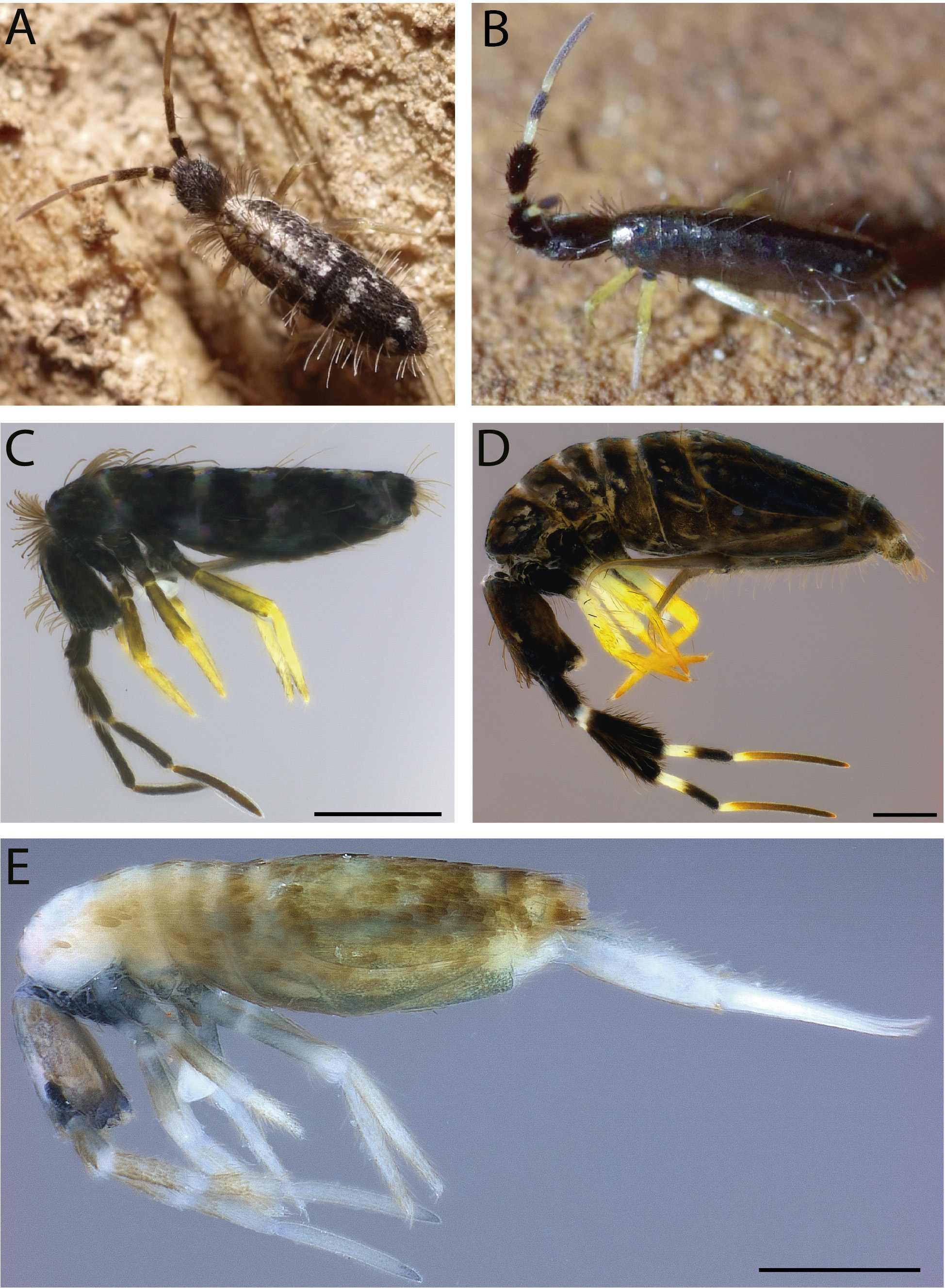
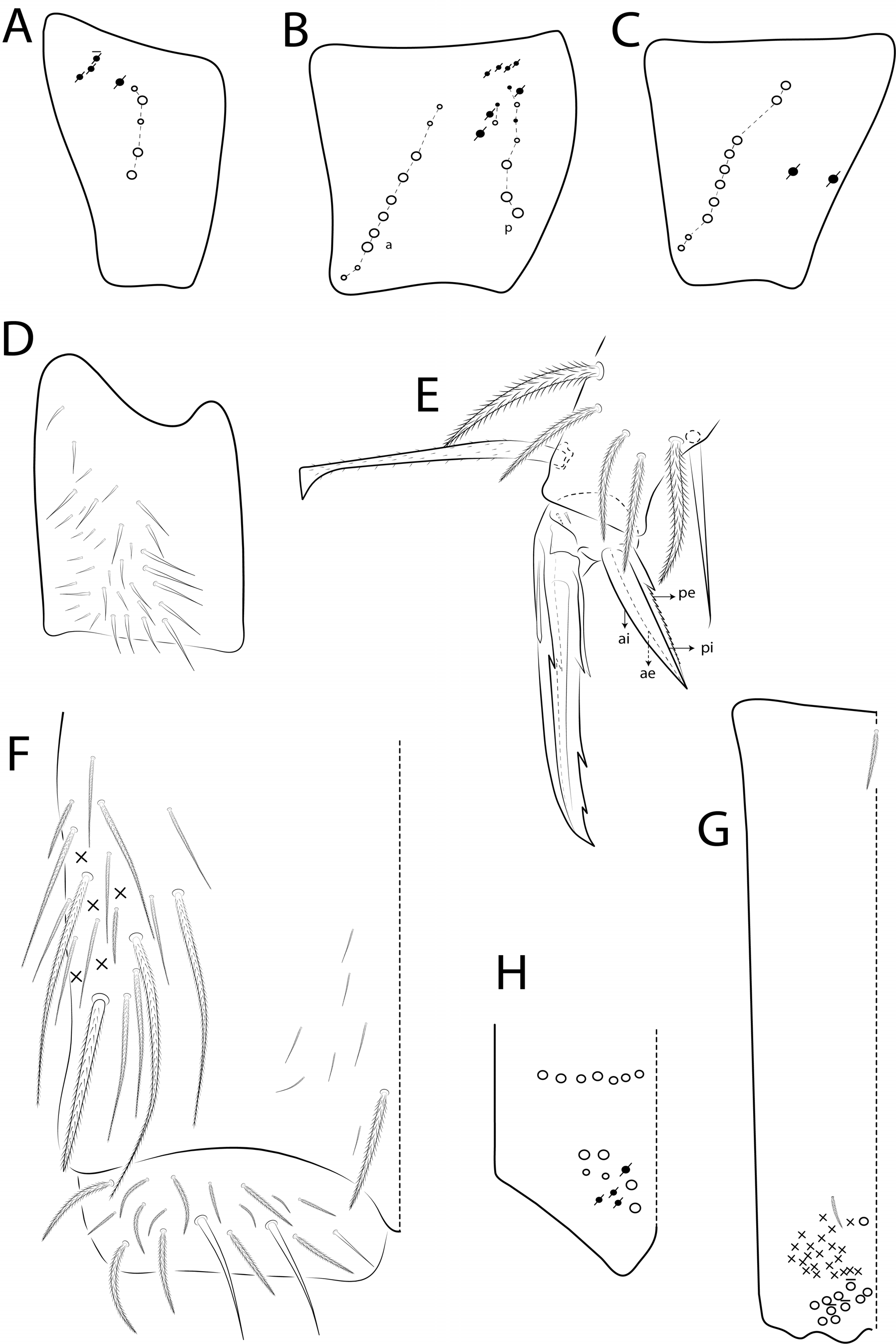
Figs 3 A, E View FIGURE 3 , 30‒34 View FIGURE 30 View FIGURE 31 View FIGURE 32 View FIGURE 33 View FIGURE 34 , Tables 1‒2
Diagnosis. Body with two longitudinal bands on Th II‒Abd II, and Abd III‒ IV with irregular transverse spots or restricted on head and thorax laterally when preserved in alcohol ( Figs 3 A, E View FIGURE 3 ) ; head mac M 4i, S4, Pa 4 and Pp 6 absent, eyepatches with 4 interocular chaetae (q absent) ( Fig. 31E View FIGURE 31 ) ; labral papillae conical, outer papillae reduced to conical projections ( Fig. 31C View FIGURE 31 ); Th II with 5 medio-central mac (m1‒ 1i 2, m2‒ 2i) and PmA‒PmC groups with 7‒9 , 3 and 6 mac respectively ( Fig. 32 A View FIGURE 32 ); Th III ‒ Abd III with 15‒17 , 6 , 5 and 1‒2 ( rarely a3 as mac) central mac, respectively ( Figs 32 B‒E View FIGURE 32 ) ; Abd IV with 14 central mac, 16‒17 lateral mac and about 10 psp anteriorly ( Fig. 33 A View FIGURE 33 ) ; unguis apical tooth present ( Fig. 34E View FIGURE 34 ); unguiculus outer edge serrated and with proximal tooth ( Fig. 34E View FIGURE 34 ); manubrium ventrally with 2/2 subapical and 14‒20 apical chaetae ( Fig. 34G View FIGURE 34 ).
Type material. Holotype female on slide (EHL0005/MNCN): Spain, Huelva Province, Minas de Riotinto municipality (37°39'11''N; 06°37'10''W), on wood, 445 m, 11.x.2015, A GoogleMaps . Burgers coll. Paratypes on slides (INPA and MNCN): 3 females and 2 specimens in alcohol, same data as holotype .
Other material. 2 females and 1 juvenile on slides (EAL0001/ JIAP), Spain, Almería province, Vera municipality, Playa de Vera (37°12'09''N; 01°48'42''W), back of the beach, 1 m, 27.ix.2005, pitfall trap, J GoogleMaps .I. Arbea coll.
Description. Total length (head + trunk) of adult specimens 2.79‒3.44 mm (n=4), holotype 2.89 mm. Alive specimens with Ant III‒IV and legs brownish, Ant I‒II, head and body black (formed by scales) with two dorsal white longitudinal bands (formed by scales) that extend from Th II to Abd II, plus irregular transverse spots on Abd III and two spots on Abd IV ( Fig. 3 A View FIGURE 3 ). Specimens in alcohol pale white with dark blue pigment on apex of Ant IV, anterior head, Th I, coxae to basal half of femora, collophore, and Abd I‒IV laterally; eyepatches black ( Fig. 3E View FIGURE 3 ). Scales present on Ant I to basal half of Ant III, ventral and dorsal head, thorax and abdomen dorsally, legs (except empodia), anterior collophore, and manubrium and dentes ventrally ( Figs 30 A ‒D, I‒J View FIGURE 30 ).
Head. Antennae shorter than body length, antennal ratio as I: II : III: IV = 1: 1.6 0‒1.78: 1.57‒1.99: 2.38‒2.95 (n=4), holotype 1 : 1.69: 1.80: 2.72 ( Fig. 3 A, E View FIGURE 3 ) . Ant IV annulated, with simple apical bulb retractile and blunt sens. Ant III apical organ with two rod-like sens, 3 guard sens, one atypical rounded bulb and several blunt sens of different sizes ( Fig. 31 A View FIGURE 31 ) . Clypeal formula with 4 (l1‒2), 4 (f), 3 (pf0‒1) ciliate chaetae, l1 acuminate, l2 largest, and 2 outer frontal smaller ( Fig. 31B View FIGURE 31 ). Two inner labral papillae conical, outer papillae reduced to conical projections ( Fig. 31C View FIGURE 31 ). Maxillary palp with smooth apical appendage (a. a.) and basal chaeta (b.c.) weakly ciliated, thicker and 1.08 longer than apical ( Fig. 31D View FIGURE 31 ) . Eyes A and B larger, G smaller, with 4 interocular chaetae (v, p, r, t) ; head dorsal chaetotaxy ( Fig. 31E View FIGURE 31 ) with 10 ‘An’ mac (An 1, An 2‒3), 4 ‘A’ mac (A 0, A 2‒3, A 5), 3 ‘M’ mac (M1‒2, M4), 7 ‘S’ mac (S0‒3, S5‒6), 1 ‘Ps’ mac ( Ps 2, rarely as mic), 4 ‘Pa’ mac ( Pa 1‒3, Pa 5), 2 ‘Pm’ mac ( Pm 1, Pm 3), 4 ‘Pp’ mac ( Pp 1‒3, Pp 5) , and 4 ‘Pe’ mac (Pe2‒4 plus Pe3p). Basomedian and basolateral labial fields with M1‒2, R (smaller), E, L1‒2. Postlabial ventral chaetotaxy with about 17 ciliate chaetae, postlabial formula 4 (G1‒4), 3 (H2‒4), 4 (J1‒4), basal chaeta (b.c.) largest ( Fig. 31F View FIGURE 31 ).
Thorax chaetotaxy ( Figs 32 A ‒B View FIGURE 32 ). Th II, series ‘a’ with 4 mac (a5ip‒5p) ; series ‘m’ with 8 mac (m 1i 2‒2, m 4i ‒4p); series ‘p’ with 17‒19 mac (p 1i 2p2‒1p, p2a–2ep2, p3‒3p, p5), p 1i 2p2 and p1ip2p present or absent. Th III, series ‘a’ with 7 mac (a 1i ‒6); series ‘m’ with 1 mac (m6); series ‘p’ with 11‒13 mac (p1ip2‒1p2, p2a‒2ea, p3, p5‒6), p1ip and p1ip2 present or absent. Th ratio as II : III = 1: 0.49‒0.61 (n= 4), holotype 1: 0.49.
Abdomen chaetotaxy ( Figs 32 C‒E View FIGURE 32 , 33 A ‒B View FIGURE 33 ) . Abd I, series ‘a’ with 1 mac (a3); series ‘m’ with 5 mac (m 2i ‒4). Abd II, series ‘a’ with 2 mac (a2‒3); series ‘m’ with 4 mac (m3‒3e, m5). Abd III, series ‘a’ with a3 rarely as mac; series ‘m’ with 3 mac (m3, am6, pm6) ; series ‘p’ with 1 mac (p6). Abd IV with 14 central mac of series ‘A’ to ‘T’ (A 3a‒6, Ae 7, B1‒6, C1, T1) , and 16‒17 lateral mac of series ‘E’ to ‘Fe’ (E2‒4p, Ee 7, Ee 10, F1‒3, Fe 2‒5 and one unnamed as mac or mic); 10 psp ( atypical) anteriorly, at least 5 sens (as and ps type I and 3 type II) , and posteriorly with 7 mes present. Abd V, series ‘a’ with 1 mac (a5); series ‘m’ with 4 mac (m2‒3, m5‒5e); series ‘p’ with 7 mac (p1, p3‒5, ap6–6e, pp6); series ‘pp’ with 2 mac (p1p, p3pe). Abd ratio as III: IV = 1: 4.40‒5.43 (n= 4) , holotype 1: 4.98.
Legs. Subcoxa I with 5 chaetae and 3‒4 psp; subcoxa II with an anterior row of 10 chaetae, posterior row with 7 chaetae, 2 anterior chaetae and 7 psp; subcoxa III with one row of 10 chaetae and 2 posterior psp ( Figs 34 A ‒C View FIGURE 34 ) . Trochanteral organ with about 36 spine-like chaetae ( Fig. 34D View FIGURE 34 ). Unguis with basal and median teeth with the same length, apical tooth smaller. Unguiculus with all lamellae acuminate, pe lamella serrated and with small proximal tooth, other lamellae (ai, ae, pi) smooth; ratio unguis: unguiculus = 1: 0.49 ( Fig. 34E View FIGURE 34 ) . Tibiotarsus III distally with inner smooth chaeta 1.15 larger than unguiculus; and outer tenent hair capitate, discretely ciliate, and 0.81 smaller than unguis.
Collophore ( Fig. 34F View FIGURE 34 ). Anterior side with 17 ciliate chaetae, including 1 distal mac and 3 large acuminate long chaetae; posterior side with 8 ciliate chaetae, of which one distal thickest; lateral flap with 3 smooth chaetae (one smaller posteriorly) and 15 ciliate chaetae.
Furcula ( Figs 34G‒H View FIGURE 34 ). Manubrium ventrally with formula 1, 0, 0, 2/2 (subapical), 14‒20 (apical) ciliate chaetae and approximately 22 elongated apical scales per side; manubrium dorsally with 7 subapical ciliate chaetae, manubrial plate with 6 ciliate chaetaeand 4 psp.
Etymology. The species is named after the Betica Region (approximately corresponds to modern Andalucia) where it has been found.
Remarks. Seira betica sp. nov. resembles S. dagamae Dallai, 1973 and S. stachi Loksa, 1990 by color pattern of body on two longitudinal white bands on Th II‒Abd II, Abd IV with irregular transverse spots, and most dorsal macrochaetotaxy pattern (at least of S. dagamae ). However, S. betica sp. nov. differs from S. dagamae by Th II with 5 medio-central mac (4 in S. dagamae ) and PmA and PmC groups with 7‒9 and 6 mac, respectively (7 and 5 in S. dagamae ); Th III with 7‒9 mac inner to psp (6 in S. dagamae ); and Abd IV with mac B1‒2 (absent in S. dagamae ). Seira betica sp. nov. differs at this time from S. stachi more clearly by unguiculus outer edge serrated (smooth in S. stachi ). The original description of S. stachi does not present the dorsal macrochaetotaxy, as well as several other characteristics, which makes difficult to compare the species (see Loksa 1990: 274). In addition, in the description of unguis 3 internal teeth are reported (proximal, median and apical), probably wrongly, since the proximal tooth is always paired in Seira (Christiansen & Bellinger 2000; Barra 2004 a, 2004b; Bellini et al. 2010; Cipola et al. 2014 a, 2014b; Godeiro & Bellini 2014, 2015), therefore this can not be used to compare the species. The presence of a subapical rounded bulb on Ant III ( Fig. 31 A View FIGURE 31 ) and anterolateral psp in Abd IV ( Fig. 33 A View FIGURE 33 ) of S. betica sp. nov. are exclusive features registered here for the first time in Seira , which can help distinguish this species from other related taxa. Other comparisons among species are presented in Tables 1 and 2.


No known copyright restrictions apply. See Agosti, D., Egloff, W., 2009. Taxonomic information exchange and copyright: the Plazi approach. BMC Research Notes 2009, 2:53 for further explanation.
|
Kingdom |
|
|
Phylum |
|
|
Class |
|
|
Order |
|
|
Family |
|
|
Genus |