
Simulium
|
publication ID |
https://doi.org/ 10.5281/zenodo.171413 |
|
DOI |
https://doi.org/10.5281/zenodo.6261291 |
|
persistent identifier |
https://treatment.plazi.org/id/8E591D7D-FFAE-8F73-FED1-BF68A2E2FB13 |
|
treatment provided by |
Plazi |
|
scientific name |
Simulium |
| status |
|
Simulium View in CoL View at ENA (Psaroniocomps a) guaporense PyDaniel
( Figs. 1–33 View FIGURES 1 – 15 View FIGURES 16 – 24 View FIGURES 25 – 29 View FIGURES 30 – 33 )
Simulium guaporense PyDaniel, 1989: 502 View in CoL 508. HOLOTYPE pupa (on slide, INPA 58491), BRAZIL: Rondônia State, Igarapé Ponte de Pedra, km 27 da rodovia RO399, Fazenda Régis; 14.vi.1981 (V. PyDaniel) (INPA) [Examined]
Simulium (Psaroniocompsa) siolii PyDaniel, 1988: 294 View in CoL –300. [In part]
Female. General body color black. Body mean length (specimens pinned) 1.67 mm (SD = 0.05, n = 5), wing mean length 1.7 mm (SD = 0.09, n = 5), wing mean width 0.7 mm (SD = 0.03, n = 5).
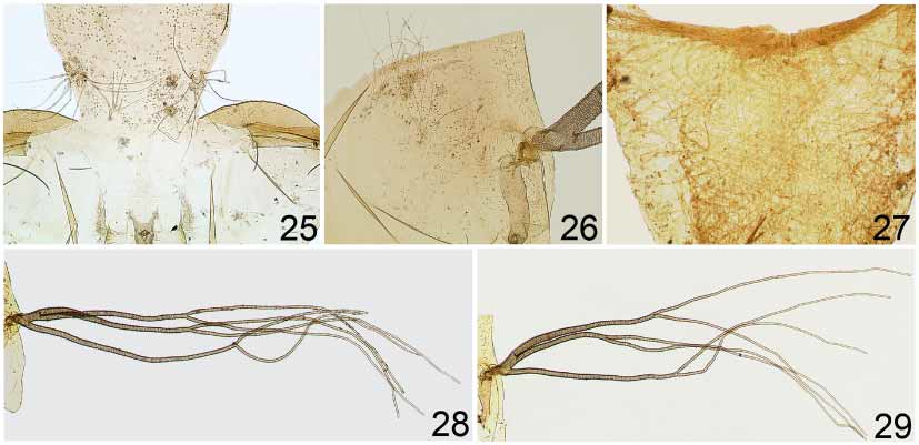
Head dichoptic with dark eyes and nudiocular area small ( Fig. 3 View FIGURES 1 – 15 ). Frons, clypeus, and occiput pollinose with metallic reflections; clypeus, frons, and occiput bare in all specimens examined. Antenna length 0.25 mm; scape and pedicel yellowish brown, flagellum dark brown ( Fig. 1 View FIGURES 1 – 15 ). Palpus brown ( Fig. 2 View FIGURES 1 – 15 ); sensory vesicle occupying less than half length of third palpomere, with short neck; palpomere V approximately 2.5 times as long as palpomere III and 1.9 times as long as palpomere IV. Mandible with 68 external serrations and 2830 internal teeth. Lacinia with 21–23 retrorse teeth. Cibarium with sclerotised cornuae and prominent teeth in central trough [triangular small teeth are in membrane of pharynx and in anterolateral margin of cornuae] ( Fig. 4 View FIGURES 1 – 15 ).
Thorax with scutum black covered by recumbent, brasscolored scalelike setae, with greenish and bluish reflections with some light. Scutum, independent of light incidence, black without pattern, covered with evenly arranged scalelike setae forming 5 longitudinal lines, 1 median and 2 + 2 submedian, on central region of scutum extending from anterior to near posterior margin of scutum, and groups of setae in clumps that form broken lines diverging from anterior to lateral margins; posterior margin with single, recumbent, unevenly arranged whitish hairs ( Figs. 9, 10 View FIGURES 1 – 15 ); humeri silver pruinose [best seen in specimens devoid of setae]; lateral and posterior margins silver pruinose [best seen with light incidence posterior and specimens tilted laterally]. Scutellum dark brown, devoid of hairs in few specimens examined. Postnotum dark brown to black with distinct silver pruinosity. Pleura and sterna black with silver pruinosity. Costa of wing with sparse distribution of spines and setae ( Fig. 20 View FIGURES 16 – 24 ); subcosta without hairs or spines ( Fig. 20 View FIGURES 16 – 24 ); radius with single row of spines toward apex, basal section of radius bare ( Fig. 20 View FIGURES 16 – 24 ); basal tuft of long, dark setae. Coloration and proportions of legs as in Figs. 5–7 View FIGURES 1 – 15 . Foreleg ( Fig. 5 View FIGURES 1 – 15 ) with coxa, trochanter, femur, and dorsal margin of tibia yellowish brown; ventral margin of tibia whitish; basitarsus and tarsomeres I–IV black. Middle leg ( Fig. 6 View FIGURES 1 – 15 ) with coxa, apical onethird of basitarsus and tarsomeres I–IV dark brown; trochanter, femur and tibia yellow; twothirds of basitarsus whitish. Hind leg ( Fig. 7 View FIGURES 1 – 15 ) with coxa, twothirds of femur and tibia, apical onethird of basitarsus and tarsomeres I–IV dark brown; trochanter, basal onethird of femur and tibia yellow; basal twothirds of basitarsus whitish [silver pruinose in pinned specimens]. Femur and tibia of hind legs covered with lanceolate scalelike setae interspersed with erect black hairs. Calcipala as broad as long, reaching pedisulcus. Tarsal claws curved with basal tooth ( Fig. 8 View FIGURES 1 – 15 ). Halters lemon yellow with dark brown base.
Abdomen ( Fig. 15 View FIGURES 1 – 15 ) with tergites I, III–V velvet dark brown, VI–IX shiny black; tergite II brown on median region with silver pruinosity on lateral margin; basal fringe with long, brown hairs. Tergal plates developed. Sternites greyish; genitalia dark brown to black. Hypogynial lobes (= gonapophyses) subtriangular, covered by small microtrichia, mainly membranous but sclerotised on inner margins ( Fig. 13 View FIGURES 1 – 15 ). Cercus suboval, covered with long, brown setae; anal lobe (= paraproct) about 1.5 times longer than wide at mid point, covered with long hairs ( Fig. 11 View FIGURES 1 – 15 ). Genital fork with slender, weakly sclerotised stem; termination of lateral arms with anterior margin nearly straight and well developed; anterior processes well developed and rounded apically; posterior processes poorly developed ( Fig. 12 View FIGURES 1 – 15 ). Spermatheca ( Fig. 14 View FIGURES 1 – 15 ) oval without external sculpturing; internal spicules arranged in groups of 2 or 3; area of insertion of spermathecal duct membranous, nearly onethird maximum width of spermatheca.
Male. General body color black. Body mean length (specimens pinned) 1.61 mm (SD = 0.03, n = 3), wing mean length 1.5 mm (SD = 0.02, n = 3), wing mean width 0.7 mm (SD = 0.03, n = 3).
Head holoptic with dark red eyes. Antenna mean length 0.25 mm, scape and pedicel yellowish brown, flagellum dark brown ( Fig. 16 View FIGURES 16 – 24 ). Palpus ( Fig. 17 View FIGURES 16 – 24 ) brown, palpomere V approximately 2.3 times as long as palpomere III and 2.1 times as long as palpomere IV; sensory vesicle small, subspherical. Rest of head and scutum coloration ( Figs 18, 19 View FIGURES 16 – 24 ) as in female; humeri weakly pruinose; lateral margins black. With light source posterior to specimen, humeri and lateral margin silver pruinose. Scutellum dark brown with long, brown, erect setae on posterior margin. Postnotum dark brown with silvery grey pruinosity. Wing setation as in female ( Fig. 20 View FIGURES 16 – 24 ). Leg coloration and setation as in female ( Figs 5–7 View FIGURES 1 – 15 ), but tarsal claw without basal tooth.
Abdominal tergites dark brown, basal fringe with thin, long, brown hairs and golden highlights; tergite II with silver pruinosity; tergites III–VIII with silver pruinosity on lateral margin, extent of pruinosity on tergites VI and VII being larger. Gonocoxite subquadrangular nearly as long as wide; gonostyle subtriangular, 1.5 times shorter than gonocoxite, terminating in 1 distinct apical spine and bearing 1 longitudinal ridge ( Fig. 23 View FIGURES 16 – 24 ); gonocoxite and gonostyle covered with long setae ( Figs. 23, 24 View FIGURES 16 – 24 ). Ventral plate ( Fig. 22 View FIGURES 16 – 24 ) subrectangular, weakly sclerotised, with welldeveloped body, distinctly raised at its center; lateral arms developed and wide apically; main body of ventral plate covered by numerous setae. Median sclerite ( Fig. 21 View FIGURES 16 – 24 ) suboval. Paramere ( Fig. 21 View FIGURES 16 – 24 ) with welldeveloped and sclerotised basal process and numerous long spines.
Pupa. Cocoon length dorsally 2.1–2.4 mm (n = 2), ventrally 2.42.6 mm (n = 2); pupa length 1.3–1.8 mm (n = 2); gill length 2.1–2.4 mm (mean = 2.3 mm, SD = 0.10, n = 10). Cocoon slipper shaped, pale brown, composed of thick coalesced fibers, without anterior projection; anterior margin reinforced ( Fig. 27 View FIGURES 25 – 29 ).
Gill configuration with short petiole and (2 + 2) + 2 filaments ( Figs. 28, 29 View FIGURES 25 – 29 ). The dorsal branch divides into 2 secondary branches on basal onethird, 1 anterior and 1 posterior, each bifurcating at different heights at some distance from gill base. The ventral primary branch bifurcates into 2 secondary branches nearly at midpoint of gill length, often nearly at same level as anterior secondary branch. Variation in this pattern occurs with ventral primary branch bifurcating more apically than anterior and posterior secondary branches, and anterior and secondary branches bifurcating at same level. Filaments slender, with rounded ends, edges crenate, covered by small, dark, spicules; all filaments approximately of same length.
Head with 2 + 2 long, quadrifid frontal and 1 + 1 long, quadrifid, dorsal (= facial) trichomes; frontoclypeus with faint group of platelets mesally and 1 + 1 dorsolaterally in frontal region, respectively; rounded and pointed tubercles sparsely to densely distributed over entire surface ( Fig. 25 View FIGURES 25 – 29 ).
Thorax with 5 + 5 dorsal bifid and quadrifid trichomes located near dorsal cleft and 1 + 1 bifid trichome on central region; tubercles rounded (some pointed), densely distributed anteriorly and scarce on posterior region of thorax ( Fig. 26 View FIGURES 25 – 29 ).
Abdominal tergite I with 1 + 1 lateral, long, simple or bifid trichomes; tergite II with 3 + 3 median, simple, small trichomes in longitudinal row, 3 + 3 simple, small, trichomes to outmost trichomes unevenly arranged and 1 + 1 sublateral, small, simple trichomes; tergites III and IV with 4 + 4 submedian, simple hooks in longitudinal row, 1 + 1 small, simple trichomes anterior to outer hooks, and 2 + 2 submedian and 1 + 1 sublateral, simple or bifid trichomes on posterior margin; tergites V, VI, and VII with spine combs on anterior margin, sometimes developed into sclerotised spines centrally and with 1 + 1 or 2 + 2 small, simple, trichomes on posterior margin; sternite VIII with small spine combs; sternite IX weakly sclerotised and 1 + 1 small terminal spine. Abdominal sternite III with 2 + 2 sublateral, simple trichomes; sternite IV with 2 + 2 submedian, simple or bifid, and 2 + 2 lateral, single trichomes; sternite V with 2 + 2 median, close, bifid or quadrifid hooks in longitudinal row, 2 + 2 simple, small trichomes anterior to outer hooks; sternite VI and VII with 4 + 4 wellseparated hooks, innermost trifid and outermost on right side bifid and on left side simple; sternite VIII with single, small, simple trichomes on posterior margin; sternite IX weakly sclerotised. Spine combs distributed on anterior margin of abdominal sternites III–VIII.
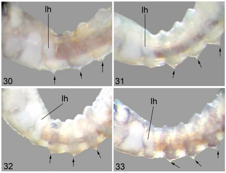
Larva. In general, as in original description ( PyDaniel, 1989), but first pair of dorsal tubercles located on third thoracic segment, not on first abdominal segment ( Fig. 30 View FIGURES 30 – 33 ); abdominal segments I–IV with 2 pairs of tubercles, 1 dorsal and 1 lateral; abdominal segment V with 1 pair of dorsal tubercles. Subesophageal ganglion not pigmented. Anal papillae with 3 branches, each with 14–16 digitiform lobes.
Taxonomic Discussion. Simulium guaporense was described by PyDaniel (1989) from larvae and pupae collected in the state of Rondônia, Brazil. In his 1989 paper, Py Daniel mentioned that in the original description of S. siolii ( PyDaniel 1988) , one larva he collected in Rondônia State, Igarapé da Cachoeira, Bacia do rio Guaporé matched the morphology of S. guaporense . The second author (LMH) of the present paper examined the holotype of S. guaporense , which is housed in INPA.
Simulium guaporense View in CoL was placed in the siolii View in CoL species group of the subgenus Psaroniocompsa by PyDaniel (1989), based on the presence of dorsal and lateral tubercles and multiple branched scalelike setae on the larval body. However, the presence of these tubercles (or protuberances) is apparently not restricted to this species group in the Neotropical Region. Shelley et al. (2002) described similar tubercles in two populations of the relatively wellstudied species Simulium guianense Wise View in CoL (species complex) collected in Rio Verdão and Rio Doce, Goiás State, Brazil. Similar variation in tubercles is also seen in larvae of the S. damnosum View in CoL complex in Africa, in which variation in tubercle size ranges from absent to well developed. In some cases, these tubercles may be associated with cytotype or occur in populations of a single cytotype ( Shelley et al. 2002).
Adults reared from pupae collected near the type locality of S. guaporense View in CoL do not resemble those of the siolii View in CoL group. The females in the siolii View in CoL group are distinguished by having the thorax black with a pattern consisting of 1 + 1 silver pruinose bands that extend from the anterior to the posterior margins [light source anterior]. In males, the thorax is also black with a pattern consisting of 1 + 1 sublateral, silver pruinose cunae [light source anterior] [BMNH Digital Images Archive; Hamada 2000]. The adults of S. guaporense View in CoL externally resemble species in the auristriatum View in CoL species group by having the thorax covered with brasscolored, scalelike, recumbent setae arranged in lines in the central region of the scutum and grouped in clumps forming broken lines from the anterior to lateral margins ( Figs. 9, 10 View FIGURES 1 – 15 , 18, 19 View FIGURES 16 – 24 ). The morphology of the female and male genitalia of S. guaporense View in CoL agrees with the characters given by Coscarón & Wygodzinsky (1984) and Coscarón (1987, 1991) for species in the subgenus Psaroniocompsa . At this stage, we prefer to regard S. guaporense View in CoL as a “species unplaced to group”, but near the auristriatum View in CoL species group in Psaroniocompsa .
The thoracic pattern of females and males of S. guaporense resembles that of other species in the auristriatum species group ( S. auristriatum Lutz ; S. anamariae Vulcano ; S. brevifurcatum Lutz ; S. schmidtmummi Wygodzinsky ; and S. stellatum GilAzevedo, Figueiró & MaiaHerzog ), from which the species can be separated by the number and configuration of gill filaments and the cocoon shape. The pupa of S. guaporense has six filaments, which separate it from S. auristriatum (two filaments), S. schmidtmummi , and S. stellatum (both with four filaments). Simulium anamariae and S. brevifurcatum also have six filaments; however, S. brevifurcatum can be identified by having all filaments originating from a small stem and bifurcating basally, whereas S. anamariae has a distinct bifid protuberance on the anterolateral margin of the cocoon. In S. guaporense , all filaments bifurcate at some distance from the gill base and the cocoon lacks the distinct bifid protuberance on the anterolateral margin.
The females of S. guaporense might also be confused with females of S. incrustatum by the thorax having numerous hairs forming a longitudinal line running the length of the scutum. However, S. incrustatum can be identified by the scutum having only one median longitudinal line of hairs and 1 + 1 silver pruinose cunae on the anterior onethird [light incidence posterior] ( Shelley et al. 2000: 192, Figs. 42–43). The males of S. guaporense have an arrangement of hairs similar to that of the females. The males are reliably separated from those of S. incrustatum by the structure of the ventral plate and the gonostyle. The ventral plate in S. guaporense is subrectangular and weakly developed centrally. The gonostyle is triangular, terminating in a welldeveloped spine and bearing one longitudinal ridge. In S. incrustatum , the ventral plate is subtriangular, with the main body not developed centrally, and the gonostyle is rectangular, terminating in a poorly developed spine ( Shelley et al. 2000: 200–201, Figs. 103, 113). Both species cannot be reliably identified based on the pupal gill configuration. Another species with similar branching pattern is S. angrense Pinto. However , S. angrense can be recognized by the considerably longer gill filaments (length 4.3 mm). In S. guaporense , the gill length ranges from 2.1 to 2.4 mm.
Variation in the pupal gill configuration and the setation on abdominal sternites V, VI, and VII was seen in specimens of S. guaporense we examined. The filaments of the primary and dorsal branches can bifurcate at the same level or at different heights ( Figs. 28, 29 View FIGURES 25 – 29 ), whereas the hooks on abdominal sternites V–VII can be simple to trifid.
In the original description of S. siolii , S. damascenoi , and S. lourencoi, PyDaniel (1988) reported the presence of one pair of tubercles, on the dorsal region of the first larval abdominal segment. However, when he described S. guaporense ( PyDaniel 1989) , he changed his description of the location of these paired dorsal tubercles in S. siolii , S. damascenoi , and S. lourencoi to the second larval abdominal segment. He believed that S. guaporense larvae had their first paired dorsal tubercles on the first abdominal segment. Hamada (2000) also placed the first pair of dorsal tubercles on the second abdominal segment in her description of S. tergospinosum . After close examination of larvae of S. guaporense , S. siolii , S. damascenoi , and S. tergospinosum , we realized that the location of the first pair of dorsal tubercles of these species needs to be reassigned. We did not examine larvae of S. lourencoi . In the larvae of S. guaporense , the first pair of dorsal tubercles is located on the third thoracic segment ( Fig. 30 View FIGURES 30 – 33 ). Figure 30 View FIGURES 30 – 33 also shows the first dorsal tubercles on the same segment as the hind leg histoblast. Figures 3133 View FIGURES 30 – 33 show the location of the first pair of dorsal tubercles on the first abdominal segment of the larvae of S. damascenoi , S. siolii , and S. tergospinosum , respectively.
Distribution. S. guaporense has been recorded only from Brazil in the state of Rondônia ( Crosskey & Howard, 1997, 2004). In this paper, we report this species for the first time in Mato Grosso State.
Biology. The immature stages of S. guaporense are found in sandybottomed streams in forest or open areas that were formerly forested, with mean width of 2.1 m (SD = 0.63, n = 4), mean water temperature of 22.5 oC (SD = 1, n = 4), mean pH of 5.1 (SD = 0.24, n = 4), and electrical conductivity of less than 10 S/cm. Larvae and pupae were collected from deciduous leaves and trailing vegetation. More than one hundred females were collected biting humans in Igarapé da Roda d’Agua, Mato Grosso State, during early morning (about 8:00–9:00 AM).


No known copyright restrictions apply. See Agosti, D., Egloff, W., 2009. Taxonomic information exchange and copyright: the Plazi approach. BMC Research Notes 2009, 2:53 for further explanation.
|
Kingdom |
|
|
Phylum |
|
|
Class |
|
|
Order |
|
|
Family |
Simulium
| Hamada, Neusa, Hernandez, Luis M. & Luz, Sergio Luiz Bessa 2006 |
Simulium guaporense PyDaniel, 1989 : 502
| Py-Daniel 1989: 502 |
Simulium (Psaroniocompsa) siolii PyDaniel, 1988 : 294
| Py-Daniel 1988: 294 |