
Stygothelphusa antu, Ng, Peter K. L. & Grinang, Jongkar, 2014
|
publication ID |
https://doi.org/ 10.11646/zootaxa.3774.1.7 |
|
publication LSID |
lsid:zoobank.org:pub:3A63AA4B-9ED6-46B2-A848-2C4BB26F76A0 |
|
DOI |
https://doi.org/10.5281/zenodo.6125439 |
|
persistent identifier |
https://treatment.plazi.org/id/038B87B2-FFE2-2B32-FF34-D2962A7CFF7E |
|
treatment provided by |
Plazi |
|
scientific name |
Stygothelphusa antu |
| status |
sp. nov. |
Stygothelphusa antu View in CoL new species
( Figs. 1–4 View FIGURE 1 View FIGURE 2 View FIGURE 3 View FIGURE 4 )
Material examined. Holotype: male (19.6 × 16.0 mm) ( ZRC 2014.0012), muddy passage, Rembus Cave, Temurang, Sarawak, 1o12’34.4”N 110o16’10.1”E, 63 m asl, Padawan-Penrissen limestone formation, coll. J. Grinang et al., 17 August 2009. Paratype: 1 male (19.2 × 15.6 mm) ( ZRC 2014.0013), same data as holotype.
Comparative material. See list of comparative material of Stygothelphusa bidiensis ( Lanchester, 1900) and S. cranbrooki Ng, 2013 , in Ng (2013).
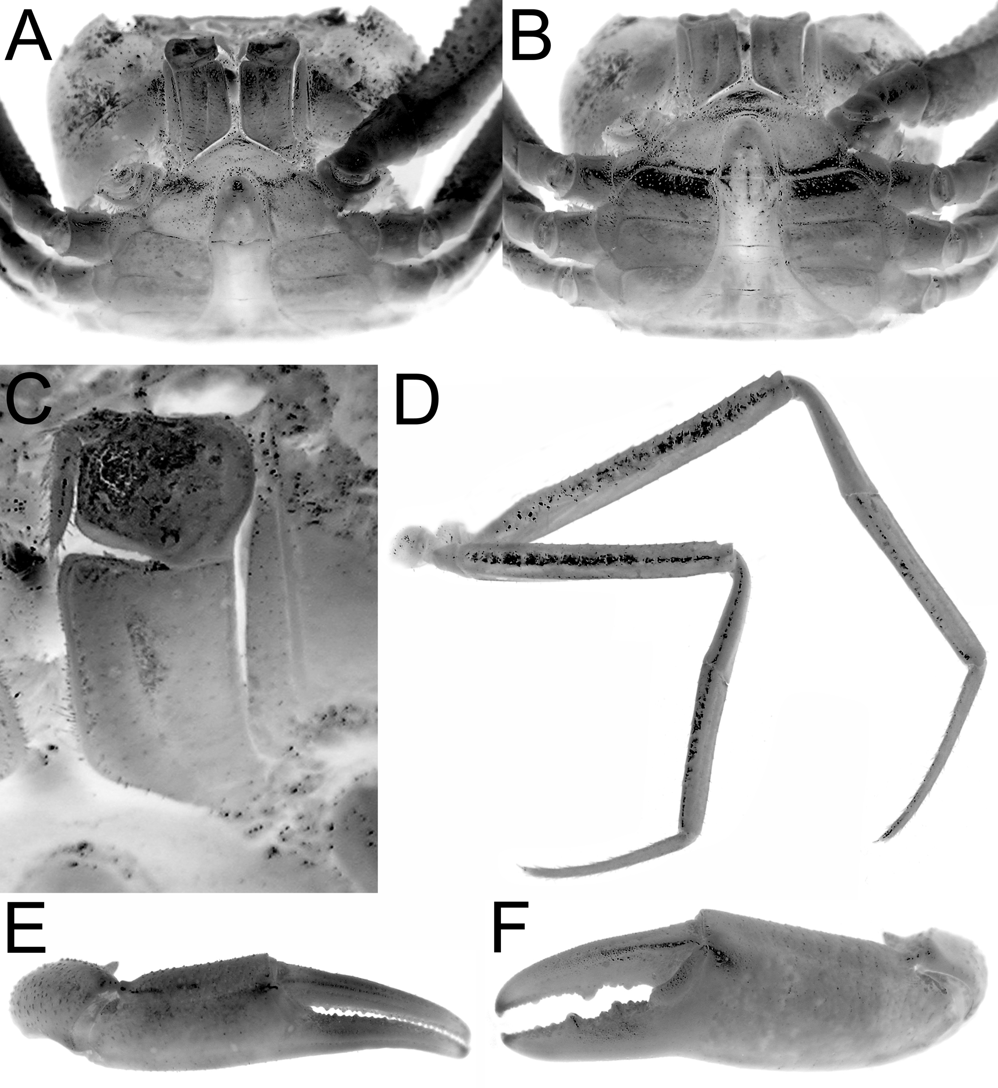
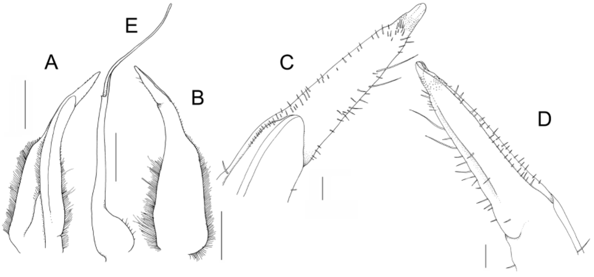
Diagnosis. Carapace subovate, distinctly broader than long; antero- and posterolateral regions covered with strong oblique striae, granules ( Fig. 1 View FIGURE 1 A, B). Branchial regions not prominently inflated when viewed frontally ( Fig. 1 View FIGURE 1 C); frontal margin conspicuously broad, almost straight in dorsal view ( Fig. 1 View FIGURE 1 C); anterolateral margin strongly convex; epibranchial tooth small but distinct; external orbital angle broadly triangular, outer margin ca. 3 times length of inner margin, epigastric cristae distinct but not sharp; postorbital cristae low, not clearly demarcated ( Fig. 1 View FIGURE 1 A, B). Eye reduced, filling only about half of orbit; distal part of ocular peduncle tapering; cornea rounded, relatively small, pigmented ( Fig. 1 View FIGURE 1 C); chelipeds distinctly elongated ( Fig. 1 View FIGURE 1 A); ambulatory legs very long, distal edge of merus of fourth leg extends beyond frontal margin when folded against carapace ( Figs. 1 View FIGURE 1 A, 2D); male abdomen distinctly T-shaped; G1 subterminal segment with concave inner margin, outer margin with distinct hump on distal third; terminal segment conical, tapering towards rounded tip, ca. 0.3 times subterminal segment, distal part with numerous minute scale-like spinules ( Fig. 3 View FIGURE 3 A-D); G2 ca. 1.6 times length of G1, distal segment slightly shorter than basal segment ( Fig. 3 View FIGURE 3 E).
Description of holotype male. Carapace subovate, distinctly broader than long; regions distinct; dorsal surfaces gently convex, rugose to smooth; antero-, posterolateral regions covered with strong oblique striae, granules; cervical grooves broad, shallow ( Fig. 1 View FIGURE 1 A, B). Pterygostomial, suborbital, subhepatic, sub-branchial regions distinctly rugose to granulose ( Fig. 1 View FIGURE 1 C). Branchial regions gently inflated dorsally, laterally, not prominently inflated when viewed frontally ( Fig. 1 View FIGURE 1 C). Frontal margin very broad, distinct, almost straight in dorsal view, below level of tip of external orbital tooth, gently deflexed downwards, margin forming very narrow, poorly defined median pseudo-frontal triangle ( Fig. 1 View FIGURE 1 C). Anterolateral margin strongly convex, clearly demarcated from posterolateral margin; epibranchial tooth small but well demarcated, separated from external orbital angle by small but distinct cleft; external orbital angle broadly triangular, outer margin ca. 3 times length of inner margin, lined with small granules; striae on anterolateral regions strong; epigastric cristae distinct, raised, rugose but not sharp, separated by deep median Y-shaped groove, just anterior of postorbital cristae; postorbital cristae low, not well demarcated, separated from epigastric cristae by end of cervical groove ( Fig. 1 View FIGURE 1 A, B). Posterior margin of carapace gently convex ( Fig. 1 View FIGURE 1 A, B). Epistome wide; posterior margin with distinct median triangular lobe with rounded tip, lateral margins gently sinuous ( Fig. 1 View FIGURE 1 C). Eye distinct but reduced, filling only about half of orbit; distal part of ocular peduncle tapering; cornea rounded but relatively small, pigmented ( Fig. 1 View FIGURE 1 C). Antennular fossa transversely narrow, rectangular in shape; flagellum folding transversely ( Fig. 1 View FIGURE 1 C). Third maxilliped quadrate; ischium quadrate, with shallow oblique median sulcus; merus quadrate, length and width subequal, with slightly auriculiform anteroexternal margin; exopod reaching beyond distal edge of ischium to submedian part of merus; flagellum distinct, longer than width of merus ( Fig. 2 View FIGURE 2 C).
Thoracic sternum relatively smooth, relatively narrow transversely, sternites 1–4 completely fused, without distinct median sutures visible; sternoabdominal cavity reaching to the level of the junction between sternites 2, 3, on imaginary line connecting anterior margins of coxae of the chelipeds ( Fig. 2 View FIGURE 2 A, B).
Chelipeds distinctly elongated; left larger; outer surfaces, margins gently rugose, without sharp spines or spinules; dorsal margin of long, slender merus with low, uneven granules; carpus with low inner distal spine, posterior margins lined with small granules; outer surface of palm gently rugose ( Fig. 1 View FIGURE 1 A). Chelae unequal; minor chela with fingers subequal to palm, cutting edges of fingers with numerous denticles ( Fig. 2 View FIGURE 2 E); major chela with fingers shorter than palm, cutting edge of pollex with 2 large teeth, numerous denticles, cutting edge of dactylus with submedian tooth, denticles ( Fig. 2 View FIGURE 2 F).
Ambulatory distinctly long, slender; second pair longest; distal edge of merus of fourth leg extends beyond frontal margin when folded against carapace; merus unarmed, dorsal margin gently serrated but not spiniform, with low subdistal angle; outer surfaces in first to third pairs rugose; carpus elongated with subdorsal groove on outer surface; propodus conspicuously long, with row of short ventral spines; dactylus elongated, gently curved on distal half, tip corneous, with 2 rows of spines on dorsal, ventral margins ( Figs. 1 View FIGURE 1 A, 2D).
Male abdomen distinctly T-shaped; telson elongated, longer than broad, lateral margins gently concave, tip rounded, as long as somite 6 ( Fig. 2 View FIGURE 2 A, B); somites 3–6 progressively narrower, trapezoidal ( Fig. 2 View FIGURE 2 B); somite 3 widest, covering most of thoracic sternite 8 ( Fig. 2 View FIGURE 2 B); somites 1, 2 longitudinally narrow, reaching coxae of last pair of ambulatory legs.
G1 curved; subterminal segment with concave inner margin, outer margin with distinct hump on distal third; terminal segment conical, tapering towards rounded tip, ca. 0.3 times subterminal segment, minute, scale-like spinules ( Fig. 3 View FIGURE 3 A–D). G2 ca. 1.6 times length of G1; basal segment long, distal segment flagelliform, slightly shorter than basal segment ( Fig. 3 View FIGURE 3 E).
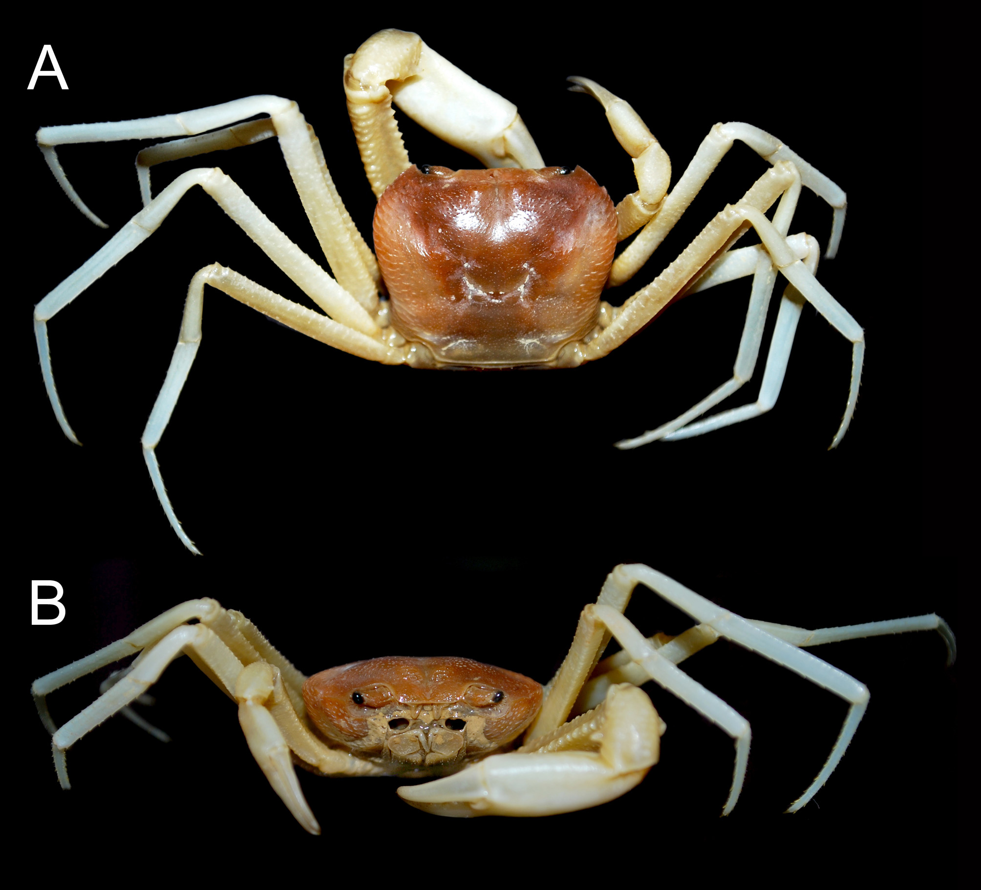
Life colour. The carapace is pale to yellow-orange, with the chelipeds and ambulatory legs white ( Fig. 4 View FIGURE 4 ).
Variation. The other male specimen agrees very well with the holotype male in all respects except for its smaller size.
Habitat. The type habitat, Rembus Cave, is a limestone cave with a variety of different passages. The cave entrance is about 15 m above the ground with a narrow and high passage with water flowing throughout the year. Aquatic organisms present near the entrance of the cave include fishes like Rasbora spp. ( Cyprinidae ) and Silurichthys sp. ( Siluridae ) as well as shrimps like Atyopsis moluccensis (Atyidae) and Macrobrachium sp. ( Palaemonidae ). Stygothelphusa antu new species was found deeper inside the cave where it was entirely dark and humid, with the substrate moist and/or muddy.
Etymology. The name is derived from the Iban word antu for “ghost” alluding to the pale coloration and habitat of the species. The name is used as a noun in apposition.
Remarks. Stygothelphusa antu new species is interesting as it is the most highly cave-adapted of the four species now known from the genus. Compared to the other three species, the eyes of S. antu new species are clearly reduced in size; with the distal part of the ocular peduncle relatively more slender, the cornea relatively smaller, and the eye fills only about half of the orbit ( Fig. 1 View FIGURE 1 B, C). In the other three species, the eyes are normal, with a stout ocular peduncle, large cornea and the eye fills most of the orbit (cf. Ng 2013). As has been discussed at length by Ng (1989, 2013), the normal condition of the eye argues against S. bidiensis , S. nobilii and S. cranbrooki being obligate troglobites. Stygothelphusa antu new species is a true troglobite as its eyes have started to degenerate.
Other than the condition of the eyes, S. antu new species is also very different from its congeners in several other key characters. Its carapace is proportionately the broadest, with the anterolateral margins prominently convex ( Fig. 1 View FIGURE 1 A, B) (versus margin gently convex to almost straight), the frontal margin is relatively the widest ( Fig. 1 View FIGURE 1 A, B), the external orbital tooth is broadly triangular ( Fig. 1 View FIGURE 1 A, B) (versus acutely triangular), the branchial regions are relatively the lowest, appearing almost flat from frontal view ( Fig. 1 View FIGURE 1 C), the ambulatory legs are proportionately the longest ( Figs. 1 View FIGURE 1 A, 2D), the outer margin of the G1 subterminal segment is humped ( Fig. 3 View FIGURE 3 A, B), the G1 terminal segment is more slender and evenly conical in shape ( Fig. 3 View FIGURE 3 A–D); and the distal part of the G1 terminal segment has relatively fewer and smaller scale-like spinules ( Fig. 3 View FIGURE 3 C, D) (cf. Ng 2013 for figures of the three congeners).

| ZRC |
Zoological Reference Collection, National University of Singapore |
No known copyright restrictions apply. See Agosti, D., Egloff, W., 2009. Taxonomic information exchange and copyright: the Plazi approach. BMC Research Notes 2009, 2:53 for further explanation.