
Sycon bellum, Chagas & Cavalcanti, 2017
|
publication ID |
https://doi.org/ 10.11646/zootaxa.4363.2.2 |
|
publication LSID |
lsid:zoobank.org:pub:7B5AC657-18B1-4A90-9268-FFAD49E1B9D0 |
|
DOI |
https://doi.org/10.5281/zenodo.6040382 |
|
persistent identifier |
https://treatment.plazi.org/id/5A058780-FFDF-A35D-13F7-70BDF0B9FF01 |
|
treatment provided by |
Plazi |
|
scientific name |
Sycon bellum |
| status |
sp. nov. |
Sycon bellum sp. nov.
Etymology. From Latin bellum = beautiful. The name is related to the charismatic morphology of the specimens.
Diagnosis. Sycon without stalk and fringe of trichoxeas but with osculum surrounded by membrane. The surface is hispid, with small tufts of diactines. Distal region of the cones with diactines and triactines. Both tubar and subatrial regions formed by triactines. The atrial region has several triactines and few tetractines.
Type material. UFBA 4527- POR [Holotype. Marina of the Nautical Tourist Terminal of Bahia (1258’20.8’’S, 3830’54.6’’W), Salvador, Bahia State, Brazil; collected by C. Chagas; 20/II/2016; 1m depth] and UFBA 4474 -POR (Paratype. Same locality; collected by F. Cavalcanti & E. Lanna; 06/VIII/2014; 1 m depth).
Type locality. Salvador, Bahia State , Brazil.
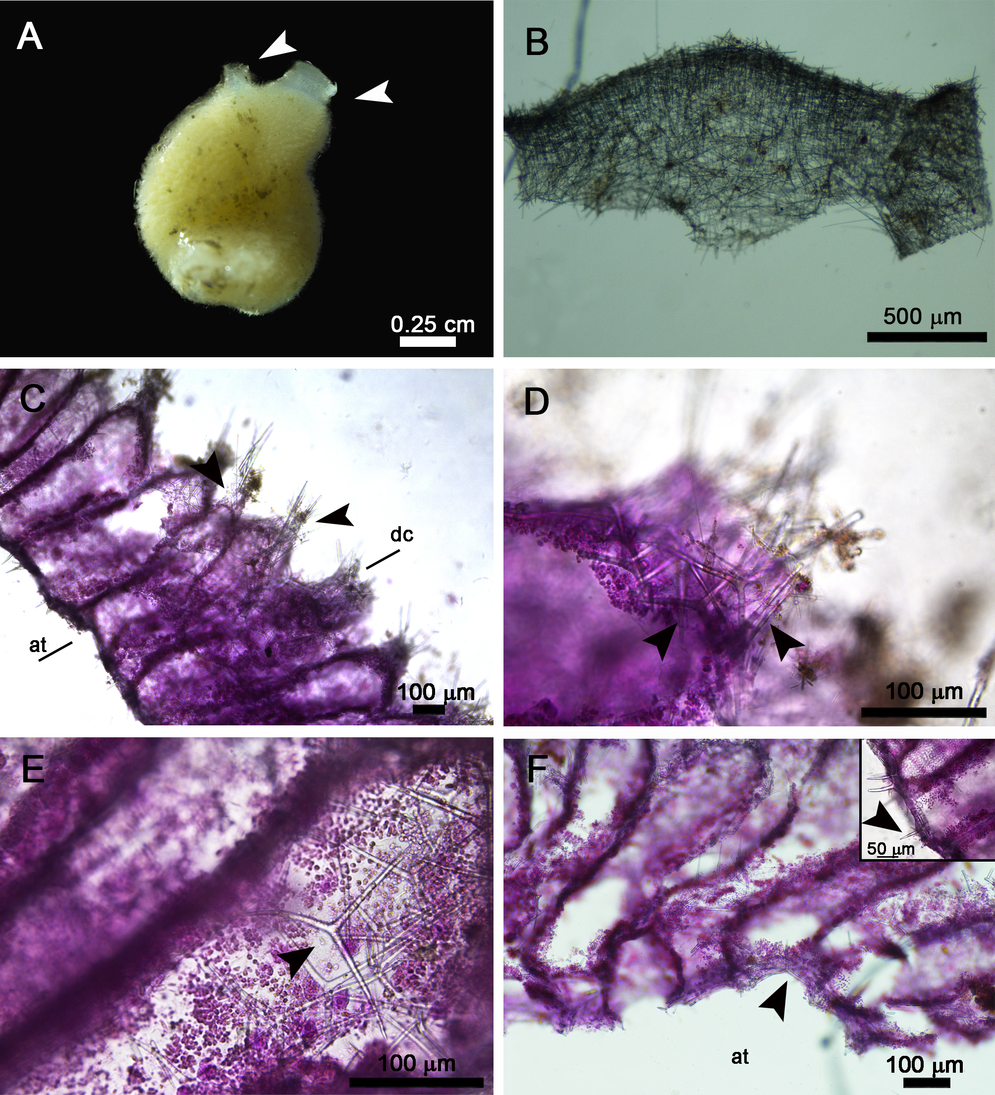
Description.: Colour is beige alive and after fixation ( Figure 6A View FIGURE 6 ). The holotype measures 0.9 x 0.5 cm (height x width). It is tubular and has two apical oscula surrounded by membranes ( Figure 6A View FIGURE 6 ). These oscular membranes have tetractines and numerous triactines, the latter with the unpaired actine larger than the paired ones and projected to the base of the sponge ( Figure 6B View FIGURE 6 ). Hispid surface, but with small tufts of diactines ( Figures 6A, C View FIGURE 6 ). The body wall is 0.15 cm thick. The aquiferous system is syconoid. The atrial cavity is wide but does not fill the whole specimen.
The distal portion of the cones is composed of triactines and is commonly ornamented with tufts of diactines and trichoxeas ( Figures 6C, D View FIGURE 6 ). In the tubar skeleton, there are triactines with the unpaired actine towards the distal region ( Figure 6E View FIGURE 6 ). These spicules form an articulated skeleton, although rare scattered spicules are observed. The subatrial region is formed by few triactines with the unpaired actine choanosome-oriented. The atrial skeleton is formed mainly by triactines and few tetractines, so its surface is slightly hispid ( Figure 6F View FIGURE 6 ).
Spicules ( Table 3; measurements obtained from two specimens):
Trichoxeas: Present only in the distal region of the cones. Thin and variable in size.
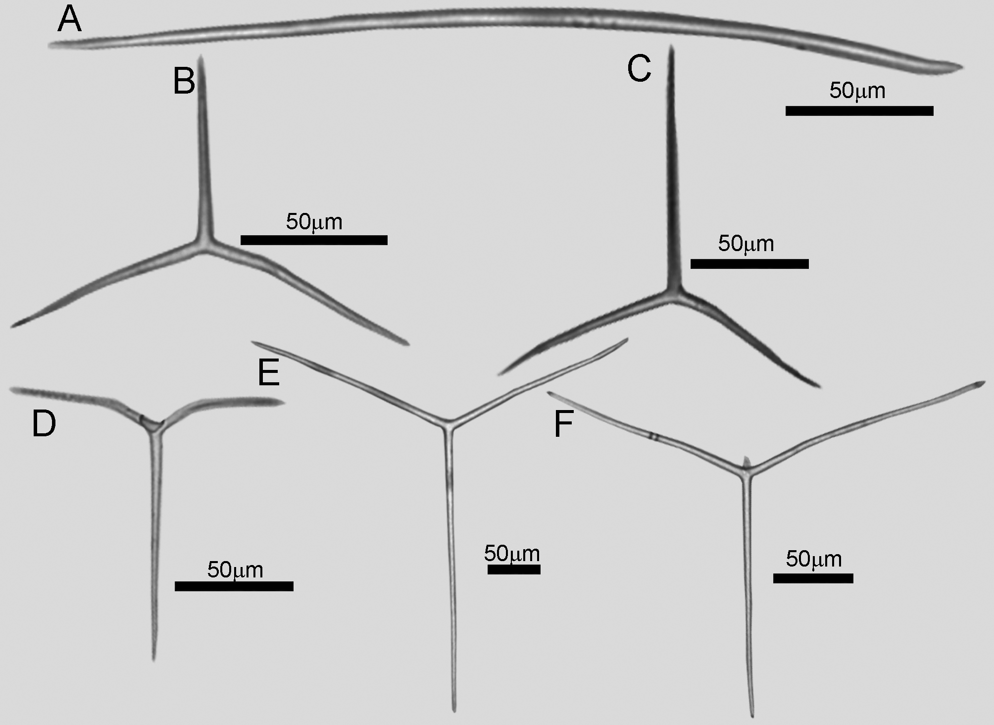
Diactines: Smooth and slightly curved, with blunt tips [118.1– 248.5 (60.9)–373.4/1.9– 5.6 (1.9)–11.4 µm; Figure 7A View FIGURE 7 ].
Triactines of the distal cones: Slightly conical, with blunt tips. Paired actines are curved down and can be slightly wavy. The unpaired actine is straight and short or is the same size as the paired ones [paired: 32.9– 67.6 (12.3)–91.5/2.5– 4.8 (1.0)–7.2 µm; unpaired: 30.4– 64.0 (17.9)–113.5/2.8– 5.0 (1.3)–9.3 µm; Figure 7B View FIGURE 7 ].
Tubar triactines: Slightly conical with blunt tips. Paired actines commonly forming an angle close to 180°. In many cases, one of the paired actines has a curvature while the other is straight and short. The unpaired actine is larger or the same size as the paired actines [paired: 51.5– 99.0 (30.2)–200.1/3.1– 5.5 (1.5)–13.0 µm; unpaired: 50.7– 105.0 (33.2)–222.7/3.3– 6.0 (1.6)–13.3 µm; Figure 7C View FIGURE 7 ].
Subatrial triactines: Rare, sagittal, cylindrical and with blunt tips. Paired actines have different sizes and are slightly curved at the base. The unpaired actine is larger than the paired ones [paired: 38.5– 61.3 (15.3)–101.7/2.9– 4.8 (1.0)–7.0 µm; unpaired: 52.5– 106.5 (23.5)–149.3/2.3– 5.8 (1.6)–8.9 µm; Figure 7D View FIGURE 7 ].
Atrial triactines: Cylindrical and sharply pointed. The unpaired actine is sometimes slightly smaller. The atrial triactines are larger than the other categories of triactines [paired: 87.7– 134.4 (22.1)–184.9/3.3– 6.1 (1.9)–10.4 µm; unpaired: 79.5– 142.7 (27.4)–198.4/3.8– 6.2 (1.8)–12.6 µm; Figure 7E View FIGURE 7 ].
Atrial tetractines: Less abundant than the atrial triactines. Cylindrical with sharp tips. Paired actines are slightly curved and the unpaired is straight. All basal actines are the same size. Apical actine is curved [paired: 88.9– 143.8 (25.1)–191.9/4.5– 6.9 (1.6)–10.0 µm; unpaired: 84.6– 137.9 (28.6)–193.4/5.4– 8.2 (1.5)–11.5 µm; apical: 8.0– 19.7 (8.3)–42.9/3.5– 6.0 (2.0)–11.9 µm; Figure 7F View FIGURE 7 ].
Ecology. The holotype was found on recruitment plates prepared with fragments of nautical cables. As they remained submerged for 2 months (at 1 meter depth), this is the maximum age of the individual. Other organisms, such as tunicates, bryozoans and macroalgae, also colonised the plates. The paratype was found on nautical cables used to dock the boats, and it is not possible to estimate its age.
Remarks. Sycon is one of the richest genera within the class Calcarea. We compared our specimens with the 89 species known to Sycon and concluded that their skeletal composition, with many triactines and few tetractines that are exclusive of the atrial region, in addition to differences in the sizes of spicules, makes S. bellum sp. nov. a new species to science. In a straight comparison with species from the Atlantic Ocean ( Table 4), the most similar species considering the skeletal composition are S. ampulla ( Haeckel, 1870) , S. brasiliense Borojevic, 1971 , S. barbadense ( Schuffner, 1877) , S. elegans ( Bowerbank, 1845) , S. protectum Lambe, 1896 , and S. raphanus Schmidt, 1862 . Their type localities are Southern Caribbean, Brazil, Barbados, South Africa, Vancouver Islands ( Canada), and the Adriatic Sea, respectively. Considering their distributions, S. elegans and S. raphanus had never been recorded along the Western Atlantic Ocean.
Species Distal cones Tubar skeleton Subatrial skeleton Atrial skeleton (-) Not mentioned by the original description.
The most obvious difference between S. ampulla and the new species described here is the external morphology. The former is tubular, composed of several tubes united by stalks of diactines, each tube with an apical osculum ornamented by a well-developed fringe of trichoxeas ( Haeckel 1870; Burton 1963). In contrast, S. bellum sp. nov. is formed by a sole tube with two apical oscula ornamented by membranes, without a fringe of trichoxeas or stalk.
An important difference between Sycon bellum sp. nov. and S. brasiliense is that in the latter species diactines are rare and do not form tufts in the distal cones (the sponge surface is smooth). Although not evident to the naked eye, in the new species described here, these tufts of diactines are present in most of the distal cones, as observed in Figure 6C View FIGURE 6 . In relation to Sycon barbadense , S. elegans and S. protectum , the size of the apical actines of their atrial tetractines is larger than that of our new species ( S. barbadense : 80/13 µm; S. elegans : 120–160/12–16 µm; S. protectum : 85/6µm; S. bellum sp. nov.: 8.0– 19.7 (8.3)–42.9/3.5– 6.0 (2.0)–11.9 µm). Additionally, S. elegans has triactines in the distal cones that are considerably thicker (paired: 50–90/25–35 µm, unpaired: 200–400/25–35 µm; S. bellum sp. nov.: paired: 32.9– 67.6 (12.3)–91.5/2.5– 4.8 (1.0)–7.2 µm; unpaired: 30.4– 64.0 (17.9)–113.5/2.8– 5.0 (1.3)–9.3 µm). Finally, S. protectum and S. raphanus have diactines that are up to 12 times larger than the mean value observed in S. bellum sp. nov. ( S. protectum : 1000/19 µm; S. raphanus : 1000–3000/20–24 µm; S. bellum sp. nov.: 118.1– 248.5 (60.9)–373.4/ 1.9– 5.6 (1.9)–11.4 µm).
Sycon avus sp. nov.
Etymology. From Latin avus = grandfather. For Manoel Pedro das Chagas, the paternal grandfather of the first author of this work, who in life worked close to the studied area.
Diagnosis. Sycon with hispid surface. Distal cones with triactines and tufts of smooth diactines. Tubar skeleton formed by triactines in which, commonly, one of the paired actines is short and curved while the other paired actine is straight. Subatrial skeleton formed by triactines and few tetractines. Atrial region formed by few triactines and several tetractines. A long projection of the body can be present below the fringe of trichoxeas, similar to a “long neck.
Type material. UFBA 4526- POR [Holotype. Marina of the Nautical Tourist Terminal of Bahia (1258’20.8’’S, 3830’54.6’’W), Salvador, Bahia State , Brazil; collected by C. Chagas; 20/II/2016; 1 m depth] and UFBA 4656 - POR (Paratype. Same locality and sampling data).
Type locality. Salvador, Bahia State , Brazil.
Description. Colour is beige alive and after fixation. The holotype measures 0.7 x 0.5 cm (height x width) and is formed by a single tube, with apical osculum ornamented by a fringe of trichoxeas ( Figure 8A View FIGURE 8 ). The base of this fringe has a ring of sagittal spicules, mostly tetractines, but triactines are also present ( Figure 8B View FIGURE 8 ). Such spicules are positioned with the unpaired actine pointing to the base of the sponge body. It is worth mentioning that in some of the analysed specimens, two fringes of trichoxeas were observed: one of which was more closed (surrounding the osculum) and the other more open (external to the internal fringe). In addition, some specimens have a long oscular membrane similar to a “long neck”. In the holotype (UFBA 4526-POR), this membrane is short and contains sagittal spicules, mainly tetractines with the apical actines towards the osculum. The surface of the body is hispid with tufts of diactines. The body wall is 0.1 cm thick. The atrial cavity is large, and the aquiferous system is syconoid.
The distal skeleton is composed of triactines and tufts of diactines piercing the cones ( Figures 8C, D View FIGURE 8 ). In the tubar skeleton, there are triactines with the unpaired actines towards the distal region and forming an articulated skeleton ( Figures 8D, E View FIGURE 8 ). There is also a monolayer of triactines outlining the choanocyte chambers. The subatrial region is mostly formed by triactines, with the unpaired actine oriented to the choanosome. Tetractines were observed in this region, but they are not abundant. The atrial skeleton is formed mainly by tetractines and by few triactines ( Figure 8F View FIGURE 8 ).
Spicules ( Table 5; measurements obtained from two specimens):
Trichoxeas: Present in the distal cones and at the oscular fringe. Thin and variable in size.
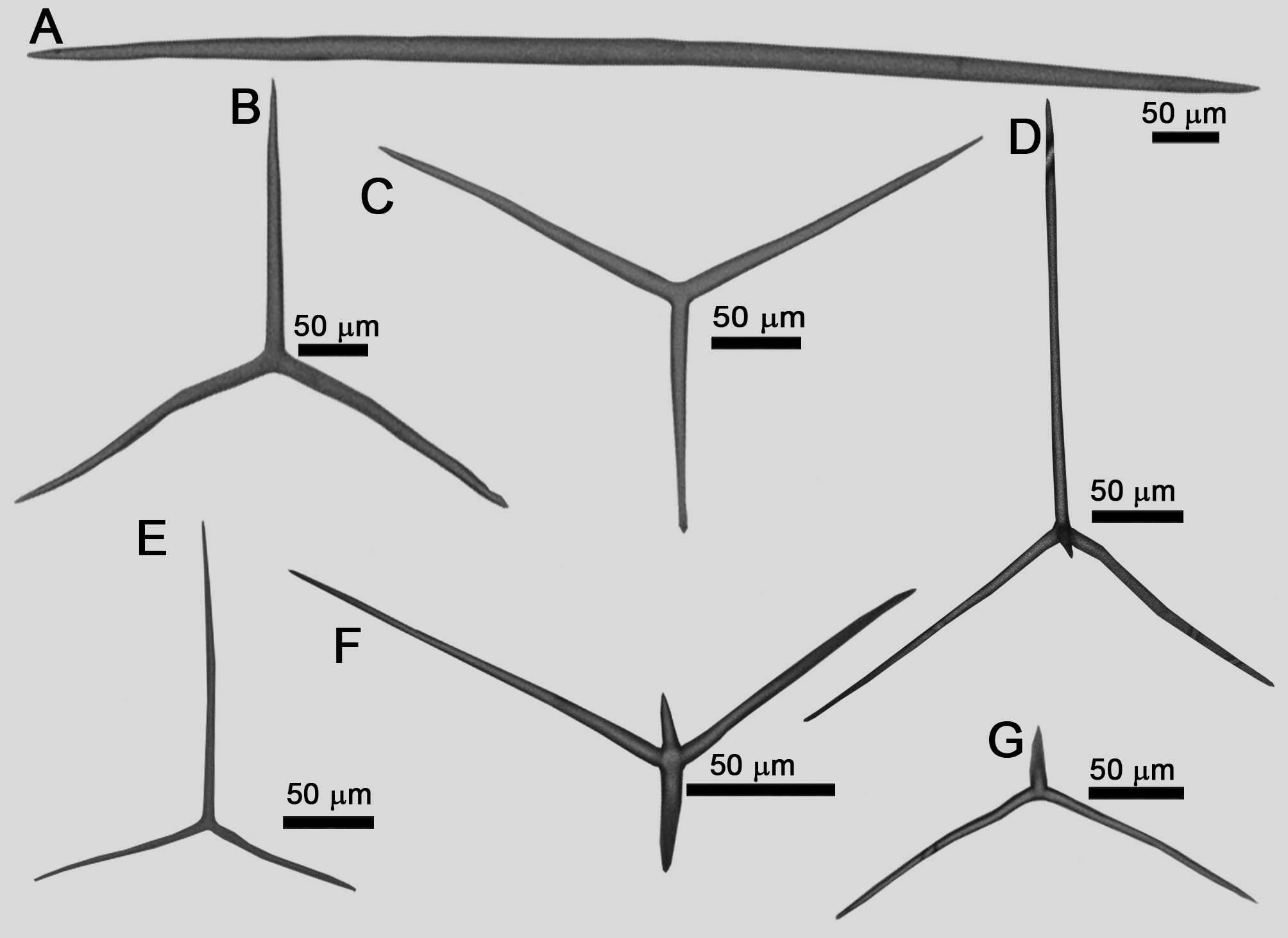
Diactines: Fusiform, sharply pointed with smooth surface. Several diactines are broken, possibly due to their large size [321.1– 807.9 (260.4)–1413.0/8.7– 12.6 (2.7)–20.0 µm; Figure 9A View FIGURE9 ].
Triactines of the distal cones: Slightly conical or cylindrical and with sharp ends ( Figure 9B View FIGURE9 ). The paired actines are curved. The unpaired actine is straight and is the same size or is slightly smaller than the paired ones [paired: 61.0– 117.5 (30.3)–194.4/3.3– 7.4 (1.8)–12.0 µm; unpaired: 58.1– 94.4 (25.6)–156.5/3.3– 7.6 (2.0)–17.7 µm; Figure 9B View FIGURE9 ].
Tubar triactines: Cylindrical with sharp tips. The unpaired actine is long, and the unpaired angle is around 160°. Commonly, one of the paired actines is short and curved while the other paired actine is straight [paired: 53.3– 117.8 (21.1)–160.5/4.2– 7.5 (1.3)–10.9 µm; unpaired 65.4– 117.2 (24.0)–176.1/4.4– 7.3 (1.8)–11.8 µm; Figure 9C View FIGURE9 ]. The spicules that form the monolayer on the walls of the chambers are similar to these triactines.
Subatrial tetractines: Similar to the subatrial triactines but with a short apical actine. They are rare [paired: 103.1– 141.3 (30.6)–191.7/3.0– 4.8 (1.1)–6.3 µm; unpaired: 180.2– 202.1 (18.8)–234.8/3.1– 5.0 (1.1)–7.0 µm; 10.2– 12.5 (1.4)–14.5/2.0– 3.7 (0.9)–5.0 µm; Figure 9D View FIGURE9 ].
Subatrial triactines: Cylindrical with sharp tips. The unpaired actine is longer than the paired ones [paired: 54.2– 94.5 (20.6)–140.1/2.3– 6.5 (2.1)–12.6 µm; unpaired: 101.7– 163.1 (26.5)–222.4/3.8– 6.7 (1.7)–10.9 µm; Figure 9E View FIGURE9 ].
Atrial tetractines: Cylindrical and with sharp tips. The apical actine is slightly curved and shorter than the basal actines [paired: 56.5– 107.9 (31.3)–176.9/3.0– 5.9 (1.7)–8.7 µm; unpaired: 57.1– 152.3 (41.3)–223.1/5.7– 7.8 (1.5)– 10.8 µm; apical: 39.7– 50.6 (4.9)–59.3/4.4– 6.4 (1.1)–8.5 µm; Figure 9F View FIGURE9 ].
Atrial triactines: Less abundant than the atrial tetractines. Cylindrical, with sharp tips and with actines thinner than those of the tubar triactines. Paired actines are slightly curved. The unpaired actine is smaller or is the same size as the paired ones [paired: 32.3– 70.5 (18.0)–92.6/2.4– 5.1 (1.6)–8.8 µm; unpaired: 53.1– 134.0 (38.6)–186.4/ 3.1– 4.6 (1.3)–8.7 µm; Figure 9G View FIGURE9 ].
Ecology. All specimens were found on the nautical cables colonised by tunicates, demosponges, bryozoans and macroalgae, at 1 meter depth. Although they have about two months of life (the period of immersion of the cables), the holotype, sampled at February 2016, has reproductive elements ( Figures 8C–E View FIGURE 8 ).
Remarks. The skeletal composition of Sycon avus sp. nov. is also observed in two other species of Sycon found in the Atlantic Ocean: S. frustulosum Borojevic & Peixinho, 1976 and S. natalense Borojevic, 1967 ( Table 4), from the coasts of Brazil and South Africa, respectively.
Sycon frustulosum is the most similar to the new species described here, but shows important differences in the shape of the diactines (which are jagged, while in S. avus sp. nov. they are smooth) and in the size of this spicule category and of the tubar triactines ( S. frustulosum : diactines: 148–480 µm, tubar triactines: paired actines 30–60 µm, unpaired 40–100 µm; Sycon avus sp. nov.: diactines: 413–1383 µm, tubar triactines: paired actines 67–161 µm, unpaired 66–176 µm). Sycon natalense can also be easily differentiated from S. avus sp. nov. In contrast to the new species, it has a discreet hispidation that is invisible to the naked eye, an osculum without a fringe of trichoxeas and abundant subatrial tetractines. The size of their diactines is variable. The longest diactines extend along the wall of the tubes, while only the smallest ones are limited to the distal cones. This arrangement is very different from that observed in S. avus sp. nov. and in most of the species that comprise Sycon , in which diactines form tufts restricted to the distal cones. The shape and size of those diactines are not similar between S. natalense and S. avus sp. nov. and make recognition of both species easy (in the former: spear-shaped, 100–300/10 µm; in the latter: smooth, 321.1– 807.9 (260.4)–1413.0/8.7– 12.6 (2.7)–20.0 µm).
Finally, the main differences between our two new species of Sycon are the presence of a fringe of trichoxeas in Sycon avus sp. nov., the size of the diactines of the distal cones ( Sycon bellum sp. nov: 138–366 µm; Sycon avus sp. nov.: 413–1383 µm), the presence of tetractines in the subatrial skeleton of Sycon avus sp. nov., and the composition the atrial skeleton (with predominant triactines in Sycon bellum sp. nov.). Despite the clear differences between these new species, and between them and the Sycon species already known, it became apparent that the genus needed a taxonomic revision, because many of the descriptions are too outdated and lack data currently considered as important for the taxonomy of Sycon .

| POR |
Universit� degli Studi di Napoli Federico II |
No known copyright restrictions apply. See Agosti, D., Egloff, W., 2009. Taxonomic information exchange and copyright: the Plazi approach. BMC Research Notes 2009, 2:53 for further explanation.
|
Kingdom |
|
|
Phylum |
|
|
Class |
|
|
Order |
|
|
Family |
|
|
Genus |