
Terebellides parapari, Lavesque & Hutchings & Daffe & Nygren & Londoño-Mesa, 2019
|
publication ID |
https://doi.org/ 10.11646/zootaxa.4664.2.1 |
|
publication LSID |
lsid:zoobank.org:pub:6F0BFDDC-99CA-4CED-9F56-B6DA226CD42D |
|
persistent identifier |
https://treatment.plazi.org/id/728A8EE4-F8FC-4723-861F-C76FC8610072 |
|
taxon LSID |
lsid:zoobank.org:act:728A8EE4-F8FC-4723-861F-C76FC8610072 |
|
treatment provided by |
Plazi |
|
scientific name |
Terebellides parapari |
| status |
sp. nov. |
Terebellides parapari View in CoL n. sp.
Figures 15–16 View FIGURE 15 View FIGURE 16 , Table 2 View TABLE 2
Type material: Holotype: MNHN-IA-TYPE 1884, complete specimen, gravid, Northeast Atlantic Ocean , Bay of Biscay, P40, 43°33’26’’N, 1°41’37’’W, 112 m depth, May 2018 GoogleMaps ; Paratypes: MNHN-IA-TYPE 1885, one complete specimen, Northeast Atlantic Ocean , Bay of Biscay, P37, 43°33’31”N, 1°43’49”W, 129 m depth, May 2018 GoogleMaps ; MNHN- IA-TYPE 1886, one complete specimen, Northeast Atlantic Ocean , Bay of Biscay, P31, 43°38’40”N, 1°35’05”W, 86 m depth, May 2018 GoogleMaps ; MNHN-IA-TYPE 1887, one complete specimen, Northeast Atlantic Ocean , Bay of Biscay, P40, 43°33’26’’N, 1°41’37’’W, 112 m depth, May 2018 GoogleMaps ; MNHN-IA-TYPE 1888, two complete specimen, Northeast Atlantic Ocean , Bay of Biscay, P40, 43°33’26’’N, 1°41’37’’W, 112 m depth, May 2018 GoogleMaps , mounted for SEM; AM W.51403, 4 complete specimens (2 of them gravid), Northeast Atlantic Ocean , Bay of Biscay, P35, 43°36’35”N, 1°40’35”W, 120 m depth, May 2018 GoogleMaps ; AM W.51404, 3 complete specimens (2 of them gravid), Northeast Atlantic Ocean , Bay of Biscay, P36, 43°35’0”N, 1°42’02”W, 125 m depth, May 2018 GoogleMaps .
Description. Small species, holotype 13.4 mm long (6.2–14.1 mm) and 0.9 mm (0.6–0.9 mm). Body tapering posteriorly with segments becoming increasingly shorter and more compact towards pygidium.
Prostomium compact; eyespots absent; upper lip with single anteriorly elongated projecting lobe surrounding mouth with many buccal tentacles ( Figs 15C View FIGURE 15 & 16A View FIGURE 16 ). Buccal tentacles of two types, uniformly cylindrical and with expanded tips, spatulate ( Fig. 16B View FIGURE 16 ). Lower lip forming an expanded structure below upper lip ( Fig. 15C View FIGURE 15 ). SG 1 and 2 short, only visible ventrally; following segments with lobes as ventral collars ( Figs 15 View FIGURE 15 A–C & 16B). Lateral lappets on SG 3–7 ( TC 1–5), continuing ventrally in TC 1–5 ( Figs 15C View FIGURE 15 & 16B View FIGURE 16 ), largest on TC 1–3 and declining in size posteriorly. No conspicuous dorsal rounded projection on anterior chaetigers. Small round glandular region on TC 3, situated latero-dorsally ( Fig. 15C View FIGURE 15 ).
Branchiae arising as a single structure from TC 1, reaching TC 4 ( TC 5), consisting of a single elongate and annulated stalk placed mid-dorsally ( Figs 15 View FIGURE 15 A–C & 16A, C), two pairs of lobes, not fused, lower pair thinner ( Figs 15B View FIGURE 15 & 16A, C View FIGURE 16 ). Upper lobes with about 20 widely spaced lamellae ( Figs 15C View FIGURE 15 & 16A, C View FIGURE 16 ). Branchial lamellae, with mid-dorsal pointed tip ( Fig. 16 View FIGURE 16 C–D), with sparse tufts of cilia. Branchiae provided without any papillar projections pointing over margins of the branchial lamellae ( Fig. 16 View FIGURE 16 C–D). Distal region of upper lobes with small terminal pointed projections, lower lobes with short filaments ( Figs 15 View FIGURE 15 A–C & 16A–C). Anterior branchial projection (5 th lobe) absent ( Fig. 16A, C View FIGURE 16 ).
Eighteen pairs of thoracic notopodia ( SG 3–20). First notopodium on TC 1 well-developed, same size than subsequent notopodia; TC 1 and TC 2 situated more dorsally; notochaetae from TC 1 longer than notochaetae from TC 2, notochaetae from TC 2 longer than following ones ( Figs 15C View FIGURE 15 & 16B View FIGURE 16 ). All notochaetae simple capillaries, arranged in two rows. Neuropodia present as sessile pinnules from TC 6 ( SG 8) to pygidium; uncini arranged in single rows from TC 7. First thoracic neuropodium ( TC 6) provided with about three to four sharply bent acute tipped, geniculate chaetae.All subsequent thoracic neuropodia with about 10–12 uncini per torus arranged in one irregular row. Uncini as shafted denticulate hooks provided with long, thin and pointed main fang, straight terminally. Two to four teeth above the main fang, surmounted by a row of three to five short teeth and an upper crest of several minute denticles ( Fig. 16E View FIGURE 16 ). About 30 abdominal neuropodia as erect pinnules paddle-shaped with entire margin provided with about 15 uncini; uncini with three or four pointed teeth above main fang, surmounted by a row of three short pointed teeth and an upper crest of minute teeth ( Fig. 16F View FIGURE 16 ).
Nephridial papillae not seen. Pygidium blunt, slightly crenulated, as a funnel-like depression.
Methyl green staining pattern. First 4 TC stain solid; from TC 5 to about TC 11, stain stripped with space between stripes pale blue. Ventral part of TC 4 stain darker ( Fig. 15C View FIGURE 15 ). Presence of transverse diagonal white lines on lateral part of first three chaetigers ( Fig. 15C View FIGURE 15 ). Glandular region whitish.
Etymology. The species is named after Dr. Julio Parapar, for his many contributions to the taxonomy of the Terebellides genus, especially in European waters and for his friendship.
Habitat. Mud to sandy mud, around 120 m depth.
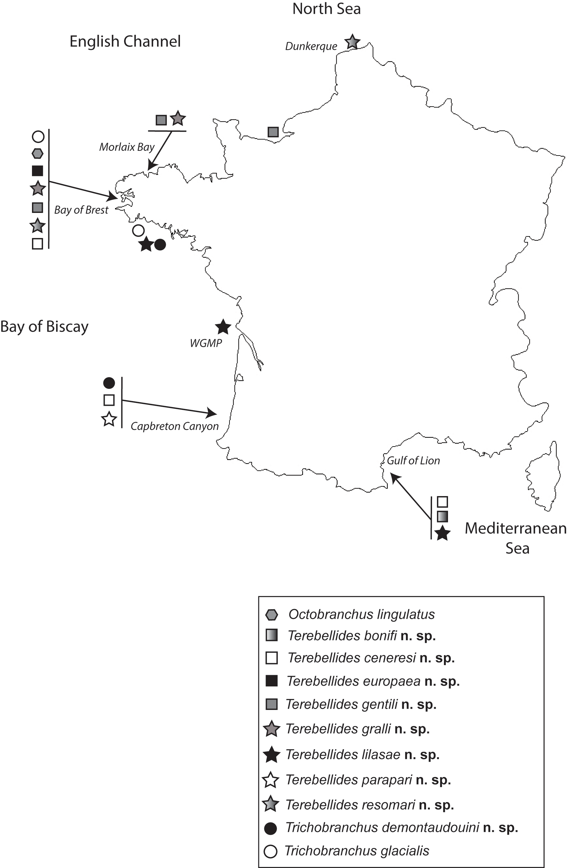
Type locality. Near Capbreton Canyon , Bay of Biscay, Northeast Atlantic Ocean, France .
Distribution. Only known from type locality ( Fig. 1 View FIGURE 1 ).
Remarks. Terebellides parapari n. sp. is similar to T. shetlandica in having a relatively small size ( T. shetlandica : 6–19 mm vs T. parapari n. sp.: 8–23 mm), lobes free from each other and filaments in each of lower lobes. Nevertheless, these filaments are much longer for T. shetlandica (about ½ of length of lower lobe for T. shetlandica , instead of less than ¼ of length for T. parapari n. sp.). These two species also differ by the presence of glandular region for T. parapari n. sp. (absent in T. shetlandica ), absence of dorsal papillae on each thoracic and abdominal chaetiger for T. parapari n. sp. (present in T. shetlandica ), by the size of notochaetae (longer on the first chaetiger for T. parapari n. sp., same length for T. shetlandica ), by the shape of branchial lamellae (rounded for T. shetlandica and pointed for T. parapari n. sp.) and absence of cilia on branchial lamellae for T. parapari n. sp. (instead of presence of rows and tufts of cilia in T. shetlandica ). Finally, these two species differ by the MG pattern (compact from CH 1–4 and striped from CH 5–11, with white latero-diagonal white lines on CH 1– CH 3 for T. parapari n. sp. vs compact from TC 1–6 and striped from CH 7–12 for T. shetlandica ) ( Table 2 View TABLE 2 ).


| AM |
Australian Museum |
| MG |
Museum of Zoology |
No known copyright restrictions apply. See Agosti, D., Egloff, W., 2009. Taxonomic information exchange and copyright: the Plazi approach. BMC Research Notes 2009, 2:53 for further explanation.
|
Kingdom |
|
|
Phylum |
|
|
Class |
|
|
Order |
|
|
Family |
|
|
Genus |