
Thamnocephalus (Thamnocephalus) chacosaltensis, Cohen, Rosa Graciela, 2016
|
publication ID |
https://doi.org/ 10.11646/zootaxa.4088.1.3 |
|
publication LSID |
lsid:zoobank.org:pub:D6438635-35F8-460A-878D-34E5AEE89446 |
|
DOI |
https://doi.org/10.5281/zenodo.5665728 |
|
persistent identifier |
https://treatment.plazi.org/id/924F1842-4E7A-FFE5-FF55-4A74FD01EFAA |
|
treatment provided by |
Donat |
|
scientific name |
Thamnocephalus (Thamnocephalus) chacosaltensis |
| status |
sp. nov. |
Thamnocephalus (Thamnocephalus) chacosaltensis View in CoL sp. nov.
( Figs. 3–4 View FIGURE 3 View FIGURE 4 )
Etymology. The specific epithet makes reference to the Argentinean region from where the specimens of the new species came.
Type locality. Temporary turbid pond in the Rivadavia Department, at the East of Salta province, Argentina, near the Teuquito River, 24º13`19.3’’S; 62º 52’14.3’’W.
Type material. Holotype. Male (FML–CRUST 01100a), coll. F. Cancino, L. Lobo, T. Van Dooren and M. Fourcade, I/10/2012.
Allotype. Female (ovigerous); (FML–CRUST 01100b), coll. F. Cancino, L. Lobo, T. Van Dooren and M. Fourcade, I/10/2012.
Paratypes. 2 males and 1 female (ovigerous); (FML–CRUST 01100c), coll. F. Cancino, L. Lobo, T. Van Dooren and M. Fourcade, I/10/2012.
Comparative material examined. Several specimens belonging to other species of Thamnocephalus were observed in order to establish comparisons. T. (T.) mexicanus was compared on the base of its descriptions in the literature. Thamnocephalus (T.) platyurus : 3 males and 3 females from Cochise County, Arizona, USA. Stock pond 3.3 miles south of Kansas settlement turnoff on the East side of Arizona highway 186. Belk Collection, DB 200. Date: IX/1/73. Col. and leg. D. Belk. Thamnocephalus (T.) venezuelensis : 4 males and 3 females. Aruba (South Caribbean Island). Gravel pit ponds south of Boton. K.A.L. Reading. D. C. Rogers Collection, DCR 319. Date: III/ 14/89. Leg. D. C. Rogers. Thamnocephalus (S.) salinarum : 3 males, 2 females. Argentina, Córdoba Province. A turbid saline temporary lagoon about 5 km2 and 30 cm deep, close to Las Toscas (30° 9’ S, 64° 56’ W), near Quilino, in Salinas Grandes. Date: XII/19/98. Col. and leg. S. Vernet.
Description. Male. Biometrics. Mean measurements of 3 specimens (holotype included). Total front to anus, lateral length: 31.7 mm; head + thorax lateral length: 20.1 mm. Pedunculate eye: 1.4 mm in diameter, 1.9 mm long. First antennae: 3.5 mm long. Basal segment of the second antennae, in lateral view: 2.1 mm maximum width, and 2.5 mm long (excluding the medial process) or 4.0 mm long (including the medial process). Distal segment of the second antenna, in lateral view: 5.7 mm long. First genital segment: lateral length (at the middle of the segment), 1.8 mm; dorsal width: 3.8 mm. Second genital segment: lateral length (at the middle of the segment): 1.5 mm; dorsal width: 3.6 mm. Total abdomen lateral length to the anus: 11.6 mm; lateral lengths of abdominal segments I– II–III–IV–V–VI–VII: 1.8 mm–1.6 mm–1.3 mm–1.3 mm–1.2 mm–2.1 mm–2.3 mm; dorsal width of abdominal segments I–II–III: 3.5 mm– 3.4 mm– 3.8 mm. Caudal fin length, in ventral view: 8.6 mm.
Head with a small neck organ. First antennae almost twice the length of pedunculate eyes (compound eye plus peduncle), ending in three sensory setae and several aesthetascs. Compound eyes almost rounded.
Second antennae typical of the genus ( Fig. 3 View FIGURE 3 A). The basal article is short and thick ( Figs. 3 View FIGURE 3 A, B, C). Distally, the basal article presents, in its lateral face, a short subapical conical spine (CS) which projects laterally, and in addition, an anterior–medial soft, laterally compressed process (SP) ( Figs. 3 View FIGURE 3 A, B, C). This process seems to arise from an incomplete hinge at almost half the length of the basal article ( Fig. 3 View FIGURE 3 A); it extends right along almost the basal quarter of the distal article, and culminates rounded at the tip. The process shows its cuticle partially ornamented with tiny buttons and presents a minute mound (M) protruding sub apically from the lateral–posterior edge ( Fig. 3 View FIGURE 3 C).
Measured in lateral view, the basal article is more than twice thicker than the basal portion of the distal article; the distal article is tusk–like, round in cross section, and it regularly curves posterior–medially tapering gradually towards the tip ( Fig. 3 View FIGURE 3 A). Both distal articles converge distally.
With a frontal appendage well developed ( Fig. 3 View FIGURE 3 D); its main trunk is long, more than twice the antenna 2 basal article length excluding the medial process. The main trunk splits in two cylindrical arms, left and right; in one male, main trunk was 5.6 mm length and 1.8 mm width, and each arm was approximately 1 mm width. Three branches, first, second and third branches ( Figs. 3 View FIGURE 3 D, E, F, G) arise from both arms. Main trunk, left and right arms, first and second branches and their sub–branches are transversely striated or annulated. First and second branches show several sub–branches which are, either straight or curved distally, and present a curved spine at the tip; besides, except for the proximal sub–branch of the first branch ( Figs. 3 View FIGURE 3 D, E) all the sub–branches and their ramifications are profusely spiny ( Table 1 View TABLE 1 ).
The first branch ( Figs. 3 View FIGURE 3 D, E: FB; Table 1 View TABLE 1 ) is composed by three sub–branches which are the largest and widest of the entire frontal appendage. The most proximal sub–branch is coiled, lacks spines all along, gradually thins in distal direction and shows the peculiarity of being flattened in the distal half; the distalmost flattened portion is folded several times over itself and is somewhat twisted; the sub–branch does not end acutely but blunt, and bears a small spine at the tip ( Figs. 3 View FIGURE 3 D, E); sometimes, the apical spine could be preceded by a few additional small spines. The middle sub–branch, rather flattened, is the longest of the three. Except for one male in which the more distal sub–branch bifurcates distally both, in the right and left arms, the sub-branch is unbranched.
The second branch ( Fig. 3 View FIGURE 3 D: SB; Table 1 View TABLE 1 ) has six to seven dorsal sub–branches ( Fig. 3 View FIGURE 3 D: dsb 1 to dsb 6–7) numbered from proximal to distal; the more distal sub–branch (dsb 6 or 7) is apical. All the dorsal sub–branches are ramified; the number of sub–branches and their ramifications differ slightly between right and left arms. In addition, one or two isolated, spiny and unbranched ventral sub–branches ( Fig. 3 View FIGURE 3 D: vsb 1–2) arise distally from the ventral edge of each second branch.
The third branch ( Figs. 3 View FIGURE 3 F, G: TB; Table 1 View TABLE 1 ) is a broad–based triangular, thin, much flattened medio–dorsal outgrowth. This outgrowth is folded once or several times over itself; when it is stretched, it can be seen that the margins are rather undulate and that the outgrowth tapers to the apex where there is a small spine.
Maxilla 1 presents 16–17 long setae plus a single short and slender spine on the ventral tip; basal width of spine is almost half the width at the base of the long setae. Each long seta shows a basal widest portion, distally armoured with 2 longitudinal rows of 3 to 7 knife–blades–shaped setulae each (increasing the number of setulae in each row from the ventral to the more dorsal setae), and a distal slender plumose portion; the distalmost setule of the basal portion, in the limit with the distal portion of each maxillary seta, is a complex hypertrophied setular structure ( Fig. 3 View FIGURE 3 H).
Maxilla 2 is a hairy rounded lobe, somewhat flattened antero–posteriorly, with two to three medial anterior directed plumose all around setae, and an additional seta attached to the medial posterior margin and projecting backwards.
Thoracopods constitute a homonymous series; they only differ in size, the middle pairs being the larger. All the lobes of thoracopods are widened, as usual in the genus. Exopodite, epipodite, praepipodite and endopodite are similar shaped throughout the pairs 1 to 10. From pairs 1 to 9–10 the endopodite is sub–rectangular, with medial, lower (distal) and lateral borders well defined, but henceforth it become sub–oval. Exopodite is sub–triangular throughout the series of thoracopods. Exopodite and endopodite not surpass distally to each other markedly. Throughout the eleven pairs the epipodite is big, widened, rounded and with smooth border, and the unique praepipodite is big and wide, shows a marked notch or recess at the proximal 2/3 of the lobe, and the edge is continuously serrated.
The setose armature of endopodite slightly differs among thoracopods. In pairs 1 to 9, the medial border of endopodite bears proximally, straight or slightly curved pectinate setae, with small and fine setulae; the setae increase in size towards the middle of the medial border; from there, in distal direction, the setae shorten gradually becoming short and straight spines with or without a few setulae at both sides, to reach up the lower border of the endopodite. Following along the lower border, the spines elongate and become gradually plumose, being the longest plumose setae those at the middle of the border (though much shorter than the plumose setae of exopodite). Pairs 10 and 11 slightly differ from the preceding pairs in the setose armature of endopodite. In pair 10, the setae of the medial border look similar than in preceding thoracopods; instead, in pair 11, the setae in the medial border are short, spine–like and slightly longer than those of the lower border. In both posterior pairs, all the setae in the lower border are short, spine–like and similar sized. Though the setose armature of endopodite throughout the thoracopod series is quite weak, the longest and strongest setae of this lobe are the proximal finely pectinate ones of the medial border.
The endite 1 and 2 in all thoracopods bear the usual anostracan number of anterior setae (Linder, 1941). Throughout all the limb series, the endite 1 presents 3 anterior setae: the proximal one, an isolate long and slender spine–like seta; the distal two, a long, stronger setulated seta and a very short and small spine. The endite 2 bears two proximal anterior setae: one median spine and one longer setulated setae. The endites 3–5 show throughout the series of thoracopods the typical number of anterior setae proposed by Linder for the genus (Linder 1941); endites 3 and 4 each show two setae: one spine and one longer setulated seta; endite 5 shows only one setulated seta. Besides, in all the thoracopods the number of posterior setae in endites 3 to 5 is 3–2–2 long plumose setae, respectively.
Genital segments. At the first genital segment loops of vasa deferentia nearly touch at dorsal midline, as usual in the genus. In dorsal view, the genital segments are wider than the preceding thoracic segments; in lateral view both partially fused genital segments protrude due to two ventral cuticular protuberances transversely elongated ( Figs. 4 View FIGURE 4 B, C, D: TP), close to the posterior edge of the last genital segment. Genital and abdominal segments lack medio–ventral spines near their posterior margins.
Gonopods ( Figs. 4 View FIGURE 4 B, C, E, F, G) proceed ventrally, close to each other. The basal non eversible part of each gonopod is smooth and bears medially a short rounded process (RP) with minute denticles on its surface ( Figs. 4 View FIGURE 4 E, F, G). The distal eversible part of each gonopod is very rough and profusely provided with spines of varying sizes; also, three big and strong claw–like spines are found at the apex so that, seeing the gonopod from the tip ( Figs. 4 View FIGURE 4 F, G), they look like blender blades. One of those apical claw–like spines points laterally (s1), other points apical– medially (s2) and the third, slightly subapical, are directed in proximal direction of the gonopod (s3). Besides, two other large spines arising from oval, flattened and ornamented bulges, are found at the middle of the eversible part of the gonopod: one is lateral (in the described specimen, the bulge bears two spines of different size), pointing in proximal direction of the gonopod (LS), and in the same longitudinal line as the lateral claw–like spine (s1) in the apex of the gonopod; the other large spine is medial (MS), pointing medially ( Fig. 4 View FIGURE 4 F, G) and in the same longitudinal line as the apical–medially directed claw–like spine (s2) in the apex of the gonopod; in one male, instead of a single medial spine, two medial large spines longitudinally aligned as saw teeth, were observed.
The distribution pattern of smaller spines is very complex. On the lateral face of the gonopod ( Fig. 4 View FIGURE 4 E) a group of spines (ts) partially surrounds the lateral bulge–large spine (LS) association; from this bulge, in distal direction, several longitudinal rows of spines ( Figs. 4 View FIGURE 4 E, G: lrs) are inserted on cuticular folds and reach the base of the apical claw–like spine which points laterally (s1). In the distal part of the gonopod and near the base of the claw–like spine which points proximally ( Figs. 4 View FIGURE 4 E, F, G: s 3), a group of spines are arranged in transversal rows, following transversal cuticular folds ( Figs. 4 View FIGURE 4 E, F: ps). In the medial face of the gonopod a group of spines ( Figs. 4 View FIGURE 4 F, G: mfs) is distributed halfway between the claw–like spine pointing medially (s2) and the one or two medial large spines (MS). Finally, a small group of spines inserts close to the claw–like spines at the apex of the gonopod ( Fig. 4 View FIGURE 4 G: sas). Stretched, the gonopods do not surpass the first abdominal segment.
Abdominal segments. Even though the first two abdominal segments are cylindrical and slightly narrower than the genital segments in dorsal view, the abdominal segments III to VII gradually widen and became slightly flattened dorso–ventrally, in caudal direction ( Fig. 4 View FIGURE 4 A).
Caudal fin bilobed paddle–like starting at the sides of the abdominal segment III, near to its posterior articulation ( Figs. 4 View FIGURE 4 A, B, C), where the segment flattens and extends laterally; from there the caudal fin encompasses the following segments IV to VII and the uropods (or cercopods) ( Figs. 4 View FIGURE 4 A, B). At the posterior end, both lobes of the caudal fin converge subterminally in the anus. On the surface of the fin are clearly demarcated the posterior articulations of abdominal segments III, IV, V and VI ( Fig. 4 View FIGURE 4 A). The distal lobes of the fin are regularly bordered with plumose setae, from the anus forwardly to a point somewhat anterior to the posterior articulation of abdominal segment VI.
Female. Biometrics. Mean measurements from two females. Total front to anus, lateral length: 37.7 mm; head + thorax lateral length: 24.8 mm. Pedunculate eye: 1.5 mm in diameter, 1.8 mm long. First antennae: 5.3 mm long. Second antennae, in anterior view: 12.6 mm long; basal width: 1.3 mm; proximal ribbon–shaped half width: 0.6 mm; distal leaf–shaped half maximum width: 1 mm. Nuchal organ: 0.4 mm wide x 0.3 mm height. First genital segment: lateral length (at the middle of the segment), 2.5 mm; in one female, maximum dorsal width: 6.3 mm.
Second genital segment: lateral length (at the middle of the segment): 3.2 mm; in one female, dorsal width: 6.3 mm. Total abdomen lateral length to the anus: 13.2 mm; lateral lengths of abdominal segments I–II–III–IV–V–VI– VII: 2.0 mm–1.7 mm–1.6 mm–1.4 mm–1.5 mm–2.3 mm–2.7 mm. In one female, dorsal width of abdominal segments I–II–III: 5.2 mm–4.0 mm–4.2 mm. Caudal fin length, in ventral view: 9.1 mm.
Head with small neck organ as in male. First antennae is longer than in male, almost thrice the length of pedunculate eyes (compound eye plus peduncle), ending in three sensory setae and several aesthetascs.
Second antennae very long, when stretched it reaches the 7th thoracic segment; wide at the base, it narrows in the proximal half like a ribbon; in the distal half it flattens and gradually widens taking a leaf shape, and distally it tapers to the tip.
Ovaries long, uniramous, extending posteriorly to abdominal segment VII.
Brood pouch wide at the level of genital segments and narrows abruptly in caudal direction ( Fig. 4 View FIGURE 4 H); it reaches the joint between abdominal segments II and III or half the abdominal segment III. The gonopore lips are rounded, the dorsal surpasses the ventral one ( Fig. 4 View FIGURE 4 I).
Caudal fin bilobed as in the male, margined with regularly arranged short plumose setae, which extend from the anus to near the hinge between abdominal segment VII and VI.
Cysts. 380 µm range (330–400 µm) in mean diameter (n =30), with polygonal ornamentation.
Differential diagnosis. Male with frontal appendage well developed. Proximal sub–branch of the first branch is coiled, lacks spines all along, gradually thins in distal direction and flattens in the distal half; the distal flattened portion is folded and ends blunt, bearing a small spine at the tip. Second branch with 6–7 dorsal ramified sub– branches, and 1–2 unbranched ventral sub–branches. Third branch is a broad–based triangular, thin, much flattened and folded outgrowth, with a small spine at the apex. Genital and abdominal segments lack medio–ventral spines near their posterior margins. Eversible part of gonopod, very rough and spiny, bearing in the apex three big and strong claw–like spines arranged as blender blades.
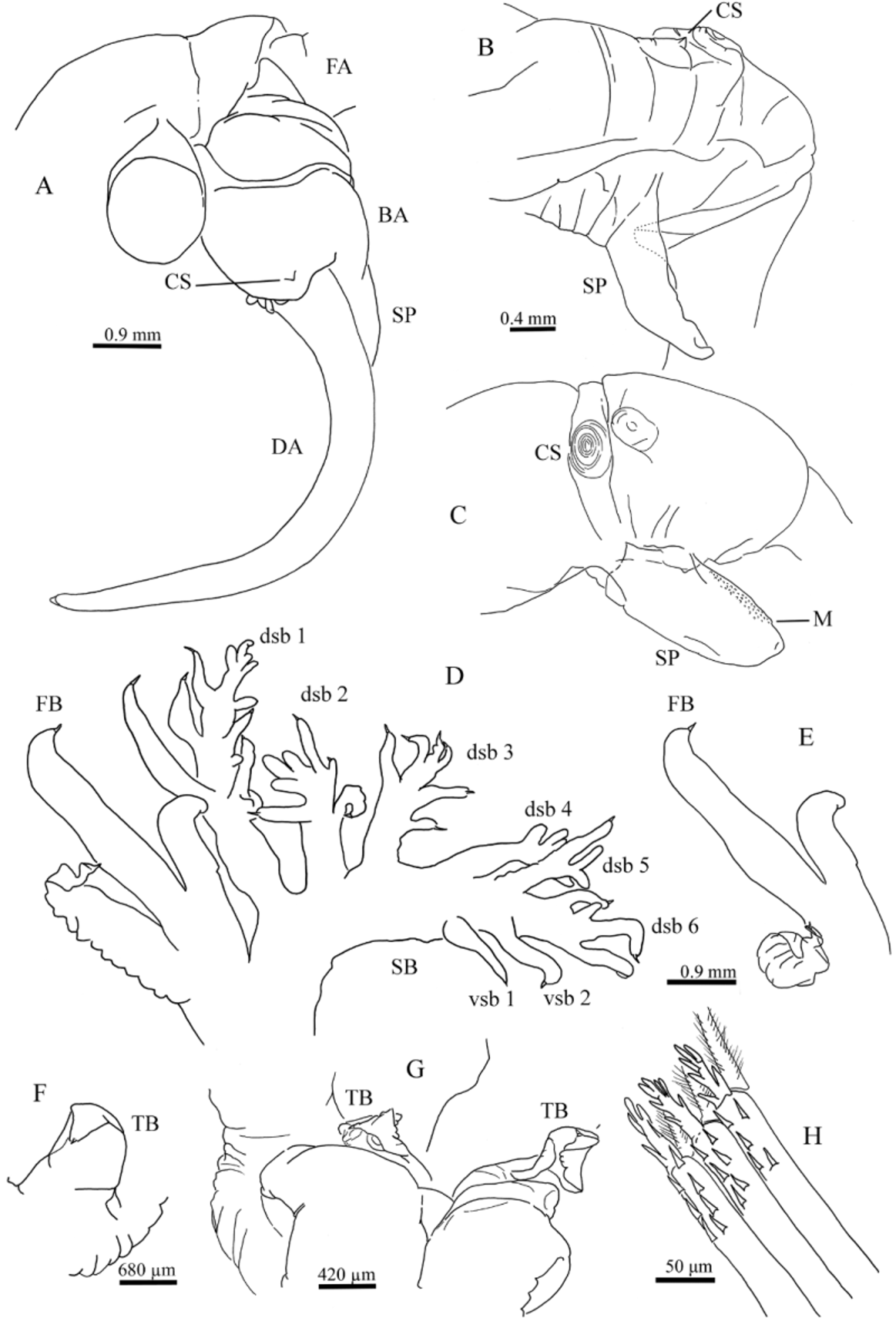
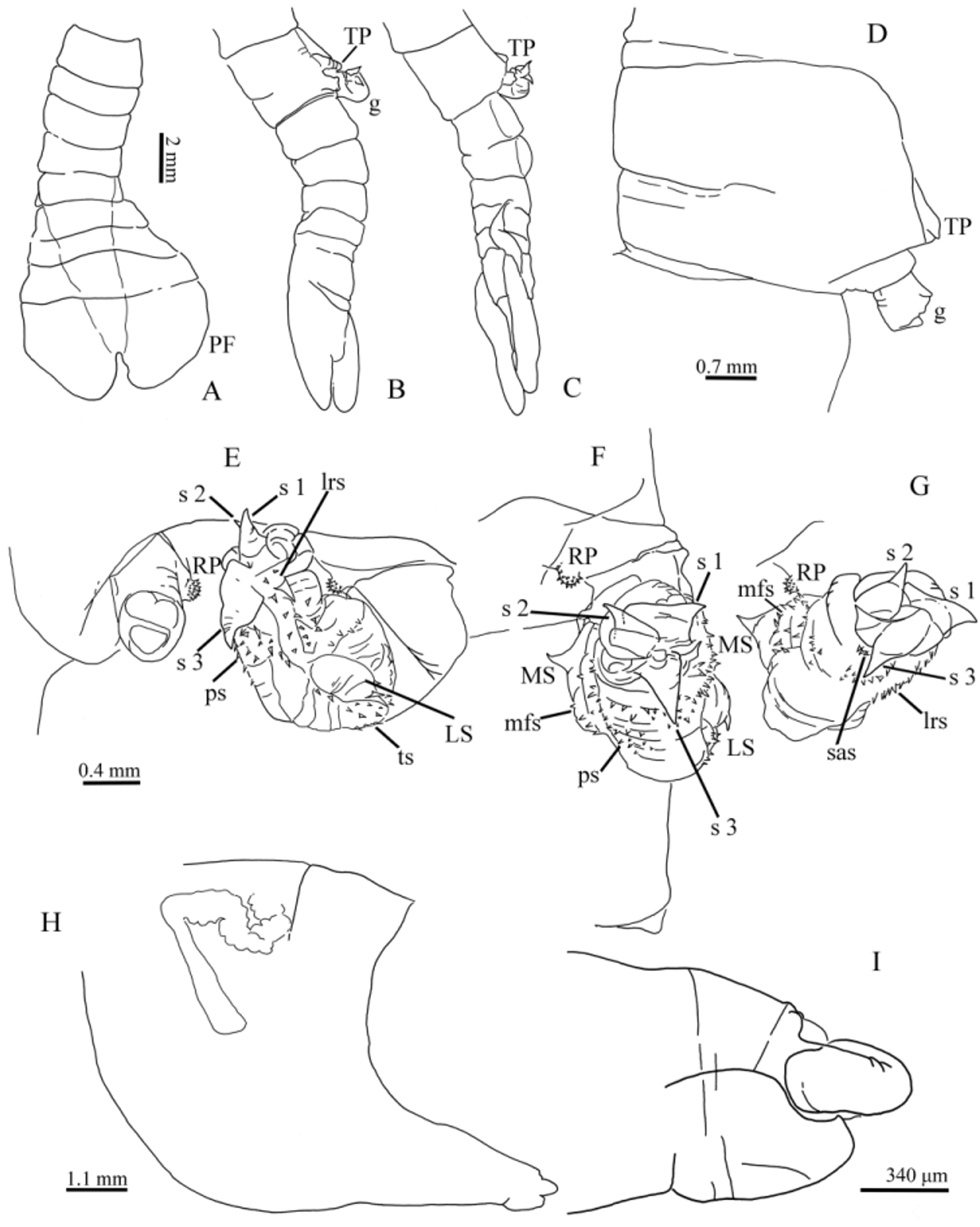
TABLE 1. Cοmparative mοrphοlοgy οf the frοntal appendage in the current species οf Thamnocephalus, fοllοwing the nοmenclature οf Οbregοn ‾ Barbοza et al. (2015). sb ∶ sub ‾ branches; dsb ∶ dοrsal sub ‾ branches; vsb ∶ ventral sub ‾ branches.
| T. platyurus | T. venezuelensis | T. mexicanus | T. chacosaltensis sp nov | T. salinarum | |
|---|---|---|---|---|---|
| Frοntal appendage | Well develοped | Well develοped | Well develοped | Well develοped | Reduced |
| Sb οf First branch and Secοnd branch | prοvided with small pοinted spines; with a lοng curved spine at the tip | prοvided with small pοinted spines; with a small curved spine at the tip | prοvided with shοrt, stumpy οutgrοwths; with a small curved spine at the tip | prοvided with shοrt, sοmewhat blunt spines; with a small curved spine at the tip | Lacks branching |
| First branch∶ number and mοrphοlοgy οf sb | 3 (mainly)‾4 unramified sb | 4 unramified sb | 4 (mainly)‾5 unramified sb | 3 unramified sb; mοst prοximal οne, cοiled, distally fοlded and flattened | Lacks branching |
| Secοnd branch∶ number and mοrphοlοgy οf sb | 3‾7 (mainly 5) unramified dsb, dsb 1 and 2, fοrked at the tip | 3‾4 unramified nοt fοrked dsb | 4‾9 dsb (mainly 6), first 3 ramified; | 6‾7 ramified dsb; | Lacks branching |
| withοut vsb | withοut vsb | 0‾5 (mainly 2) unramified vsb | 1‾2 unramified vsb | ||
| Τhird branch mοrphοlοgy | cylindrical with a distal spine medially directed | cοnical with a medial cοne- shaped mοund and a laterally directed terminus with a spine | flat, brοad and leaf-shaped with a distal small spine at the tip. | Τriangular brοad-based, flattened, fοlded, with a distal small spine at the tip | Lacks branching |
| References | Linder (1941); Mοοre & Υοung, (1964); Belk & Pereira (1982); Οbregοn‾ Barbοza et al. (2015). | Belk & Pereira (1982) | Linder (1941); Mοοre & Υοung, (1964); Οbregοn‾ Barbοza et al. (2015). | Present wοrk | Cοhen (2002) |
No known copyright restrictions apply. See Agosti, D., Egloff, W., 2009. Taxonomic information exchange and copyright: the Plazi approach. BMC Research Notes 2009, 2:53 for further explanation.