
Thelandros agama Adamson & Nasher, 1984
|
publication ID |
https://doi.org/ 10.11646/zootaxa.3852.1.2 |
|
publication LSID |
lsid:zoobank.org:pub:CE7E8E7A-073D-442A-B1D8-4CD661B59205 |
|
DOI |
https://doi.org/10.5281/zenodo.6129712 |
|
persistent identifier |
https://treatment.plazi.org/id/03AB505F-7853-A37B-1FC9-F9CCA95406DE |
|
treatment provided by |
Plazi |
|
scientific name |
Thelandros agama Adamson & Nasher, 1984 |
| status |
|
Thelandros agama Adamson & Nasher, 1984
Figs. 8–9 View FIGURE 8 View FIGURE 9
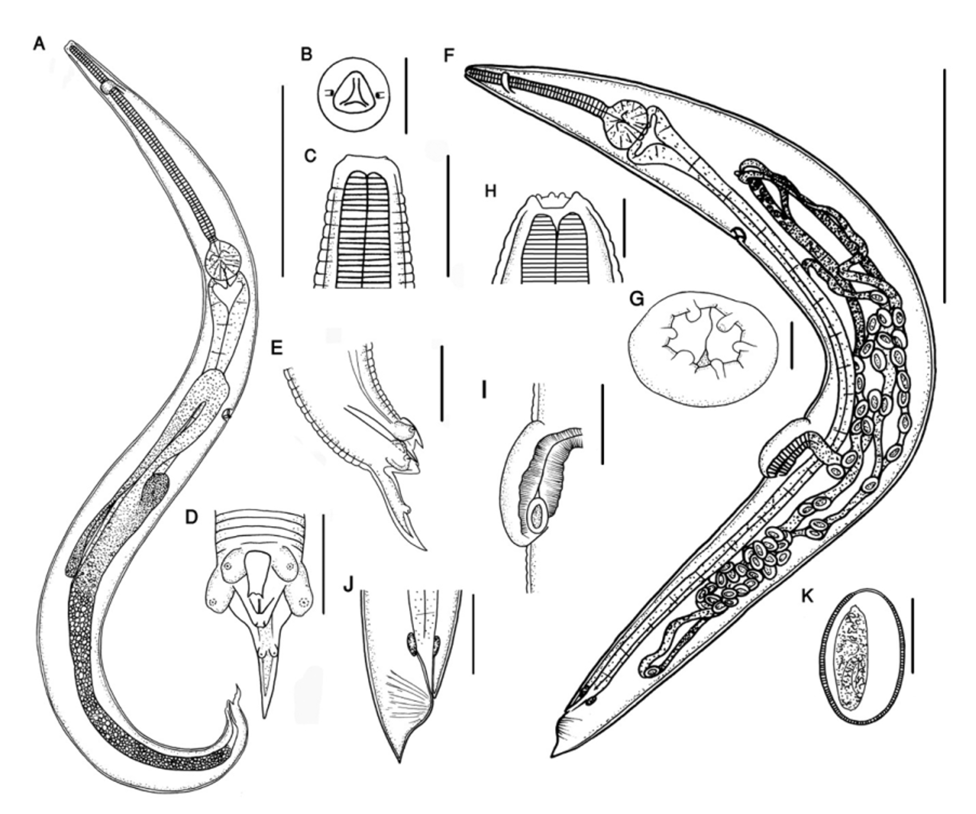
General: Cephalic extremity flattened. Mouth subtriangular with three subventral cuticular flaps projecting into mouth from sides of buccal cavity. Excretory pore posterior to oesophageal bulb, lined by ring of thickened cuticle.
Male (based on 10 specimens; mean ± SD [range]): Cylindrical worms, 2.55 ± 0.54 mm (1.99–3.67 mm) long, 179 ± 20 (155–215) wide at level of excretory pore. Cephalic sense organs consisting of 4 papillae and 2 amphids surrounding mouth ( Fig. 9 View FIGURE 9 A). Cuticle with annulations approximately 8 µm wide at mid-body. Oesophageal corpus 663 ± 70 (559–762) long; isthmus 43 ± 5 (35–50) long; bulb 89 ± 6 (78–96) long, 94 ± 7 (80–102) wide. Nerve ring and excretory pore, 149 ± 18 (129–195) and 983 ± 96 (902–1147), respectively, from anterior end. Three pairs of caudal papillae: anterior pair ventrolateral in position; second pair adcloacal, lateral in position; third pair located in middle of tail, 31 ± 3 (25–36) from tip of tail filament. Pre- and adcloacal papillae pedunculate. Tip of anterior cloacal lip serrated, with 3 serrations. Posterior cloacal lip bilobed. Caudal alae reduced, extend from base of adcloacal papillae to anterior third of tail filament. Spicule 64 ± 5 (59–72) long. Tail filament 68 ± 8 (59–83) long, terminal in position, directed posteriorly.
Female (Based on 10 specimens; mean ± SD [range]): Length 4.47 ± 0.67 mm (3.31–5.37 mm); width at level of vulva 394 ± 80 (300–558). Cephalic extremities include 6 processes, each in form of a central finger-like structure with 2 wide rectangular cuticular membranes on either side; Outer papillae and amphids raised on peduncles surrounding mouth ( Figs. 7 View FIGURE 7 C, E). Oesophageal corpus length 740 ± 115 (641–1,052), isthmus length 42 ± 7 (34–52), bulb length 178 ± 22 (134–202), width 198 ± 26 (152–234). Nerve ring, excretory pore, and vulva 142 ± 15 (127–166), 1.42 ± 0.23 mm (1.20–1.78 mm), and 2.86 ± 0.56 mm (1.71–3.49 mm), respectively, from anterior end. Vulva located postequatorially, with prominent prevulvar swelling (not present in rare instances) ( Figs. 9 View FIGURE 9 F). Reproductive structures arise posterior to oesophageal bulb. Anus 170 ± 31 (124–221) from posterior end; postanal region of body developed into short cone. Eggs oval, 98 ± 10 (72–107) long, 61 ± 4 (54–67) wide, with terminal operculum.


No known copyright restrictions apply. See Agosti, D., Egloff, W., 2009. Taxonomic information exchange and copyright: the Plazi approach. BMC Research Notes 2009, 2:53 for further explanation.