
Trichomycterus uisae, Castellanos-Morales, 2008
|
publication ID |
https://doi.org/ 10.1590/S1679-62252008000300003 |
|
persistent identifier |
https://treatment.plazi.org/id/03AFBC06-2034-DF7B-FC63-FE9AFE0CF9B4 |
|
treatment provided by |
Carolina |
|
scientific name |
Trichomycterus uisae |
| status |
sp. nov. |
Trichomycterus uisae View in CoL , new species
Holotype. CAC-CDMB 72.5 mm SL, Colombia, Departamento de Santander, Municipio de Los Santos, Vereda Mesa de Los Santos, Acuarela road 3.5 km, Cueva El Misterio (06º50’21”N, 73º05’18”W, elevation 1600 m), upper Sogamoso River basin; C. Castellanos-M, L. L. Marino-Zamudio & M. Pardo-Peñaloza, 1 Nov 2006. GoogleMaps
Paratypes. All collected at holotype locality . ANSP 187498 View Materials , 45.5 mm SL, C. Castellanos-M & L. L. Marino-Zamudio, 28 Feb 2007 . CAC-CDMB 88, 55.9 mm SL, C. Castellanos-M & L. L. Marino- Zamudio, 28 Feb 2007. IAvH-P 10806 , 2 , 52.2 -57.0 mm SL, C. Castellanos-M, L. L. Marino-Zamudio & M. Pardo-Peñaloza, 1 Mar 2007 . UIS-T 1698 , 49 mm SL, C. Castellanos-M, L. L. Marino- Zamudio & M. Pardo-Peñaloza, 1 Mar 2007 . UIS-T 1699 , 43.5 mm SL, C. Castellanos-M, L. L. Marino-Zamudio & M. Pardo- Peñaloza, 2 Mar 2007 .
Non-type material. All collected at holotype locality. CAC-CDMB 90, 2 C&S, 32.1-41.1 mm SL, 1 Mar 2007. CAC-CDMB 91, 45.5 mm SL, 2 Mar 2007. CAC-CDMB 92, C&S, 28.3 mm SL, 2 Mar 2007. UIS-T 1700, 39.8 mm SL, C. Castellanos-M. & L. L. Marino- Zamudio, 2 Mar 2007.
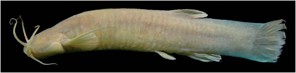
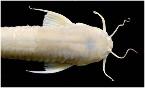
Diagnosis. Trichomycterus uisae ( Figs. 2-3 View Fig View Fig ) is distinguished from both epigean and troglomorphic species of the genus by the following combination of characters: anterior and posterior fontanels separated but connected by a narrow channel (vs. fontanels separated by epiphyseal bar); five to nine opercular odontodes (vs. 8 to 16); reduced mouth width (32.6- 41.2% of HL); relatively deep head (48.6-66.3% of HL); reduced pigmentation (except for epigean T. gorgona and caverestricted congeners T. chaberti , T. itacarambiensis , T. sandovali , T. santanderensis and T. spelaeus ) consisting of a narrow predorsal dark bluish-gray stripe from nape to origin of dorsal fin and ground coloration light-brown without spots (vs. pigmented body with dark spots); variable reduction of eyes from 7.4 to 11.1% of HL (except for epigean T. gorgona and hypogean T. chaberti , T. itacarambensis and T. santanderensis ); extended nasal, maxillary and rictal barbels (85.2-108%, 93.6-111.8% and 55.1-70.2% of HL, respectively); caudal fin with slightly convex margin, dorsal lobe of caudalfin longer than ventral lobe. Other characters shared with
SL. Cueva El Misterio, upper Sogamoso River basin, Santander, various other species of Trichomycterus yet useful for identification include origin of pelvic fin anterior to vertical through dorsal-fin origin; short caudal peduncle (19-21.9% of SL); tip of first pectoral-fin ray prolonged as a long filament (52.1- 89% of pectoral-fin length) and pelvic fins not widely separated.
Description. Morphometric data presented in Table 1. Body elongated, deeper than wide, gradually deeper from trunk toward caudal peduncle; dorsal profile of trunk convex. Ventral profile of trunk straight. Dorsal and ventral profiles of caudal peduncle slightly convex. Integument thick with lateral cutaneous folds forming vertical rings between pectoral and anal fins in specimens preserved in alcohol. Anal and urogenital openings closer to pelvic-fin base than to anal-fin origin, totally covered when pelvic fin extended. Free vertebrae 35. Pleural ribs 11-12.
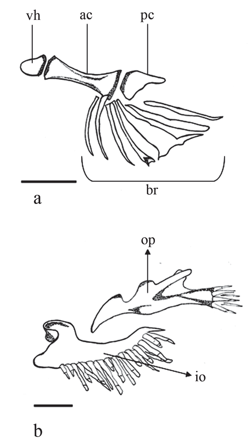
Head wide, trapezoidal, depressed in dorsal view. Dorsal profile of head straight, ventral and lateral profiles convex. Eye small, black, rounded, well defined, with variable diameter, positioned dorsally on anterior half of head. Mouth subterminal, with corners oriented backwards. Lower lip with conspicuously fleshy lateral lobes. Teeth conical, curved, arranged in 3-4 irregular rows on upper jaw and three rows on lower jaw. Jaw muscles not particularly developed and not bulging from surface of head. Neurocranium with elongate mesethmoid T-shape. Anterior fontanel small, oval in shape, located between frontals at level of infraorbital laterosensory canal exit 10-11. Posterior fontanel long and broad. Anterior one-third of posterior fontanel situated between frontal bones, with remainder in anterior portion of supraoccipital bone ( Fig. 4 View Fig ). Both fontanels connected by a narrow channel. Branchial membranes thick, united to isthmus anteromedially and forming free fold across isthmus. Gill opening wide. Seven branchiostegal rays ( Fig. 5a View Fig ), six externally visible from below. Nasal and maxillary barbels surpassing base of pectoral fin. Maxillary barbel longer than nasal barbel. Anterior nostril surrounded by slightly raised thick integument, continuous with nasal barbel, both forming a tubular-shaped structure around nostril. Posterior nostril oriented transversally, its anterior edge delimited by thin and long flap of integument. Interopecular patch of odontodes well developed, with 27-32 conical and elongated odontodes arranged in four irregular rows, with interopercular odontodes on posterior edge larger. Opercular patch of odontodes small, with 5-9 conical odontodes arranged in three irregular rows ( Fig. 5b View Fig ).
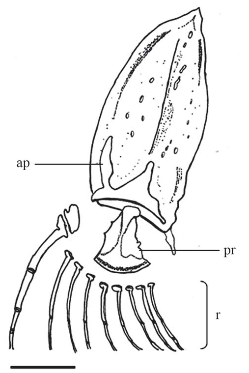
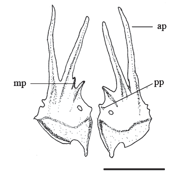
Pectoral fin rounded with i,8 rays. First ray thin and fragile, prolonged as a long filament. Scapulocoracoid with long anteriorly directed process, located close to first pectoral-fin ray base ( Fig. 6 View Fig ). Dorsal-fin rounded, located posterior to vertical through midbody, with iv,7 rays (only two unbranched rays externally visible). First pterygiophore inserted between neural spines of 15-16 th free vertebra. Pelvic-fin rays i,4, with a lateral splint. Pelvic-fin origin anterior to vertical through dorsal-fin origin and its posterior edge slightly surpasses urogenital opening. Pelvic-fin bases not widely separated. Basipterygium with two long anterior processes narrowing from base to distal tip, one or two medial processes and one short posterior process ( Fig. 7 View Fig ). Anal-fin similar to dorsal fin, but smaller, with ii, 5 rays, its origin at level of last dorsal-fin ray. First pterygiophore inserted between hemal spines of free vertebrae 20-21. Caudal-fin rays i, 5+6, i. Parhypural and hypurals 1+2 associated with eight rays, hypural 3 with 3 rays, and hypurals 4+5 with 3 rays. 17-18 dorsal and 12 ventral procurrent rays. Caudal-fin edge slightly convex, uppermost rays larger. Caudal skeleton with neural spine of preural centrum 2 well developed. Hypurals 1 and 2 fused to parhypural; hypural 3 partially fused to hypurals 4+ 5 in young specimens ( Fig. 8a View Fig ), and separated in adult specimens ( Fig. 8b View Fig ).
Coloration in live specimens. Body color light-brown-(M 10YR - 5/6). Base of all fins with yellow tones from base to edge (M 5YR – 6/8 to M 2.5Y – 7/6). One predorsal narrow dark bluish gray band (M G-5PB).
Coloration in alcohol. All specimens preserved in alcohol with ground color yellowish (M 10YR - 5/6). Base of all fins with yellow tones (M 2.5Y – 7/6), with exception of two nontype specimens with ground color brown-yellowish.
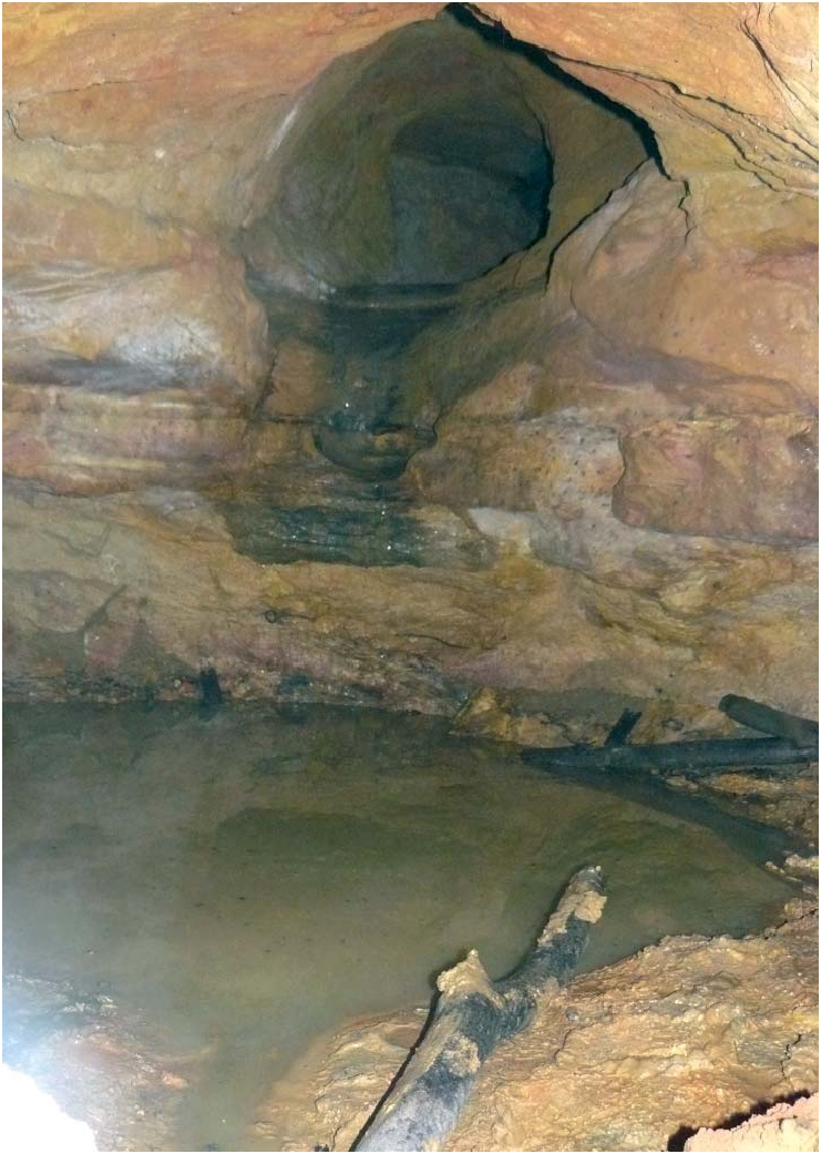
Ecological notes. The Cueva El Misterio is located at the east side of a plateau known as Mesa de los Santos, in the Municipio de los Santos. The plateau has Cretaceous sedimentary rocks and limestones of the Rosablanca Formation ( Williams, 1990); reaches 1800 meters above sea level and is located on the oriental versant of the Chicamocha Canyon, in the Colombian Andes, of Santander Department. The cave, with a total of 110 m of explored passages, is isolated from the epigean stream and is oriented longitudinally with galleries formed by gentle slope tunnels and narrow passageways. The cave has small wells interconnected by reduced descending channels where water infiltration was observed. In dry months, an isolated sump pool was observed at each gallery. The bottom of each well is rocky and contains much sediment composed chiefly of bat excrement. Cydnid bugs (Hemiptera: Heteroptera) were found inside the wells. Diptera, crayfish and bats were observed in the interior of the galleries ( Fig. 9 View Fig ). Water temperature, when the holotype was collected, was 20°C and cave temperature was 18.5°C.
Etymology. The specific epithet “ uisae ” refers to the acronym UIS corresponding to the Universidad Industrial de Santander, in the Departamento de Santander, Colombia. The name is used as a noun in apposition.
Common name. Trepador


No known copyright restrictions apply. See Agosti, D., Egloff, W., 2009. Taxonomic information exchange and copyright: the Plazi approach. BMC Research Notes 2009, 2:53 for further explanation.