
Trimma blematium, Winterbottom & Erdmann, 2018
|
publication ID |
https://doi.org/ 10.11646/zootaxa.4444.4.7 |
|
publication LSID |
lsid:zoobank.org:pub:D943FB45-6B57-430D-9B77-3B0FB0F647DE |
|
DOI |
https://doi.org/10.5281/zenodo.5997051 |
|
persistent identifier |
https://treatment.plazi.org/id/F43E8FB7-3F8E-4885-A0B8-128E740277CA |
|
taxon LSID |
lsid:zoobank.org:act:F43E8FB7-3F8E-4885-A0B8-128E740277CA |
|
treatment provided by |
Plazi |
|
scientific name |
Trimma blematium |
| status |
sp. nov. |
Trimma blematium new species
Blue-eyed Pygmygoby
Figs. 1–4 View FIGURE 1 View FIGURE 2 View FIGURE 3 View FIGURE 4 .
No published names pertain to this species.
Material examined. Holotype. ROM 102761, 22.0 mm SL male, Papua New Guinea, Milne Bay Province, Normanby I., Whampus, 09° 58.122' S, 150° 50.350' E, 65 m, 29 May, 2016, field # MVE-16-023, M.V. Erdmann.
Paratypes. ROM 101301, 4(15.9–22.8), collected with the holotype. ROM CS 1972, (16.2 male), collected with the holotype (now cleared and stained). ROM T20892, (13.8), collected with the holotype, tissue specimen.
Diagnosis. A species of Trimma with scales present on cheeks and opercle, 8–9 scales in predorsal midline, 16 unbranched pectoral fin rays, a branched 5th pelvic fin ray 45–50% length of 4th ray, 20–22 total gill rakers, a broad interorbital (46–63% pupil width) with narrow crease-like postorbital trench ending at posterior-most papilla in row p, nasal apparatus small and situated on anterior one-third of snout with posterior nares forming posterodorsal margin of nasal sac, and 7 papillae in row p (with a single papilla below row n). When live or freshly collected, dorsal surface of eye dark blue; preserved specimens with fairly evenly distributed melanophores over dorsal surface of snout.
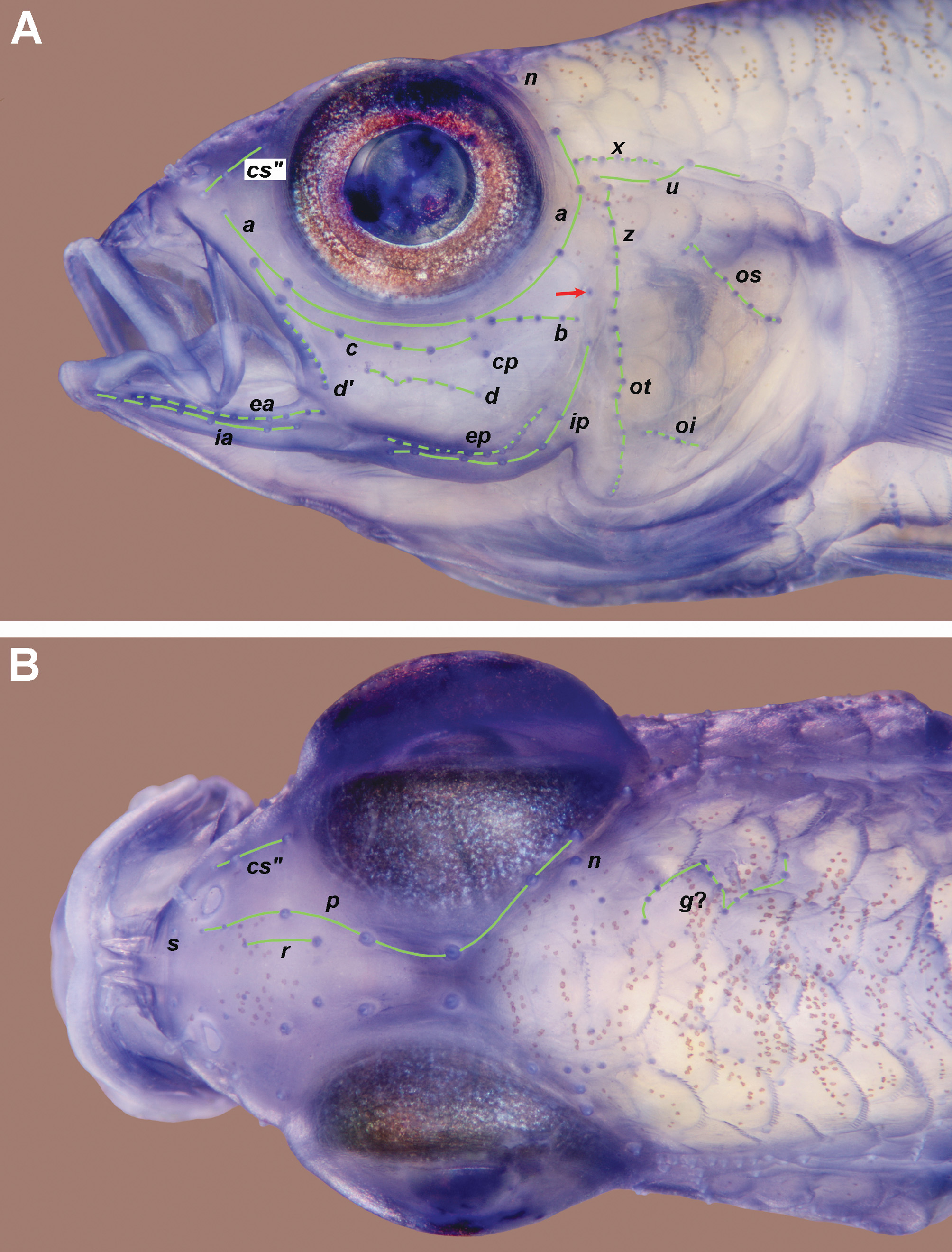
Description. The description is based on the holotype and 5 paratypes. Dorsal fin VI + I 8– 9 (8.2), second spine elongated ( Fig. 1 View FIGURE 1 ), reaching posteriorly when adpressed to between base of 4th ray to 1 st scale behind last dorsal fin ray (mean = base of 7th ray), first ray of second dorsal fin branched (unbranched in one), remaining fin rays branched except for posterior element of last ray, fin reaches posteriorly 39– 41 –50% (44%) distance between base of last ray and first exposed dorsal procurrent caudal fin ray; anal fin I 8, first ray unbranched, fin reaches posteriorly 31 –42% (35%) distance between base of last ray and first exposed ventral procurrent caudal fin ray; pectoral fin 16, all rays unbranched, fin reaching posteriorly to region above urogenital papilla; pelvic fin I 5, fifth ray with single dichotomous branch point and 40– 42 –45% (43%) length of fourth ray, which reaches posteriorly to between bases of anal spine to 2nd anal ray, pelvic rays 1–4 with single sequential branch point; basal membrane forming fold across midline above last pre-pelvic scale; no fraenum. Lateral scales 23; anterior transverse scales 8– 9 (8.1); posterior transverse scales 7– 8 (7.5); cheek with two cycloid scale rows, upper row of 1 –3 and lower row with 8 –9 (1.8 and 8.8 respectively, n = 5), midline of predorsal with 8 –9 (8.5) scales, anterior 1–4 rows may be cycloid, otherwise ctenoid; anteriormost scales on sides (may be ctenoid or cycloid) and top of nape reaching almost to posterior margin of eye; opercle fully scaled above papilla row oi in 4– 5 horizontal rows, dorsalmost row of 4, then 3 -4, 3, 2– 3, 2 –3 and 0- 2 mostly cycloid scales, although larger scales in mid-upper region may be ctenoid, up to 2 small auxiliary scales may be present above upper row; 3 vertical rows of cycloid scales on pectoral fin base with 1– 2 – 3 in anteriormost row, 3– 4 in second row and 4 in outer row (n = 5); 7 –8 (7.2) cycloid scales in midline anterior to pelvic fin base; area between pelvic spine and ventral margin of pectoral fin base with cycloid (smaller specimens) or ctenoid (larger specimens) scales; anterior few rows of scales in midline of belly cycloid, those behind ctenoid; circumpeduncular scales 12, scales rows in midline between base of last anal ray and first ventral procurrent caudal fin ray 9 –10 (9.2). Description of teeth based on ROM 1972CS (16.2 mm SL female, jaws excised). Upper jaw with outer row of spaced, enlarged curved canines which decrease slightly in height and reach posteriorly 4/5ths of length of premaxilla, two irregular inner rows of small conical teeth (about half height of outer teeth) becoming reduced to single row at bend of premaxilla and continuing posteriorly to 3/5ths length of premaxilla. Lower jaw with short row of about 3 enlarged, spaced, curved canines from symphysis almost to bend of dentary, about 2 irregular rows of slightly curved smaller (2/3rds height of outer teeth) teeth at symphysis, grading to single row, decreasing slightly in size posteriorly, and reaching to base of coronoid process of dentary. Tongue broadly rounded, may have small central tip. Gill opening extending anteroventrally to below mid-pupil; gill rakers 4 –5 + 16 –17 = 20 –22 (4.2 + 16.5 = 20.7). Nasal apparatus small, situated on anterior one-third of snout, anterior naris short tapering tube reaching anteriorly to above anterior margin of upper lip, posterior opening a large, porelike opening with slightly raised rim covering posterodorsal width of nasal sac, transverse width of pore about 65% length of nasal capsule, posterior margin of posterior naris well separated from bony front of orbit by 4– 5 –6 times its transverse width (mean = 5.0), nasal sac only very slightly raised above surrounding area of snout ( Fig. 2 View FIGURE 2 ). Bony interorbital width 46– 57 –63% (56.4) pupil diameter; profile of snout gently convex, with shallow concave depression between eyes (between 4th and 5th papillae of row p); epaxialis reaching anteriorly in midline to vertical above posterior margin of pupil; no narrow ridge of skin in midline of nape extending anteriorly from origin of first dorsal fin. Caudal peduncle depth as percentage caudal peduncle length 37.9– 39.6 –41.5 (39.4); head length as percentage SL 31.4– 33.8 –35.0 (33.5); as percentage head length: horizontal eye diameter 34.5– 36.4 –37.9 (36.1); snout length 21.4– 28.7 (25.3); cheek depth 14.3– 18.2 –19.6 (17.2). Cephalic sensory papillae as in Fig. 3 View FIGURE 3 . Number of papillae in each row: a = 6; b = 4– 5 (4.7); c = 6; cp = 1; d = 5– 7 (6.7); dʹ = 6– 8 –10 (7.8); e-anterior = 11– 12 –15 (13.2, n = 5); e-posterior = 12– 15 –17 (14.8, n = 5); i-anterior = 6– 7 –8 (7.0, n = 5); i-posterior = 7 –8 (7.2, n = 5); p = 7, with 2 papillae just medial to posterior naris and one papilla below row n; r = 2; f = 3– 4 –5 (4.0, n = 5); cs" = 3 (n = 5); g = apparently absent, but may be represented by zig-zag line of 11 papillae ( Fig. 3 B View FIGURE 3 ); n = 1; x = 7– 8 –9 (7.8); u = 4 –5 (4.2); z = 5– 8 (6.8, n = 4); ot = 9– 13 –16 (13.2, n = 5); os = 8– 9 –11 (9.0, n = 4); oi = 4– 6 –7 (5.4, n = 5). Note that posteroventralmost papilla of row p is not visible in Fig. 3 A View FIGURE 3 , being covered by edge of eye, but is visible in Fig. 3 B View FIGURE 3 . Abdominal/caudal vertebral transition (see Winterbottom, 2011:130, for definitions) unusual for the genus (based on single cleared and stained specimen, ROM 1972CS, which unfortunately did not take up the alizarin well). No haemal canal present on 9th abdominal vertebra but canal present on 10th vertebra. Two foramina present ventrally on 1 st caudal vertebra, with small basal canal and enlarged foramen distally formed by haemal arches joined only at tips (similar to arch found in Type A, but not as well developed).
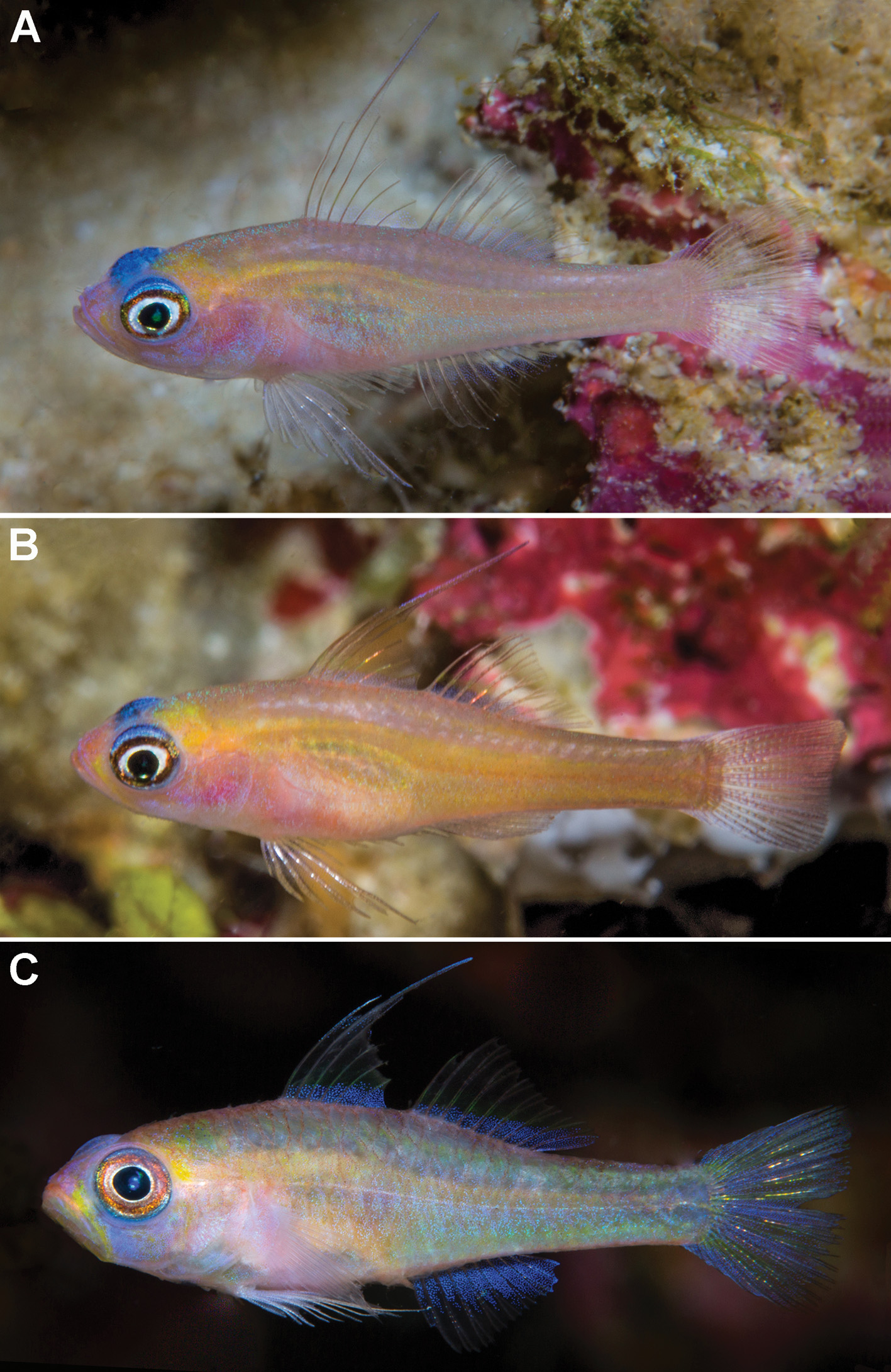
Colour pattern. Live, based on 10 images from Normanby Island (see Fig. 1 View FIGURE 1 , A & B for examples). Head and body translucent light pink or yellow-brown, with large yellowish area posterodorsal to eye over posterior half of braincase, spinal cord yellow but fading into background below end of second dorsal fin, abdomen and lower half of head lighter, snout pink. Iris with white inner margin around pupil followed by dark brown ring, dorsomedial surface of eye dark to medium blue (usually lighter at lateral margins). Basal half of dorsal and anal fins, and pelvic fin membranes heavily invested with iridocytes ( Fig. 1 A View FIGURE 1 ). Freshly collected, based on image of 22.4 mm SL male paratype, ROM 101301 ( Fig. 1 C View FIGURE 1 ). Upper and lower midlines reddish, body semi-translucent greenish/bluish with scale margins diffusely outlined with dark red, spinal cord and posterior region of cranium dull yellow, medial half of upper lip orange, region of snout below anterior margin of eye greenish-yellow, anterior cheek blue-grey. Dorsal fins with basal stripe of bluish chromatophores, stripe half pupil-width anteriorly to full pupil-width posteriorly, fin elements yellow above this, grading to red more distally, first two spines of first dorsal fin white at extremities; anal fin with similar but much wider (2 X pupil-width) basal stripe; caudal with bluish margins and 3 equidistant blue streaks in body of fin, the middle medial in position, rest of fin greenish-yellow; membranes of pelvic fin with iridocytes, rays off-white, membranes of pectoral fin hyaline with rays light pink. Preserved. Holotype pale straw coloured, lighter on head. Melanophores outlining first 1.5 vertical scale rows along dorsum from first predorsal scale almost to end peduncle. A few scattered light brown melanophores on dorsal surface of snout (visible in Fig. 3 B View FIGURE 3 ), a few similar melanophores behind eye and along dorsal part of opercle, and basally in membranes of posterior half of second dorsal fin. Iris dark brown with narrow light circle around pupil ( Fig. 3 A View FIGURE 3 ). No other pigmentation discernible. Paratypes generally somewhat more heavily pigmented, with scale pockets outlined on whole dorsal half of body, and more pigmentation on dorsal surface of snout and upper half of opercle. Thin line of melanophores may be present between base of last anal ray and first ventral procurrent caudal ray. Body pigmentation may extend below midlateral scale row, and include dorsal part of abdomen.
Etymology. The specific name ‘blematium’ is a compound word derived from the Greek µπλε (ble) = blue, and µάτι (máti) = eye, in allusion to the distinctive blue dorsal surfaces of the eyes of the new species. This species has been informally referred to as Trimma RW sp. 106.
Distribution and Habitat. Currently recorded only from off the southwestern coast of Normanby Island, Milne Bay Province, Papua New Guinea. The species was observed in a mixed rubble and sand habitat on a deep reef slope (60–70 m depth) exposed to significant current and cold-water upwelling.
Comparisons. There are 8 other described species of Trimma which possess predorsal scales in the midline, a branched 5th pelvic fin ray, and 16 or more unbranched pectoral fin rays. These are: T. annosum Winterbottom, 2003 , T. emeryi Winterbottom, 1985 , T. fasciatum Suzuki et al., 2012 , T. flavicaudatum ( Goren, 1982) , T. fucatum Winterbottom & Southcott, 2007 , T. kardium Winterbottom et al., 2015 , T. pentherum Winterbottom & Hoese, 2015 , and T. randalli Winterbottom & Zur, 2007 . The only one of these species in which the second dorsal spine is elongated posterior to the base of the second dorsal fin ray, and in which at least one row of cheek scales is present is T. randalli , which differs from T. blematium in having 5 papillae in row c (vs. 6), 6 (vs. 7) papillae in row p, with 1 (vs. 2) papillae just medial to the posterior naris, a narrower bony interorbital (<35% pupil width vs.> 45%), a basal membrane between the 5th pelvic fin rays that extends 20–40% the distance to the tip of the rays (vs. vestigial and only forming a narrow shelf across the ventral midline), fewer anterior transverse scale rows (7 vs. 8–9), a single row of cheek scales (vs. 2 rows), and in having a black or dark red caudal fin (vs. hyaline). In addition to T. randalli , only T. emeryi and T. fasciatum have been recorded as possessing scales on the cheek, but there are a maximum of two such scales (and often none) and they never form two discreet rows totalling 9–11 scales as in T. blematium .
This species is superficially most similar to T. meityae , described below, especially in overall colouration and the blue dorsal surface of the eye. However, T. blematium differs from T. meityae in the number of papillae in row p (7 vs. 8, with the former species having a single papilla, vs. 2, below papilla row n); in having 16 pectoral fin rays (vs. 17–18), in having a branched fifth pelvic fin ray (vs. unbranched), and in generally having a more extensive distribution of melanophores over the snout (reaching posteriorly as far as posterior papilla of row r vs. confined to the region medial to the posterior nares).
Discussion. An analysis of the partial COI gene of the two species described here (based on a single specimen of each species, Fig. 4 View FIGURE 4 ) revealed that they are separated by 7.7% of the base pairs of that gene. Coupled with the morphological differences described above, and a geographical separation of some 2000 kilometres (straight-line) suggests to us the validity of recognizing two distinct species. The two species are phenetically closest to each other, and fall out within the T. tevegae grade as defined by Winterbottom et al. (2014b).
Both species have been found only in deeper, mesophotic reef habitats (60–70 m for T. blematium , 50–60 m for T. meityae ), although T. blematium was found on clean outer reef sand and rubble exposed to significant current, while T. meityae was found on silty sand and rubble on a nearshore reef with minimal current.


| ROM |
Royal Ontario Museum |
No known copyright restrictions apply. See Agosti, D., Egloff, W., 2009. Taxonomic information exchange and copyright: the Plazi approach. BMC Research Notes 2009, 2:53 for further explanation.