
Xetadrilus aphanus, Schmelz, Rüdiger M., Collado, Rut & Römbke, Jörg, 2011
|
publication ID |
https://doi.org/ 10.5281/zenodo.203260 |
|
DOI |
https://doi.org/10.5281/zenodo.5611833 |
|
persistent identifier |
https://treatment.plazi.org/id/520EAA7D-D65E-4517-FF40-86EFFE74F962 |
|
treatment provided by |
Plazi |
|
scientific name |
Xetadrilus aphanus |
| status |
sp. nov. |
Xetadrilus aphanus View in CoL sp. nov.
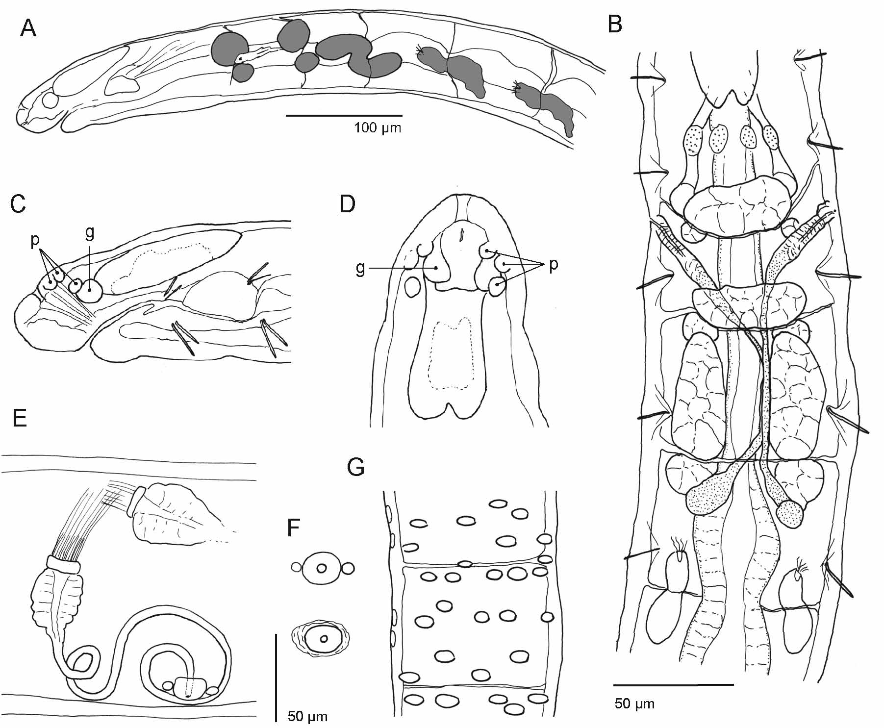
( Figs 6 View FIGURE 6 , 8 View FIGURE 8 F, Table 3 View TABLE 3 )
Holotype. MZUSP 1209, adult specimen, stained and whole-mounted in Canada-Balsam, Cachoeira, Antonina, 25°18'25"S, 48°40'24"W, 70 m a.s.l., pasture on Cambisol [site 2], Mar 2004, leg. B. Förster, R. M. Schmelz.
Paratypes. MZUSP, 32 specimens, stained whole mounts:
MZUSP 1210, 2 specimens, Antonina, Cachoeira , 25°15'22''S, 48°40'14''W, 40 m a.s.l., young secondary forest on Cambisol [site 7], May 2003, leg. J. Römbke, R. M. Schmelz.
MZUSP 1211, 11 specimens, same data as holotype.
MZUSP 1212, 10 specimens, Antonina, Cachoeira , 25°18'56"S, 48°39'29"W, 20 m a.s.l., pasture on Gleysol [site 20], Mar 2004, leg. B. Förster, R. M. Schmelz.
MZUSP 1213, 7 specimens, Antonina, Cachoeira , 25°18'32"S, 48°39'38"W, 50 m a.s.l., pasture on Cambisol [site 3], Oct 2004, leg. B. Förster, R. M. Schmelz.
MZUSP 1214, 2 specimens, Curitiba, UFPR University Campus, Agrarias, 25°24'37''S, 49°14'56''W, 909 m a.s.l., grassland, Mar 2004, leg. R. M. Schmelz.
UFPR, 30 specimens, stained whole mounts, Guarequeçaba, Itaqui:
UFPR OL-20, 8 specimens, 25°14'16.44''S, 48°29'52.1''W and 25°18'49.5''S, 48°27'10.4''W, 8 and 13 m a.s.l., respectively, abandoned ("herbaceous") pasture on Cambisol [sites 31, 33], Oct 2007, leg. P. Heine, R. M. Schmelz.
UFPR OL-21, 6 specimens, 25°18'31.8''S, 48°27'2.3''W, 28 m a.s.l., medium old secondary forest on Cambisol [site 37], Sep 2007, leg. P. Heine, R. M. Schmelz.
UFPR OL-22, 16 specimens, appr. 25°13'30''S, 48°27'40''W, 4 m a.s.l., pasture near SPVS guesthouse in Tagaçaba, Jan 2008, leg. J. Römbke, R. M. Schmelz.
Additional material. Ca. 15 specimens, several sites in Cachoeira and Itaqui, examined in vivo, not preserved.
Etymology. 'Aphanus' is a latinized form of the Greek 'aphanes', meaning inconspicuous. The name refers to the small body size of the worms.
Description. Specimens inconspicuous, among the smallest enchytraeids in all samples. Body dimensions. living adult specimens ca. 2–3 mm long and 0.07–0.1 mm wide; fixed adult specimens 2–2.2 mm long and ca. 0.1 mm wide, 0.08–0.12 mm at V, 0.1–0.14 mm at XII. Segment number of adult individuals (19)-23-28 (N = 35). Chaetae 2 per bundle, formula 2,0–0: 2–2. Lateral chaetae present in II–VII, absent from VIII on. Ventral chaetae from II on, absent in XII. Chaetae distally straight, or slightly bent to same side as proximal bend. Terminal chaetae enlarged, twice as large as anterior chaetae, not sigmoid. Ventral anterior chaetae ca. 2 µm thick, in II 10–16 µm long, gradually increasing in size posteriad to about 20–26 µm. Ventral chaetae in hindmost segments 34–40 µm long and 2.5–3 µm thick. Epidermal gland cells ( Fig. 6 View FIGURE 6 G) variable; entire epidermis glandular to varying degrees, gland cells pale and evenly distributed, or epidermal gland cells indistinguishable in living and preserved material. In specimens with glandular epidermis body surface often covered with foreign substance (humus particles). Clitellum saddle-shaped, not developed ventrally; cells in separate (viv) or dense (fix) rows; dorsally in ca. 30 transverse rows; hyalocytes and granuloctyes alternating, granulocytes isolated; ventro-laterally only granulocytes. Clitellum extending posteriorly to chaetae of XIII.
Prostomium ( Fig. 6 View FIGURE 6 A,C,D) with head pore in mid-dorsal position. Frontal prostomial epithelium thickened, with a vesicle-like recess or cleft at the frontal tip. Dorso-laterally several inner papillae in bilateral-symmetrical order, papillae also on peristomium. Prostomial musculature present. Body wall less than 5 µm thick (viv); fix: 4– 9 µm. Cuticle thin, indistinguishable (viv), or <2 µm thick (fix), visible at x400 magnification. Septa 4/5–6/7 (- 9/ 10) thicker than the rest. Brain in I–III, 2–2.5x as long as wide, incised posteriorly, sides converging anteriad ( Fig. 6 View FIGURE 6 D). A pair of ganglia on prostomial nerves. Suboesophageal ganglion in III–IV, perikarya of ventral nerve cord in segmental ganglia from V on. Pharyngeal glands ( Fig. 6 View FIGURE 6 A,B) with unpaired dorsal lobes in IV and V; primary ventral lobes in V, elongate; secondary ventral lobes in V and VI, spherical, smaller than primary ventral lobes. In VI–VII a pair of separate elongate lobes, consisting of an anterior dorsal part in VI and a posterior ventral part in VI–VII; both parts broadly connected in Z-like fashion, ventral portion extending into VII to varying degrees, without constriction at septum. Oesophageal appendages and intestinal diverticula absent, intestine from VII, here epithelium thickened in VII and parts of VIII ( Fig. 6 View FIGURE 6 B); transition from oesophagus to intestine at 6/7, widening abrupt here, or gradual over several following segments. Chloragocytes sparse, inconspicuous, small, first cells from VI. Dorsal vessel from 1/ 2 XII – XIII, difficult to see in living specimens. Pars tumida of midgut from 1/2 XVI – XXIII, extending over 1.5–4 segment lengths. Preclitellar nephridia ( Fig. 6 View FIGURE 6 B) two pairs, at 7/8 and 8/9, not constricted at septum, ca. 50–70 µm long (fix). Anteseptale with parts of nephridial body, longer than wide, funnel attached in obliquely upright position. Postseptale larger than anteseptale, length ratio ca. 2:1, with a dorsal bump in mid-section, usually without vescile, gradually tapering into short efferent duct; nephridial canal apparently with an up- and down zig-zag course; nephroporus inconspicuous, anterior to ventral chaetal bundles, no terminal vesicle. A small dorsal vesicle seen in the postseptale a few specimens, similar to the one in Guaranidrilus . First postclitellar nephridia at 13/14 or 14/15, longer than in anterior segments (ca. 70–90 µm, fix), in following segments only few positions occupied. Coelomocytes elongate, pale, broadly oval, ca. 15–20 µm long (viv, fix), filled with distinct, pale, spherical vesicles; a few vesicles may be refractile; cells of some specimens dark in aggregations but without colour or tint.
Seminal vesicle absent. Spermatozoa ca. 42 µm long, heads ca. 18 µm long in vivo. Sperm funnel ( Fig. 6 View FIGURE 6 E) small, less than half as long as body diameter and only slightly longer than wide (1.3–1.5x), collar distinct, thickwalled, slightly narrower than widest funnel body diameter, canal comparatively wide. Fix: length 35 µm diameter 20–22 µm, almost cylindrical in cross section (i.e. only slightly flattened). Vas deferens ca. 4 µm wide (viv, fix), in 2–3 wide coils in XII, of same diameter throughout. Male copulatory organ ( Fig. 6 View FIGURE 6 E) small, glandular bulb spherical or oval in top view, longest diameter ca. 20 µm (viv, fix), not more than a swelling of body wall in side view, pierced centrally by vas deferens, occasionally accompanied by a pair of small extra bulbs (diameter <10 µm), one anterior, one posterior ( Fig. 6 View FIGURE 6 E,F). No bursa, no bursal slit, male pore on body surface. Accessory glands absent. Spermatheca most often small and inconspicuous, often difficult to see in living specimens. Ectal duct ca. 25 µm long and ca. 8 µm wide (fix), short and simple, as if absent in living specimens (viv); distal part of ampulla as wide as or slightly wider than ectal duct, spherical or ovoid, diameter 10–15 µm, thin-walled, with sperm aligned in parallel in longitudinal axis of spermatheca; connecting tube thinner than ectal duct, diameter 4–5 µm, ental reservoir reaching VII or VIII when fully extended (reaching X in 1 specimen), often coiled either in VI or V; diameter 10– 12 µm proximally; ental reservoir not always developed; in these cases the proximal end as narrow as the connecting tube and difficult to distinguish in living and preserved material. Eggs. One or two mature eggs at a time.
Habitat. X. aphanus was found in grazed and abandoned pastures and in early and medium-growth stages of forest regeneration. It was absent in old-growth forest and almost absent at the agroforestry sites (1 specimen found).
Remarks. Xetadrilus aphanus worms were among the smallest enchytraeids in all samples; adult specimens were often considered as juveniles or hatchlings of other species. The most conspicuous trait is the Z-shape of the posterior pharyngeal gland in VI and VII, unique in the genus and to our knowledge also in the family. Further differentiating traits are listed in Table 3 View TABLE 3 . The species is variable with respect to (1) length of spermatheca, (2) presence and conspicuousness of epidermal gland cells, (3) size or length of pharyngeal glands in VII, (4) dorsal vesicle in the nephridial postseptale (mostly absent, seen in a few specimens), and (5) conspicuousness of male glands and copulatory body muscles.
| MZUSP |
Museu de Zoologia da Universidade de Sao Paulo |
No known copyright restrictions apply. See Agosti, D., Egloff, W., 2009. Taxonomic information exchange and copyright: the Plazi approach. BMC Research Notes 2009, 2:53 for further explanation.