
Ruizantheda aerugineus, Coelho, Beatriz W. T., De Seixas Felizardo, Sherlem P. & Engel, Michael S., 2014
|
publication ID |
https://doi.org/ 10.11646/zootaxa.3889.1.3 |
|
publication LSID |
lsid:zoobank.org:pub:6D07A7C6-C941-4093-95C8-19E79D639BB4 |
|
DOI |
https://doi.org/10.5281/zenodo.6125098 |
|
persistent identifier |
https://treatment.plazi.org/id/335F1E28-4AD5-46AD-AE9C-F500BB6EF5C6 |
|
taxon LSID |
lsid:zoobank.org:act:335F1E28-4AD5-46AD-AE9C-F500BB6EF5C6 |
|
treatment provided by |
Plazi |
|
scientific name |
Ruizantheda aerugineus |
| status |
sp. nov. |
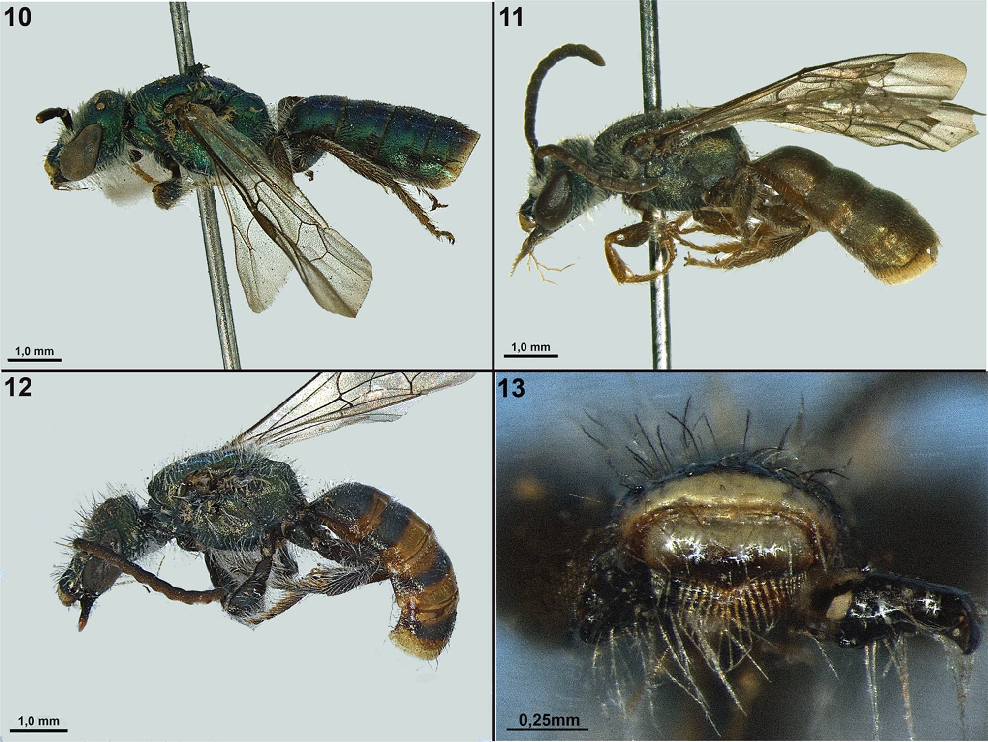
Ruizantheda aerugineus n. sp.
( Figures 11 View FIGURES 10 – 13 , 15, 18, 21, 24, 27, 30, 33, 36, 38)
Diagnosis. Ruizantheda aerugineus differs from R. kallos by its dark green coloration, head slightly wider than long, setae of compound eyes slightly shorter than an ocellar diameter, gradulus of S6 poorly invaginated medially, median apical process of S8 broadly convex, and rvl with many short setae on a conspicuously elevated semicircleshaped region on dorsal surface near apex.
Description. Male. Size. Total body length approximately 8 mm; forewing length 1.56–1.60. Structure. Head wider than longer, head width to length ratio 1.12–1.13; malar area linear; mandible simple; labral distal process in the form of a minute inverted triangle; epistomal sulcus obtuse; clypeus projecting about half of its total length below the lower tangent of compound eyes; compound eyes emarginated above level of antennal torulus; surface between compound eye and lateral ocellus straight; vertex not expanded behind ocelli; preoccipital ridge rounded; F2 about 2x length of F1, flagellomeres without glabrous areas, flagellum straight; pronotal lateral angle obtuse; anterior border of mesoscutum rounded; metapostnotum about 1.5x length of metanotum medially; legs straight; S4 and S5 unmodified; metabasitibial plate rounded, flat; gradulus of S6 with small median projection of rectangular shape, covered by membrane; pygidial plate large with rounded margin; S7 with median apical process short, without setae; S8 with median apical process broad, apical margin convex and rounded lobe medially in ventral or dorsal view, but apical margin bearing weak, lateral, membranous lobes separated by thin and transversely-elongate surface in posterior view. Genitalia. Gonobase. Length about 1/2 that of gonocoxite. Gonocoxite. Outer margins straight and divergent, sometimes strongly divergent; surface smooth; basal portion of inner dorsal margins parallel with large, semicircular excavated area; apical portion of inner dorsal margins strongly concave. Gonostylus. Basal region with rvl membranous, short, about ½ of length of gonocoxite, rounded apically, with a few short setae at extremity and many setae on conspicuous semicircle-shaped region on dorsal surface near apex; mgl a crescent-shaped lobe with scattered short setae; ogp large and nearly all membranous, ending before apex of mgl, with many long setae near apex and free margin extending to ventral region; clump of median setae at base of ogp. Penis valve. Widely angled in lateral view; main dorsal ridge central; apex narrow and parallel-sided, pointed at apex; outer lateral expansion with extremity not differentiated; ventral surface with prong narrow and parallel-sided, slightly pointed at apex, extending past volsella posteriorly. Volsella. Inner apical corner rounded; medio-apical margin convex; micro-convexity on margin between inner apical corner and medio-apical margin. Sculpture. Upper paraocular area and frons densely punctuate, with very fine, contiguous punctures, surface slightly roughened; lower paraocular area with coarse oblique punctures separated by about 1–2 puncture widths, surface between punctures microreticulate; supraclypeal area with punctures finer than those of lower paraocular and irregular in spacing in lower half, surface between punctures strongly microreticulate; clypeus with coarse punctures separated by 2–3 puncture widths, surface between punctures with microreticulation; mesoscutum densely punctuate, with fine, oblique punctures, and several scattered coarser setal bases intermixed, about threequarters anterior finely strigose; mesoscutellum with fine punctures separated by 1–3 puncture widths, becoming more densely punctured in anterior and posterior margins, surface between punctures smooth and bright, microreticulate near posterior margin; metanotum more densely punctate than mesoscutellum, surface between punctures microreticulate; mesepisternum and metepisternum densely punctate with scattered coarser setal bases intermixed; triangular area of metapostnotum microareolate with fine rugae extending about half of length of surface medially; propodeal lateral and posterior surfaces microareolate and scattered coarser setal bases intermixed; dorsal surfaces of T1–T4 densely punctate, punctures contiguous, posterior margin microreticulate.
Color. Metallic dark green, except: mandible brown with reddish apex; about apical half of clypeus with yellow transverse band, sometimes small triangular area medially in its upper margin; labral basal process yellow; labral distal process brown; antenna dark brown, flagellum lighter below than above; legs brown to light brown, pro- and metacoxa with faint green highlights, protibia inner surface yellowish; sterna brown, S1 with faint green highlights; marginal posterior zones of metasomal terga brown; pygidial plate brown. Pubescence. Dorsal surface with very short, fine, plumose whitish setae; long, simple or branched, white or light brown setae on nearly all body parts, predominantly yellowish on inner surfaces of tarsi. Compound eyes with long and light brown setae, slightly shorter than ocellar diameter; strip of short, plumose white setae along inner margin of compound eye; triangular area of metapostnotum without setae; dorsal surfaces of terga metasomal with few, simple, median setae.
Distribution. The species is found in Minas Gerais, Brazil.
Material examined. Holotype, male, BRAZIL: Minas Gerais: Barbacena, 14-15.ii.1962 (M. Alvarenga) ( DZUP). Paratypes, BRAZIL: Minas Gerais: 1 male, Barbacena, 14-15.ii.1962 (M. Alvarenga) ( SEMC); 1 male, Barbacena, 14-15.ii.1962 (M. Alvarenga) ( MPEG).
Etymology. The specific epithet is taken from the Latin term aerugineus , meaning, “dark green”, in reference to the color of the integument.
FIGURES 14–22. Male. 14–16, head; 17–19, metapostnotum; 20–22, T 1–3. 14, 17, 20, Ruizantheda kallos n. sp.; 15, 18, 21, R. aerugineus n. sp.; 16, 19, 22, R. inca n. sp.
FIGURES 23–37. Male. 23–25, genital capsule, ventral view; 26–28, genital capsule, dorsal view; 29–31, S6; 32–34, S7–8; 35–37, pygidial plate. 23, 26, 29, 32, 35, Ruizantheda kallos n. sp.; 24, 27, 30, 33, 36, R. aerugineus n. sp.; 25, 28, 31, 34, 37, R. inca n. sp. Legends: ogp, outer gonostylar plate; mgl, main gonostylar lobe; rvl, retrorse ventral lobe.
| DZUP |
Brazil, Parana, Curitiba, Universidade Federal do Parana, Museu de Entomologia Pe. Jesus Santiago Moure |
| SEMC |
USA, Kansas, Lawrence, University of Kansas, Snow Entomological Museum |
| MPEG |
Brazil, Para, Belem, Museu Paraense Emilio Goeldi |
| DZUP |
Universidade Federal do Parana, Colecao de Entomologia Pe. Jesus Santiago Moure |
| SEMC |
University of Kansas - Biodiversity Institute |
| MPEG |
Museu Paraense Emilio Goeldi |
No known copyright restrictions apply. See Agosti, D., Egloff, W., 2009. Taxonomic information exchange and copyright: the Plazi approach. BMC Research Notes 2009, 2:53 for further explanation.