
Spariolenus iranomaximus, Moradmand, Majid & Jäger, Peter, 2011
|
publication ID |
https://doi.org/ 10.5281/zenodo.277803 |
|
DOI |
https://doi.org/10.5281/zenodo.6184005 |
|
persistent identifier |
https://treatment.plazi.org/id/03C787F5-4E31-F034-EFA0-FC3D2FDBFBB5 |
|
treatment provided by |
Plazi |
|
scientific name |
Spariolenus iranomaximus |
| status |
sp. nov. |
Spariolenus iranomaximus View in CoL spec. nov.
Figs 8–19 View FIGURES 8 – 10 View FIGURES 11 – 19 , 41–42
Type material. Holotype: male, IRAN: Ilam Province: Dehloran, N 32˚44', E 51˚77', Khofash cave system, altitude 495 m, 16 April 2009, M. Moradmand, B. Fathinia and A. Keikhosravi leg. ( SMF).
Paratypes: 1 3, 3 Ƥ all with same locality data as for holotype; 1 male, 11 September 2008, M. Moradmand & B. Fathinia leg.; 1 female, 29 October 2008, B. Fathinia leg.; 2 females, 16 April 2009, M. Moradmand, B. Fathinia and A. Keikhosravi leg. ( SMF).
Additional material examined. 1 subadult Ƥ, 2 juveniles: with same data as for holotype.
Etymology. The species name is composed of two words: “ Iran ” refers to the locality where the spider is found and “maximus” (Latin: the largest) because of its impressive size; adjective.
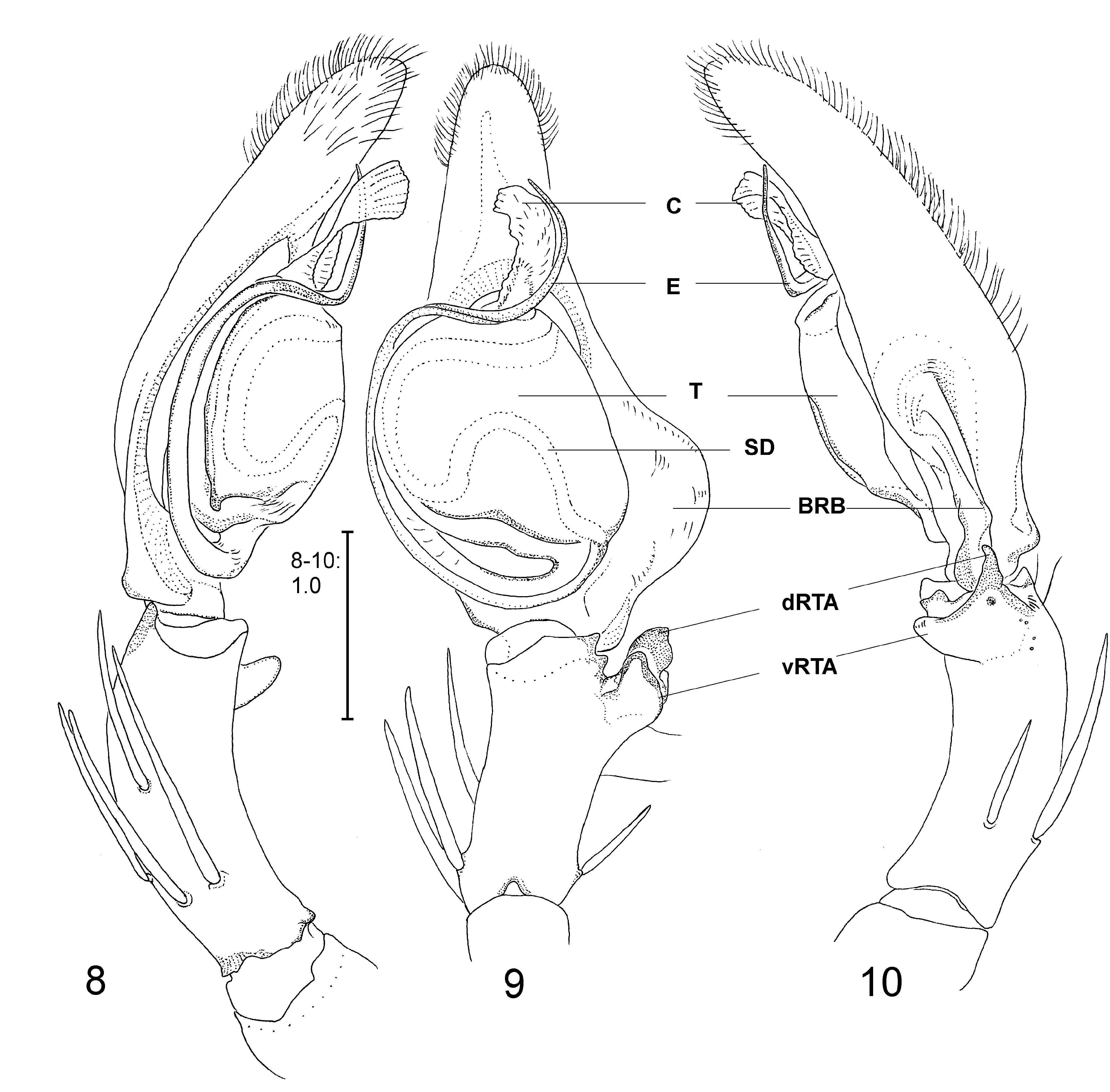
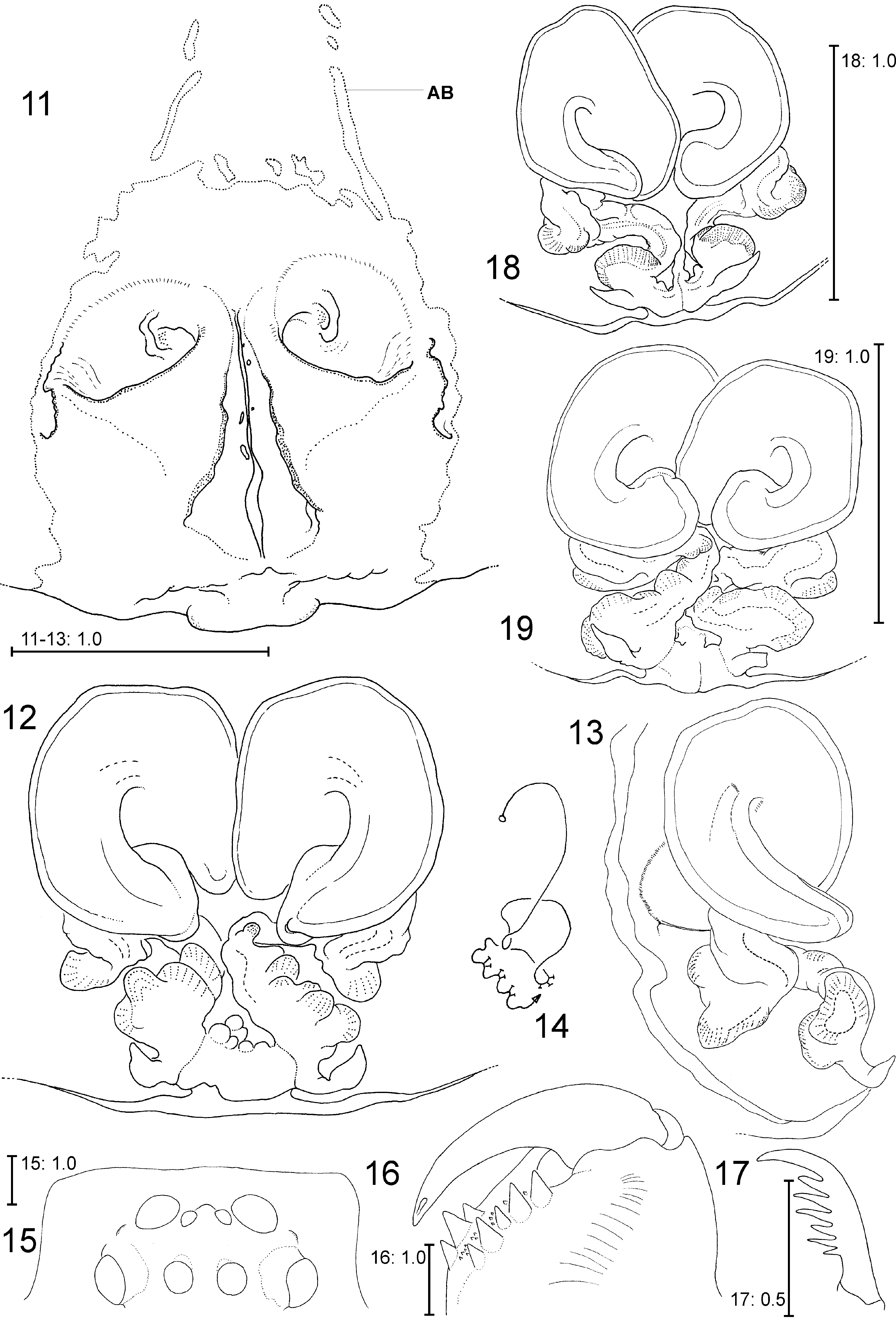
Diagnosis. The male of S. iranomaximus spec. nov. can easily be distinguished from other known males by the following characters: 1. Embolus longer than in other species, distinctly S-shaped, 2. Conductor elongated, c. three times longer than wide ( Figs 8–10 View FIGURES 8 – 10 ). Females can be distinguished from all other Spariolenus species by their prominent first coil of the internal duct system extending laterally beyond posterior parts ( Figs 12–13 View FIGURES 11 – 19 ).
Description. Male (n=2) [holotype first, with measurements of paratype in parentheses]
Males medium-sized. Total length: Prosoma length 6.3(4.6), prosoma width 5.3(4.1), anterior width of prosoma 3.8(2.3), opisthosoma length 7.4(3.8), opisthosoma width 4.8(2.6). Eyes: AME 0.30, ALE 0.65, PME 0.43, PLE 0.78, AME-AME 0.22, AME-ALE 0.05, PME-PME 0.22, PME-PLE 0.42, AME-PME 0.33, ALE-PLE 0.47, clypeus height at AME 0.75, clypeus height at ALE 0.42.
Chelicerae with 3 anterior and 5 posterior teeth. Cheliceral furrow with denticles (c. 10) but most of them close to the anterior teeth. Leg formula: 2143. Measurements of palp and legs: Palp 7.1 [3.5, 1.6, 2.0], I 42.5 [11.5, 4.0, 13.0, 11.3, 2.7], II 44.4 [12.8, 3.9, 14.4, 12.7, 2.6], III 36.2 [10.5, 3.5, 10.6, 9.6, 2.3], IV 38.6 [11.0, 3.3, 11.1, 10.6, 2.6].
Spination. Palp 131, 101, 2111; Legs: Femur I–III 323, IV 321; Patella I–II 0 0 1, III–IV 000; Tibia I–II 101(10), III 1118, IV 2126; Metatarsus I–II 0 0 0 4, III 1014, IV 3036.
Palp. palp as in diagnosis with Embolus long, tegulum broad and conductor large, cymbium longer than tibia; RTA distally bifid with branches of similar size; dRTA and vRTA subdistally broad and distally pointed ( Figs 8– 10 View FIGURES 8 – 10 ).
Female (n=3): Females very large (leg span and body length reaching 150 mm and 31.2 mm, respectively).
Prosoma length 8.4–13.0, prosoma width 7.8–12.1, anterior width of prosoma 4.6–8.2, opisthosoma length 11.5–18.2, opisthosoma width 8.3–13.1. Eyes of the largest female paratype: AME 0.46, ALE 1.11, PME 0.73, PLE 1.16, AME-AME 0.40, AME-ALE 0.12, PME-PME 0.54, PME-PLE 0.91, AME-PME 0.75, ALE-PLE 0.95, clypeus height at AME 1.59, clypeus height at ALE 0.94.
Chelicerae with 3 anterior and 5 posterior teeth; cheliceral furrow with denticles (c. 10) most of them close to the three anterior teeth ( Fig. 16 View FIGURES 11 – 19 ). Leg formula: 2143. Measurements of palp and legs: Palp 20.6 [6.4, 3.1, 4.3, 6.8], I 60.0 [16.6, 7.3, 16.9, 15.4, 3.8], II 63.8 [18.3, 7.5, 18.0, 16.5, 3.5], III 53.1 [15.7, 6.5, 14.6, 13.5, 2.8], IV 55.0 [15.3, 6.0, 15.4, 15.3, 3.0]. Female palpal claw with first tooth longer than secondary teeth, six secondary teeth present ( Fig. 17 View FIGURES 11 – 19 ).
Spination [of the largest female paratype]. Palp 131, 101, 2121, 1014; Legs: Femur I–II 423–433, III 323, IV 321–322; Patella I–II 101, III–IV 100–101; Tibia I–II 101(10)–201(10), III 1318, IV 2126; Metatarsus I–II 0 0 0 4, III 1014 –2014, IV 3036.
Epigyne/vulva. Copulatory organ as in diagnosis with epigynal field longer than wide; anterior band of epigynal field present, long and narrow may be separated or attached to the epigynal field, spirally coiled copulatory openings are broad; central epigynal rims extending laterally beyond the copulatory openings; anterior bands of epigynal field are long, narrow and attached to epigynal field; median slit long, posterior part of the median slit absent ( Fig. 11 View FIGURES 11 – 19 ); first windings of the internal duct system wide and long, flattened and with distinct margins; glandular pores present on parts of second coil but distinctly visible on the hump-like structures of third coil ( Figs 12– 13 View FIGURES 11 – 19 ).
Colouration. Yellowish brown with dark–brown spots and marking on prosoma and dorsal of opisthosoma. Legs with dark brown bands (Figs 41–42).
Variation. First windings of vulva overlapping partially each other in different ways including left copulatory duct overlapping the initial part of the left one ( Fig. 18 View FIGURES 11 – 19 ) or vice versa ( Fig. 19 View FIGURES 11 – 19 ). Third winding with only one large hump ( Fig. 18 View FIGURES 11 – 19 ) or with several small hump-like structures ( Fig. 12 View FIGURES 11 – 19 ).
Distribution. Known only from the type locality.
Life history and habitat preferences. One subadult male and several small juveniles were observed at the broad entrance passage of the cave while larger juveniles and adult spiders were encountered in the deeper parts and a huge corridor inside the cave which its walls reaching nearly 35 meters high. The spiders were observed facing downward on the walls. The cave was hot and humid. The cave floor was covered with a dense layer of bat guano. Khofash cave is an interesting cave system. It is a shelter of approximately 25–30,000 bats belonging to four different species (De Blase 1980, Sharifi personal communication). It was observed that unknown small coleopteran species feed on and live in the huge piles of bat guano carpeting the cave floor. It can be estimated that it is a good example of a “Bat Guano Ecosystem” as proposed by Paulson (1972). Several unknown species of insects belonging to the orders Blattodea and Coleoptera inhabit the cave. Representatives of Araneae were also observed on some pile of breakdowns on the cave floor, two species belong to the genera Spermophora Hentz, 1841 (Pholcidae) and Loxosceles Heineken and Lowe, 1832 (Sicariidae) . In one case a subadult S. iranomaximus spec. nov. was observed capturing and feeding on an unidentified large cockroach species (Blattodea) (Fig. 41). These large spiders might be at the end of the food chain among cave dwelling invertebrates and small guano consumers.
| SMF |
Forschungsinstitut und Natur-Museum Senckenberg |
No known copyright restrictions apply. See Agosti, D., Egloff, W., 2009. Taxonomic information exchange and copyright: the Plazi approach. BMC Research Notes 2009, 2:53 for further explanation.