
Litoblatta Hebard, 1921
|
publication ID |
https://doi.org/10.11646/zootaxa.4941.4.5 |
|
publication LSID |
lsid:zoobank.org:pub:C272B6E8-E85D-4946-8547-964588FC9A31 |
|
DOI |
https://doi.org/10.5281/zenodo.4595706 |
|
persistent identifier |
https://treatment.plazi.org/id/003B87FA-FF8A-FFC0-BCCB-F1DB2109F81E |
|
treatment provided by |
Plazi |
|
scientific name |
Litoblatta Hebard, 1921 |
| status |
|
Litoblatta Hebard, 1921 View in CoL
( Figs. 1–70 View FIGURES 1‒8 View FIGURES 9‒14 View FIGURES 15‒21 View FIGURES 22‒26 View FIGURES 27‒33 View FIGURES 34‒43 View FIGURES 44‒51 View FIGURES 52‒54 View FIGURES 55‒59 View FIGURES 60‒67 View FIGURES 68‒74 )
Ischnoptera Brunner von Wattenvyl, 1865: 130 View in CoL ( type species: Ischnoptera brasiliensis Brunner von Wattenvyl, 1865 View in CoL ) Litoblatta Hebard 1921: 237 View in CoL
Litoblatta Princis 1969: 876 View in CoL
Litoblatta Rocha e Silva Albuquerque & Aguiar 1975: 235 View in CoL
Diagnosis. Body surface scarcely coated with small hairs. Head under the pronotum, slightly exposed. Eyes and ocelli present, absent in cavernicolous species. The prominent eyes are broad dorsally near the vertex and thinner ventrally near the genae, character more conspicuous in females. Maxillar palpi with 5 segments, the 3 last articles are similar and longer than the basal two. Antennae as long as the body. Pronotum with anterior margin and pronounced posterolateral angles rounded, posterior margin convex, medially slightly projected.
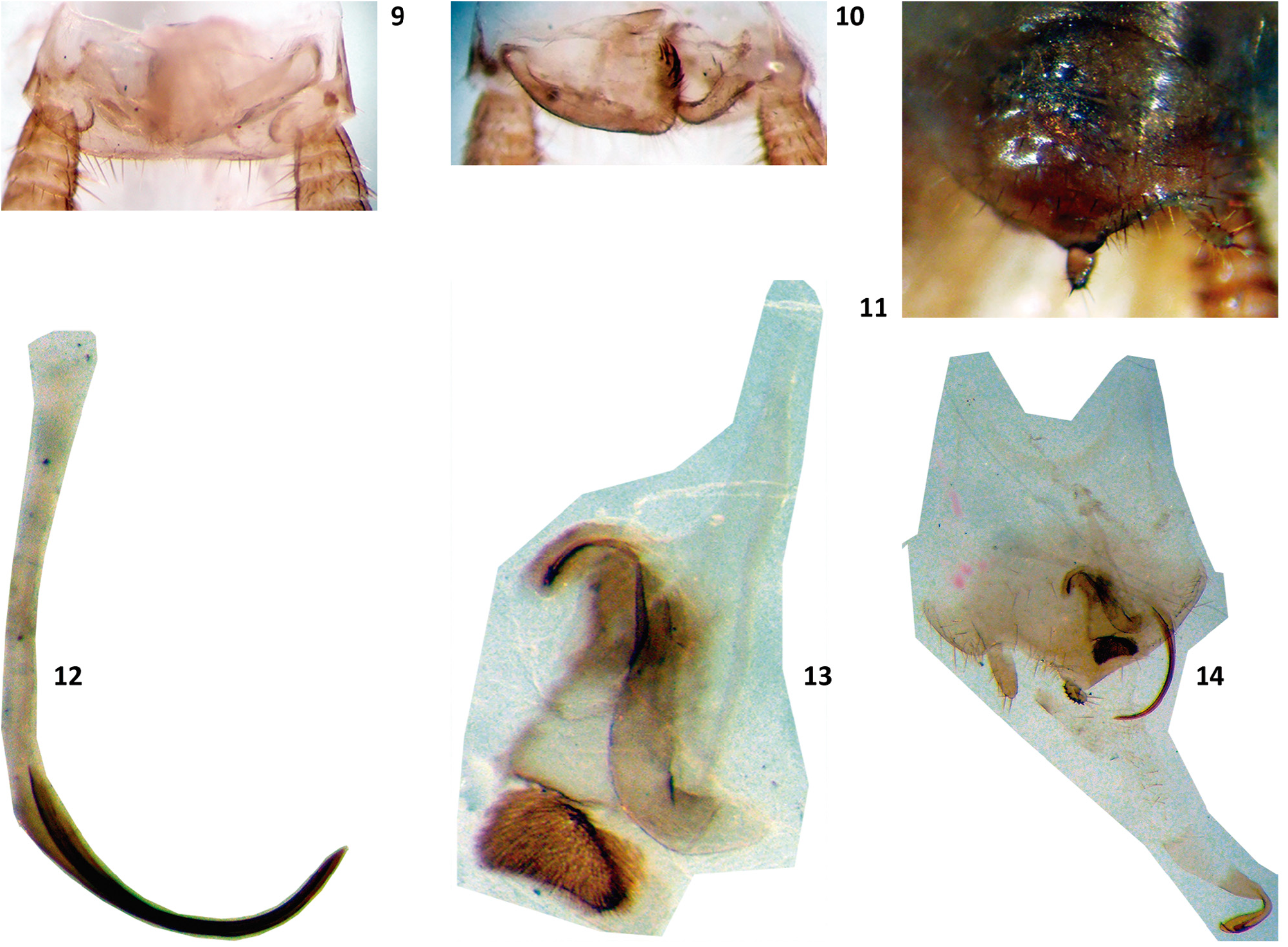
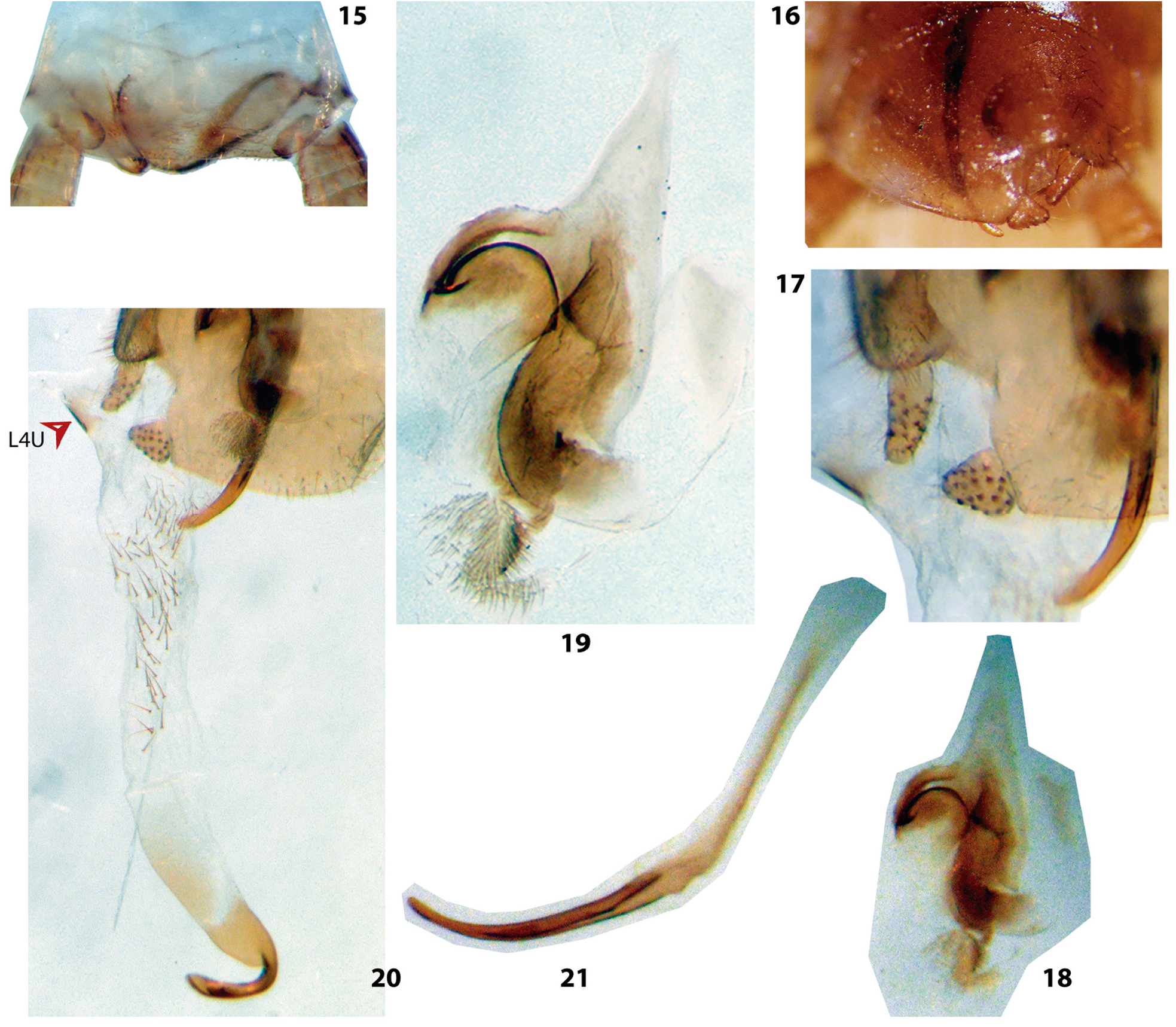
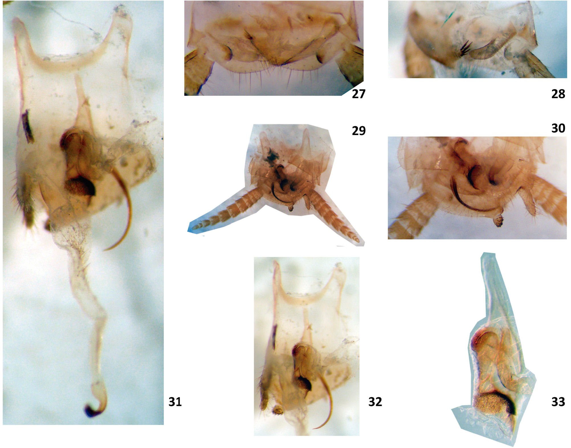
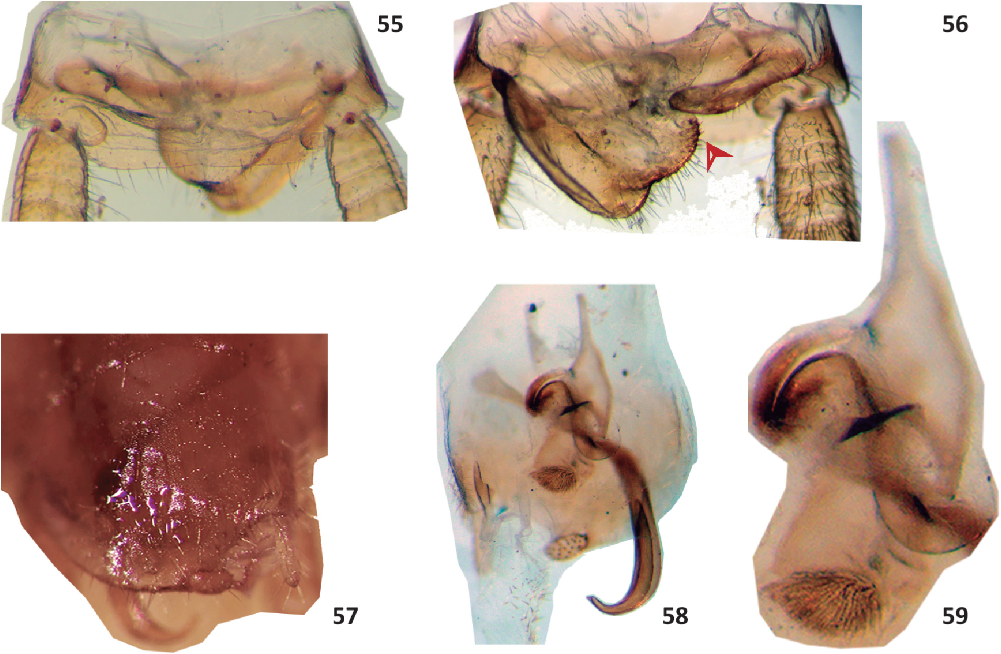
Legs very elongated and slender. Coxa with almost the length of the femur; femur 1, type A 3 – anteroventral margin with 3 long proximal spines, followed by a road of spines progressively shorter and ends with three long apical spines ( Roth 2003). Femur II and III with a genicular spine. Tibia II and III similar to the femur in length. Tarsus with four segments. Metatarsi longer or similar to the length of all apical tarsomeres added. Metatarsus, second and third tarsomeres with a double road of spines, fourth tarsomere with pulvilus. Pre-tarsus with symmetrical claws and arolium. Abdomen without specialized tergites. Supra anal plate distally may be produced, straight or sinuous. Female supra anal plate hidden under tergite VII. Paraprocts are laminar, symmetrical in females and asymmetrical in males. Male right paraproct may have short conical spines in the internal basal margin, long spines in a row along the inner border, very long and grouped spines, whose apices converge or diverge distally, placed somewhat beyond the inner basal border. Male left paraproct hook shaped .
Styli: right stylus with conical short spines with variable arrangement and the left stylus generally without spines.
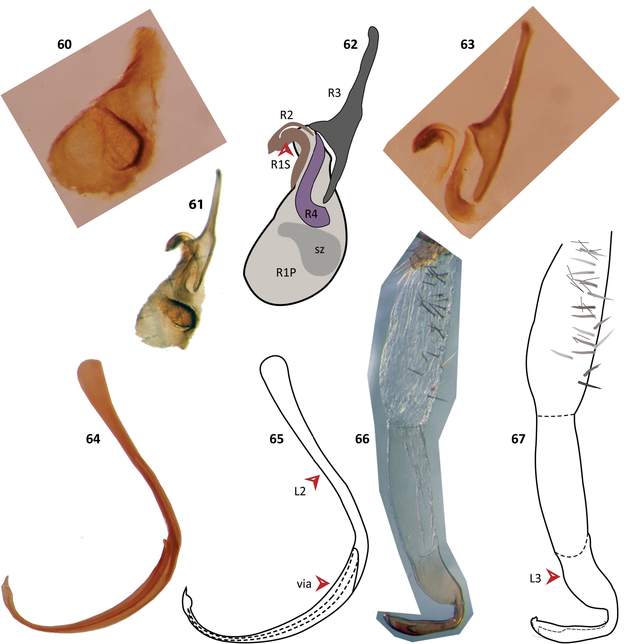
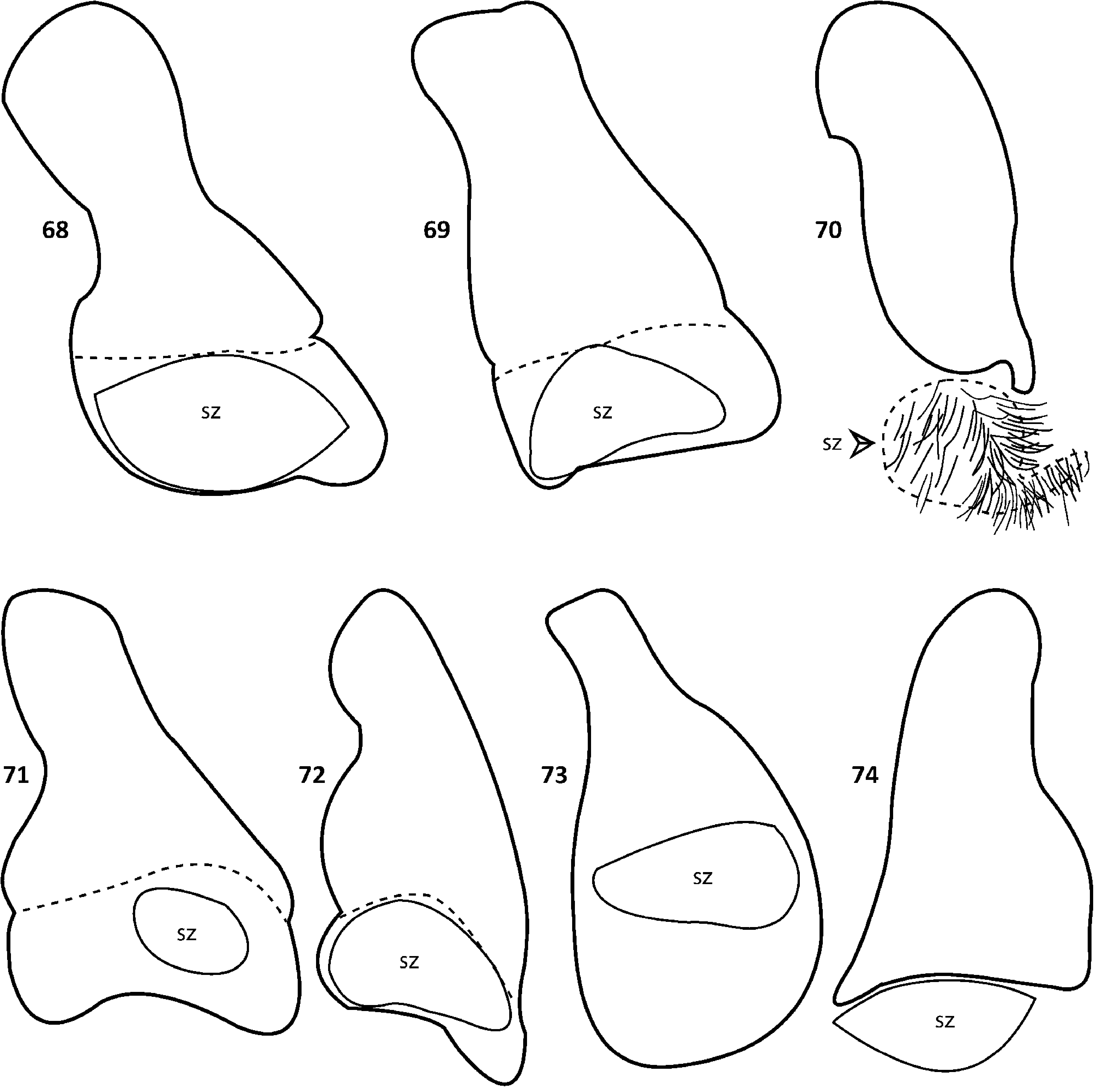
Male Genitalia ( Figs. 60–67 View FIGURES 60‒67 ). The left complex L is subdivided in L2 and L3 ( Gutiérrez 2005), L4U is also present ( Fig. 20 View FIGURES 15‒21 ). The right phallomere R is a complex of sclerites: R1P, R1S+R2, R3 and R4. R4 has the shape of a tobacco smoking pipe and articulates with R2. R1P articulates with R1S and includes caudally a convex scale zone, sz.
Female genitalia ( Figs. 52–54 View FIGURES 52‒54 ). The cerci bases are surrounded by tergum X (TX) laterally and followed by T IX. Tergal extensions of tergites VIII (tg) and IX (te) are in contact with gonocoxa VIII (first valvifer vf VIII + basivalvula bv). Gonangulum (gg) articulates with the tergal projections and the lateral part of the posterior lobe of second valvifer ring (pl) ( Klass, 1998). Internally the three pair of valves (v I, v II and v III) are observed.
The ventral chamber wall is covered by a membranous cuticle with folds. To see the spermathecal plate, the laterosternal shelf and the vestibular sclerite ( Fig. 52, 54 View FIGURES 52‒54 a-b) it was necessary to perform an additional dissection.
Oothecae ( Figs. 46–47 View FIGURES 44‒51 ). Sclerotized, lateral walls smooth, with a distinct keel along the dorsal margin. Egg chamber with 18– 20 eggs. Length 5.53–5.62 mm and height 3.44–3.61 mm. The alcohol where the oothecae were stored evaporated. Therefore, the shapes of the side walls, somehow collapsed as can be seen in the figures.


No known copyright restrictions apply. See Agosti, D., Egloff, W., 2009. Taxonomic information exchange and copyright: the Plazi approach. BMC Research Notes 2009, 2:53 for further explanation.
|
Kingdom |
|
|
Phylum |
|
|
Class |
|
|
Order |
|
|
Family |
|
|
SubFamily |
Blattellinae |
Litoblatta Hebard, 1921
| Valverde, Alejandra Del Carmen & Crespo, Francisco A. 2021 |
Litoblatta Rocha e Silva Albuquerque & Aguiar 1975: 235
| Silva Albuquerque, I. & Aguiar, G. M. 1975: 235 |
Litoblatta
| Princis, K. 1969: 876 |
Ischnoptera
| Hebard, M. 1921: 237 |