
Austroconops Wirth & Lee
|
publication ID |
https://doi.org/10.11646/zootaxa.3879.1.1 |
|
publication LSID |
lsid:zoobank.org:pub:6423894B-97D9-4286-ABB9-D4AF072B57FD |
|
DOI |
https://doi.org/10.5281/zenodo.5589199 |
|
persistent identifier |
https://treatment.plazi.org/id/027587C9-BD0A-3053-FD8A-18D84EDAE0F4 |
|
treatment provided by |
Felipe |
|
scientific name |
Austroconops Wirth & Lee |
| status |
|
Austroconops Wirth & Lee View in CoL View at ENA
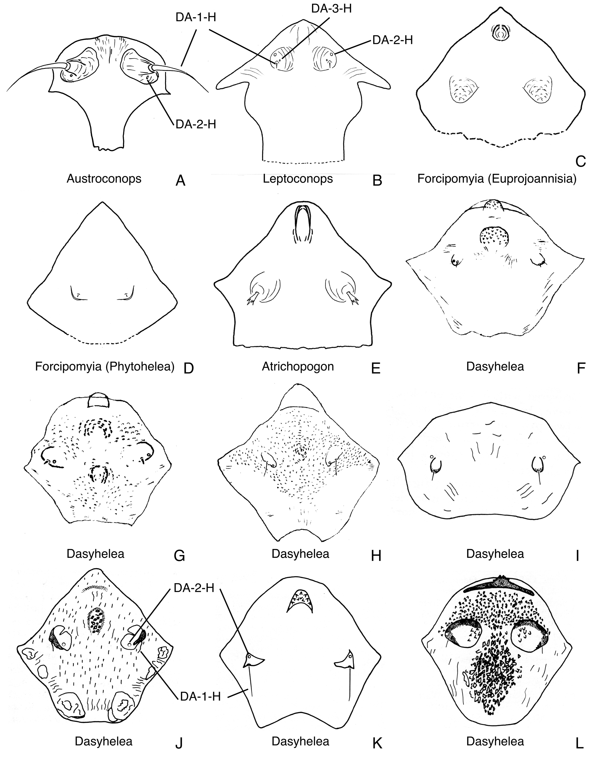
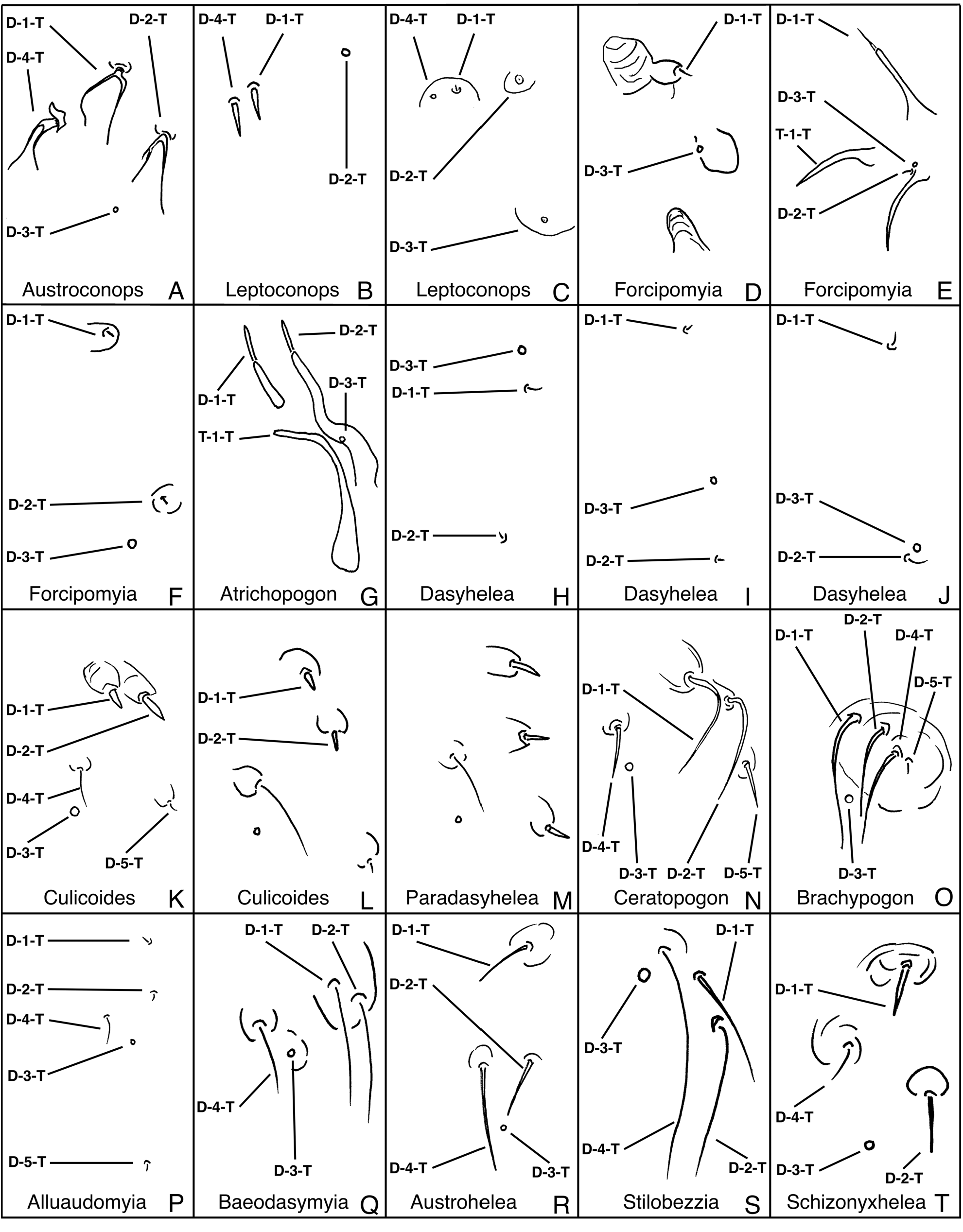
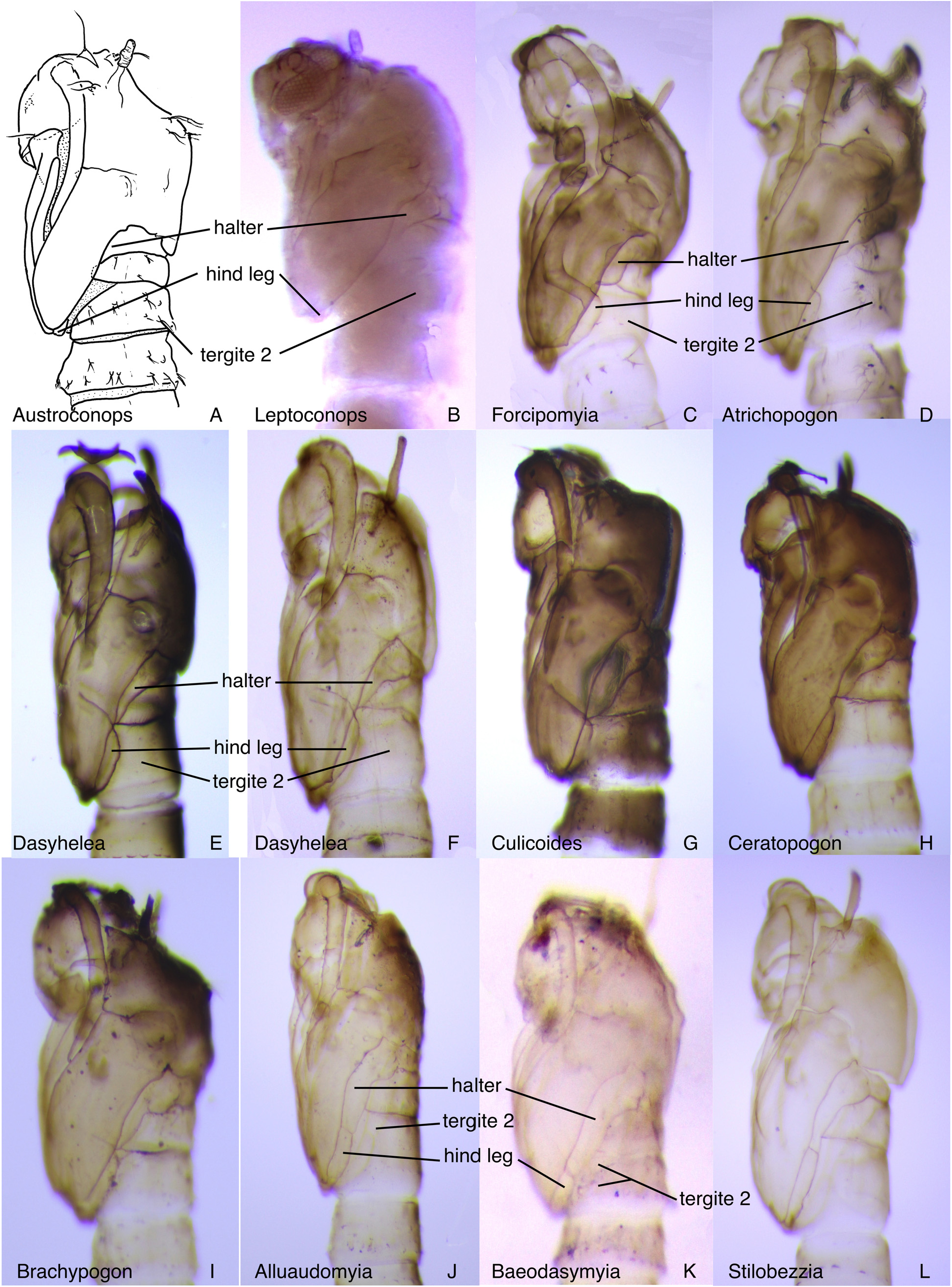
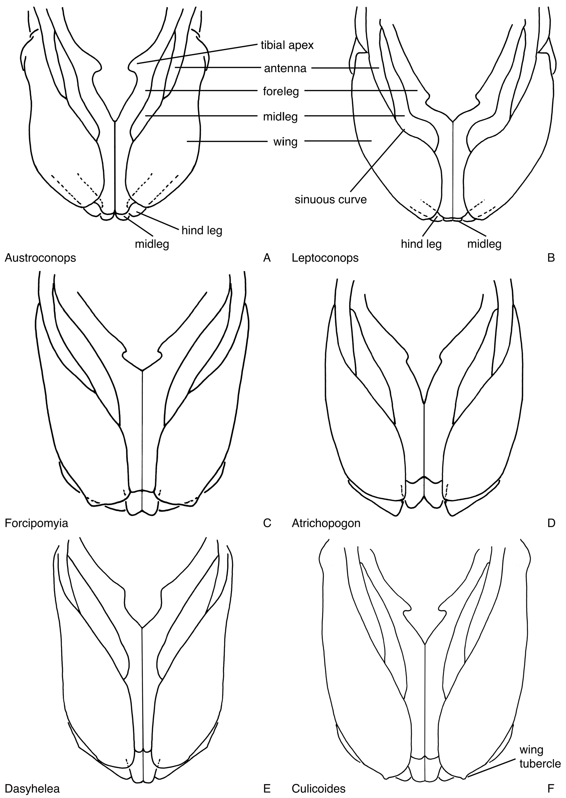
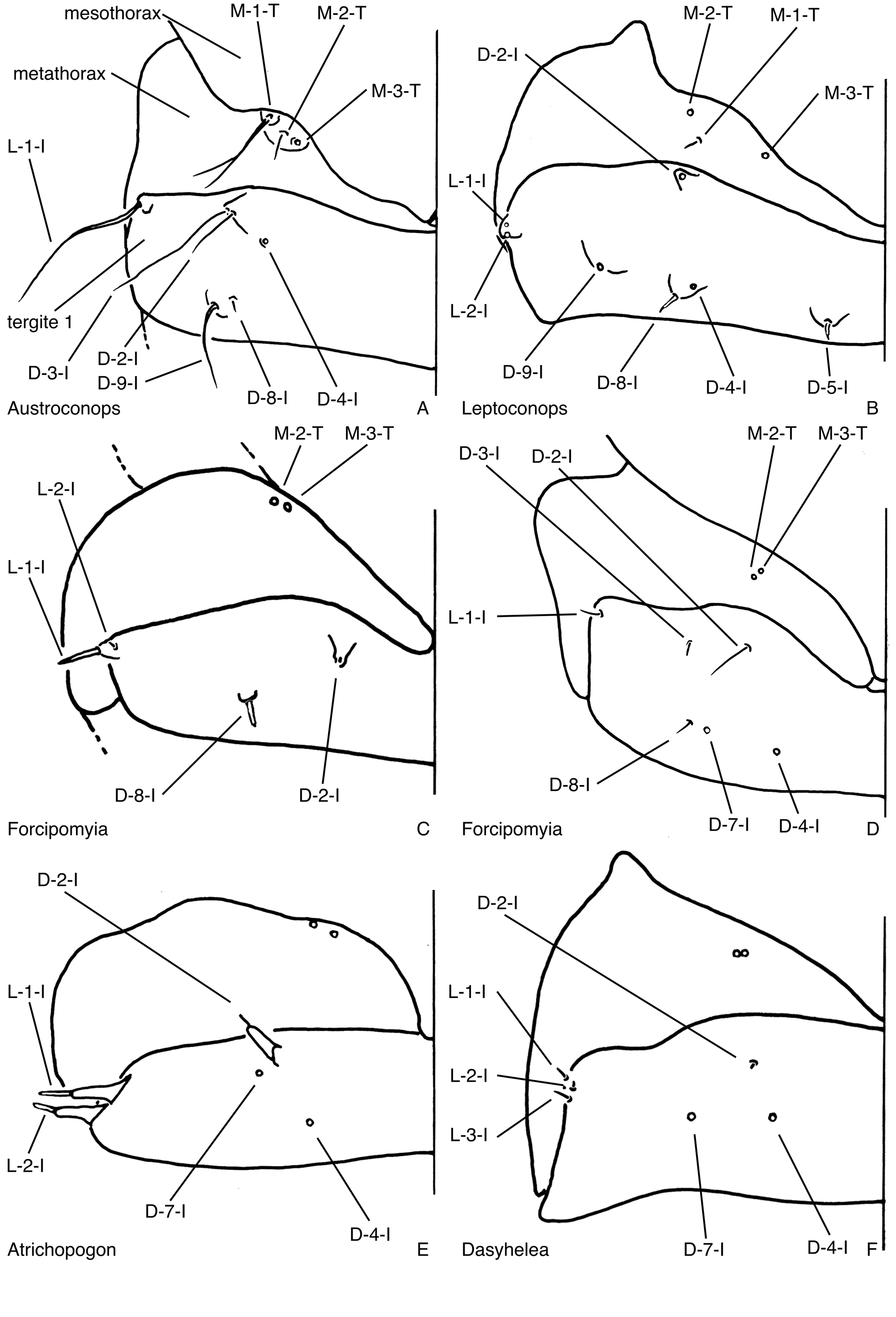
( Figs. 8A View FIGURE 8 , 14A View FIGURE 14 , 18A View FIGURE 18 , 23A View FIGURE 23 , 29A View FIGURE 29 , 32A View FIGURE 32 , 34A View FIGURE 34 , 42A–B View FIGURE 42 , 47A View FIGURE 47 , 55A View FIGURE 55 , 72A View FIGURE 72 )
DIAGNOSIS: Only pupa of Ceratopogonidae with pair of elongate dorsocephalic sclerite setae abutting the respiratory organ ( Fig. 8A View FIGURE 8 ); also unique with prothoracic extension restricted to lateral margin of palpus as narrow lobe ( Fig. 23A View FIGURE 23 ).
DESCRIPTION: Habitus as in Fig. 8A View FIGURE 8 . Total length = 1.84 mm. Without larval exuviae retained on abdomen. Exuviae with flagellum separate from lateral margin of face ( Fig. 14A View FIGURE 14 ). Ecdysial tear along anterior of and anteromedial to base of antenna, entire head capsule loose ( Figs. 14A View FIGURE 14 , 79A View FIGURE 79 ). Head: Dorsal apotome ( Fig. 18A View FIGURE 18 ), without ventral line of weakness, without dorsomedial tubercle, without central dome; dorsolateral cephalic sclerite (as in Fig. 13A View FIGURE 13 ) separated from scutum by thin cuticle, separate from scutum upon emergence, each side broadly meeting medially in whole pupa; mouthparts ( Fig. 23A View FIGURE 23 ) with mandible, lacinia well-developed, not overlapping apically; palpus extending anterior to posterolateral margin of labium; labium entire (not divided medially); apex of antenna ( Fig. 34A View FIGURE 34 ) anterior to posterior extent of midlength portion of midleg (portion lateral to mesosternum); sensilla: dorsal apotomals ( Fig. 18A View FIGURE 18 )—1 elongate seta, 1 campaniform sensillum; dorsolateral cephalic sclerite sensilla—2 elongate setae with apices laying against respiratory organ, 1 campaniform sensillum ( Fig. 8A View FIGURE 8 ); clypeal-labrals ( Fig. 23A View FIGURE 23 )—1 elongate seta (campaniform sensillum, if present, invisible on wrinkled cuticle); oculars ( Fig. 23A View FIGURE 23 )—2 elongate setae, one longer than other. Thorax: Prothoracic extension ( Fig. 23A View FIGURE 23 ) restricted to lateral margin of palpus as narrow lobe; mesonotum without tubercles, extending posteromedially, nearly dividing metathorax medially ( Fig. 47A View FIGURE 47 ); respiratory organ ( Figs. 42A–B View FIGURE 42 ) length/width = 3.27, moderately elongate, somewhat flattened laterally, with pores closely abutting at apex of respiratory organ, arranged in single row, outer surface with some wrinkles, with elongate conical pedicel, base without posteromedial apodeme, membranous base of respiratory organ elongate, annulated, tracheal tube slightly sinuous, with spirals extending just over half length, distally smooth; wing ( Fig. 34A View FIGURE 34 ) without apical tubercle or angle, separated medially by fore-, midlegs; halter apex and hind leg ( Fig. 32A View FIGURE 32 ) broadly separate; halter apex extending to about half length of tergite 1; legs ( Fig. 34A View FIGURE 34 ) with lateral margin of foreleg near midlength of wing somewhat sinuous; hind leg not visible at lateral margin of wing ( Fig. 32A View FIGURE 32 ); with apex of foreleg ventral to apex of midleg; apex of hind leg abutting apex of midleg laterally; sensilla: anteromedials—1 very minute seta; anterolaterals—2 elongate, 1 short setae; dorsal setae ( Fig. 29A View FIGURE 29 )—D- 1-T, D-2-T, D-4-T bifurcating setae, D-3-T campaniform sensillum, D-3-T, if present, posteromedial to D-4-T; supraalar 2—elongate seta ( Fig. 8A View FIGURE 8 ); metathoracics ( Fig. 47A View FIGURE 47 )—2 setae, 1 campaniform sensillum; M-3-T near anterior margin of metathorax. Abdomen: without pigmentation pattern, segments 2, 3 equally or nearly equally wide, segments with elongate, bifurcating setae, with rounded to pointed, short to moderately elongate tubercles, tergites or sternites entire, each without membranous disc; segment 9 ( Fig. 72A View FIGURE 72 ) not strongly modified, terminal processes widely separated basally, each projecting posterodorsolaterally with apex directed dorsally, tapering to pointed apex; sensilla: tergite 1 ( Fig. 47A View FIGURE 47 ) with 5 setae, 1 campaniform sensillum, including 1 lateral sensillum, D- 2-I, D-3-I on shared tubercle, D-7-I absent; segment 4 ( Fig. 55A View FIGURE 55 )—D-2-IV, D-3-IV elongate, bifid setae on single tubercle; D-5-IV, D-8-IV, D-9-IV bifurcating elongate setae on short, separate tubercles, posterior dorsal sensilla in transverse row, arranged medially to laterally: D-5-IV, D-8-IV, D-7-IV, D-9-IV; D-4-IV positioned medial to D-3- IV; L-1-IV elongate seta on short, pointed tubercle, well anterior of posterior lateral setae, L-2-IV, L-3-IV, L-4-IV bifid setae on short pointed tubercles, V-5-IV, V-6-IV, V-7-IV bifid setae, V-5-IV, V-6-IV on elongate tubercles, V- 7-IV on short tubercle; segment 8 without D-3-VIII, with L-1-VIII; segment 9 ( Fig. 72A View FIGURE 72 )—with D-5-IX, D-6-IX campaniform sensilla.
DISTRIBUTION AND HABITAT: The genus Austroconops , once broadly distributed in the Cretaceous, is now known from two species from southwestern Australia ( Borkent & Craig 2004). Although immmatures are unknown in nature, the larvae moved snake-like through very wet substrate in the laboratory, clearly indicating that their natural habitat must be subaquatic or aquatic. It would be valuable to search for larvae and pupae at Yanchep National Park, in Western Australia, where a large adult population of A. mcmillani lives with this in mind. The behaviour of the lethargic pupa was described by Borkent & Craig (2004).
TAXONOMIC DISCUSSION: Borkent & Craig (2004) described the pupa of A. mcmillani from a single pupal exuviae, reared from an egg, and this is the only known specimen of the genus. Borkent & Craig (2004) missed D-3-T and D-4-I (both campaniform sensilla), identified the closely approximated sensilla D-2-IV and D-3- IV as a single seta (as dasm ii) and missed the minute D-1-IV.
MATERIAL EXAMINED: A. mcmillani : 1 pupal exuviae, Yanchep National Park, Western Australia, Australia, reared from egg from female collected 20-XI-2001 (CNCI).


No known copyright restrictions apply. See Agosti, D., Egloff, W., 2009. Taxonomic information exchange and copyright: the Plazi approach. BMC Research Notes 2009, 2:53 for further explanation.
|
Kingdom |
|
|
Phylum |
|
|
Class |
|
|
Order |
|
|
Family |
|
|
SubFamily |
Leptoconopinae |