
Paradasyhelea Macfie
|
publication ID |
https://doi.org/ 10.11646/zootaxa.3879.1.1 |
|
publication LSID |
lsid:zoobank.org:pub:6423894B-97D9-4286-ABB9-D4AF072B57FD |
|
DOI |
https://doi.org/10.5281/zenodo.5589805 |
|
persistent identifier |
https://treatment.plazi.org/id/027587C9-BD1D-3043-FD6A-1D2149A7E3D4 |
|
treatment provided by |
Felipe |
|
scientific name |
Paradasyhelea Macfie |
| status |
|
Paradasyhelea Macfie View in CoL
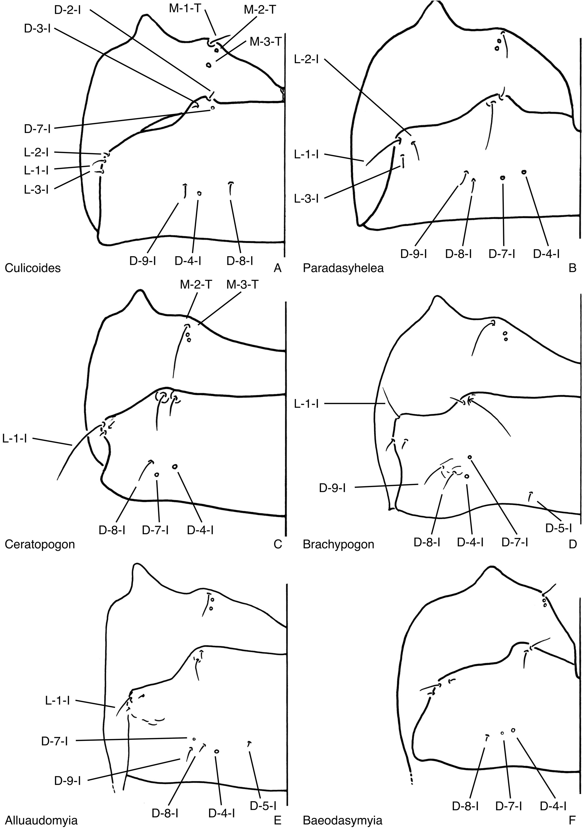
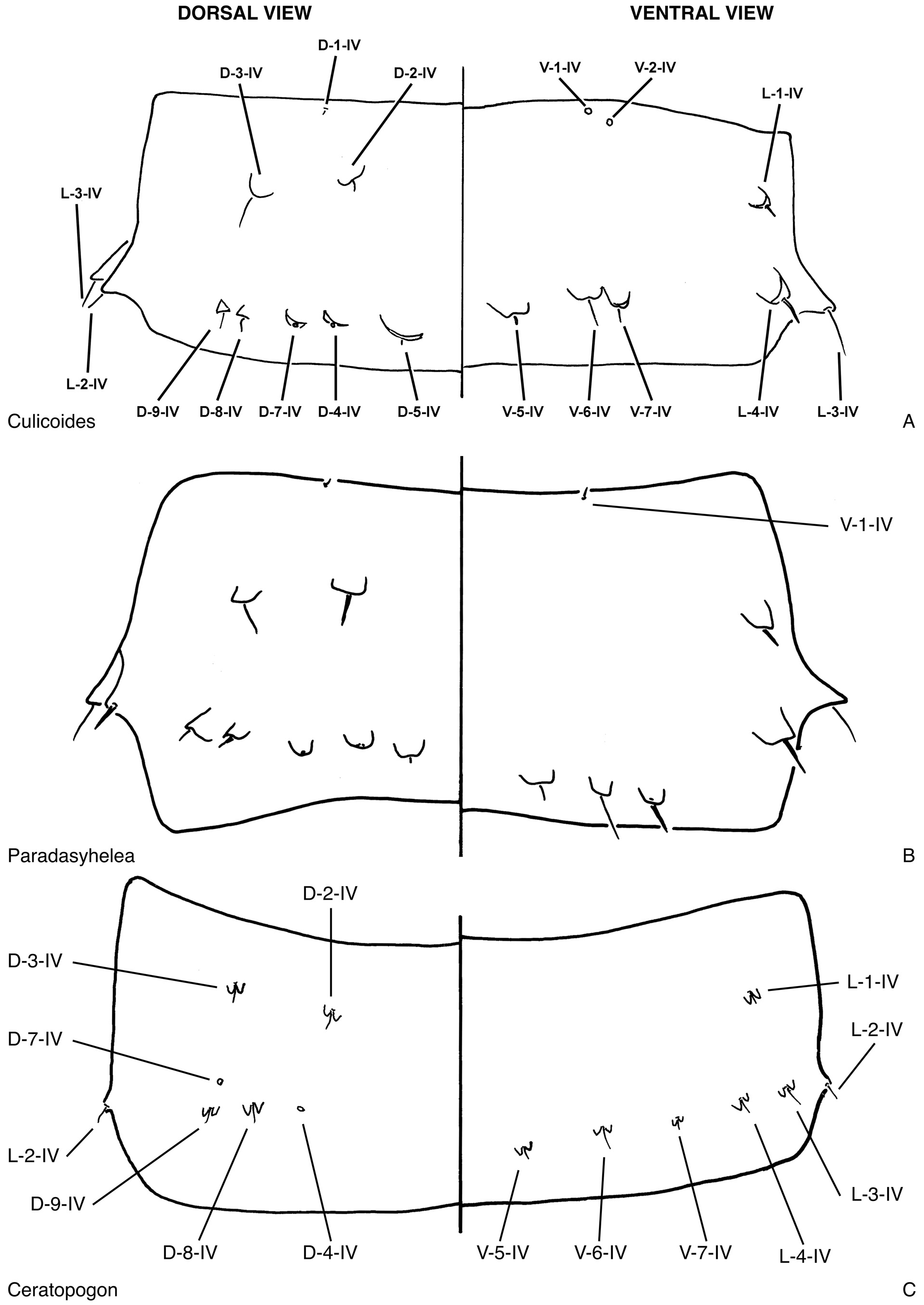
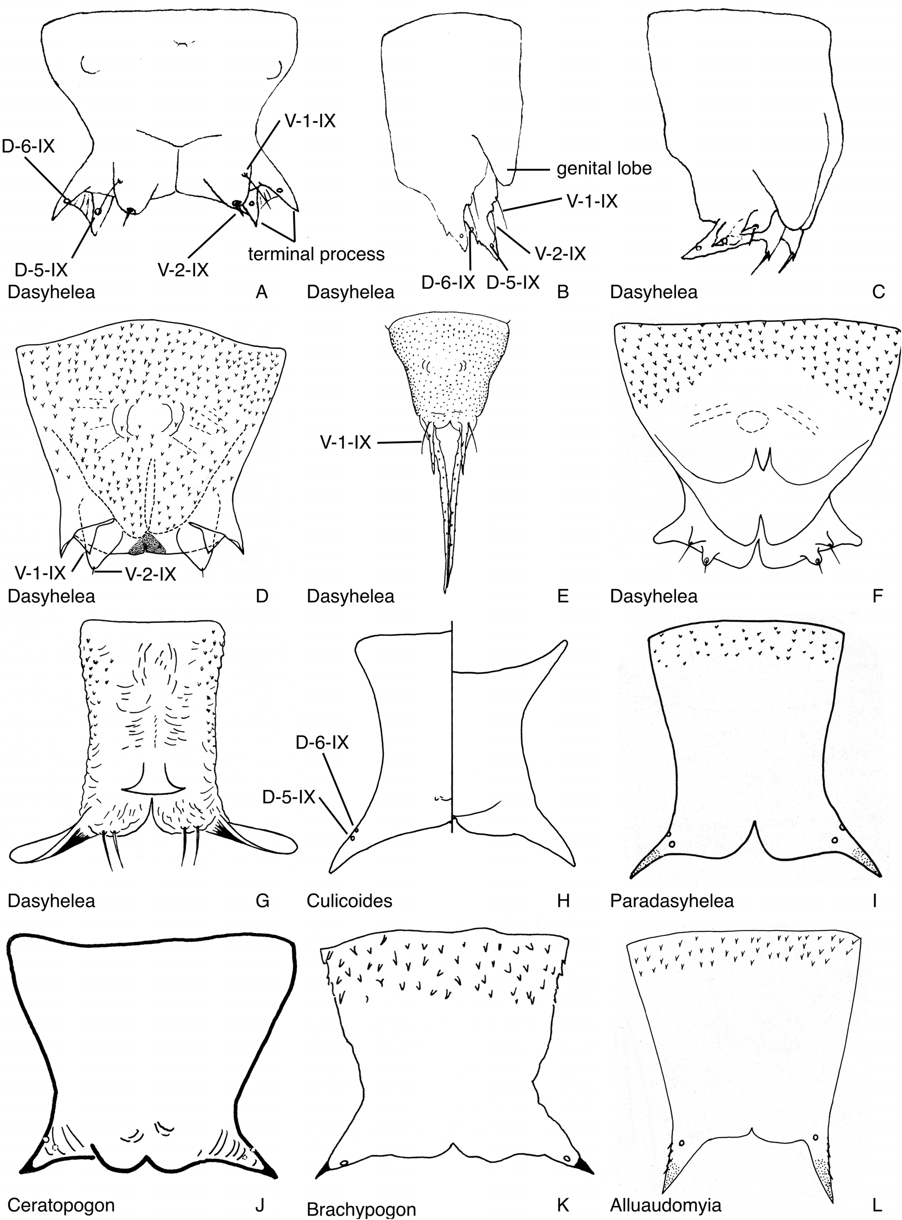
( Figs. 19E–G View FIGURE 19 , 24B View FIGURE 24 , 29M View FIGURE 29 , 35A View FIGURE 35 , 43O View FIGURE 43 , 48B View FIGURE 48 , 57B View FIGURE 57 , 73I View FIGURE 73 )
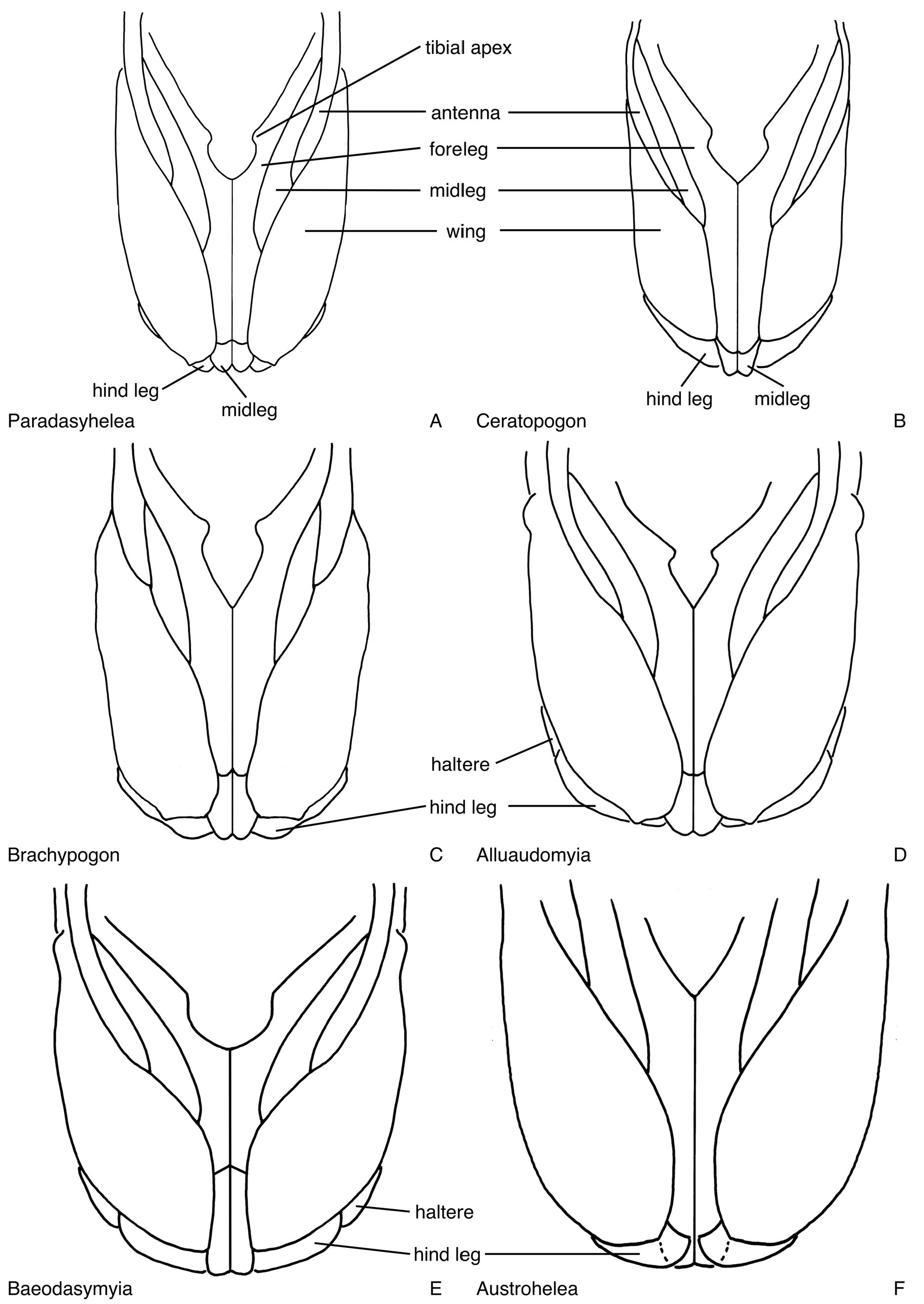
DIAGNOSIS: Only pupa of Ceratopogonidae with a prothoracic extension extending from the palpus to the antenna ( Fig. 24B View FIGURE 24 ), the halter and hind leg are slightly separated or barely touching (as in Fig. 32G View FIGURE 32 ), and the dorsal apotome ( Figs. 19E–G View FIGURE 19 ) has a lateral row of stout, pointed spicules and without further stout spicules more medially.
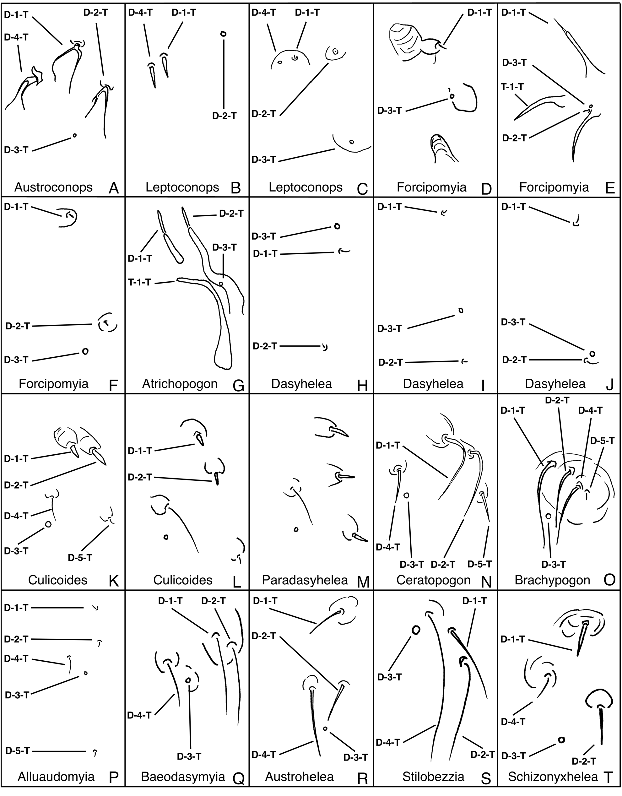
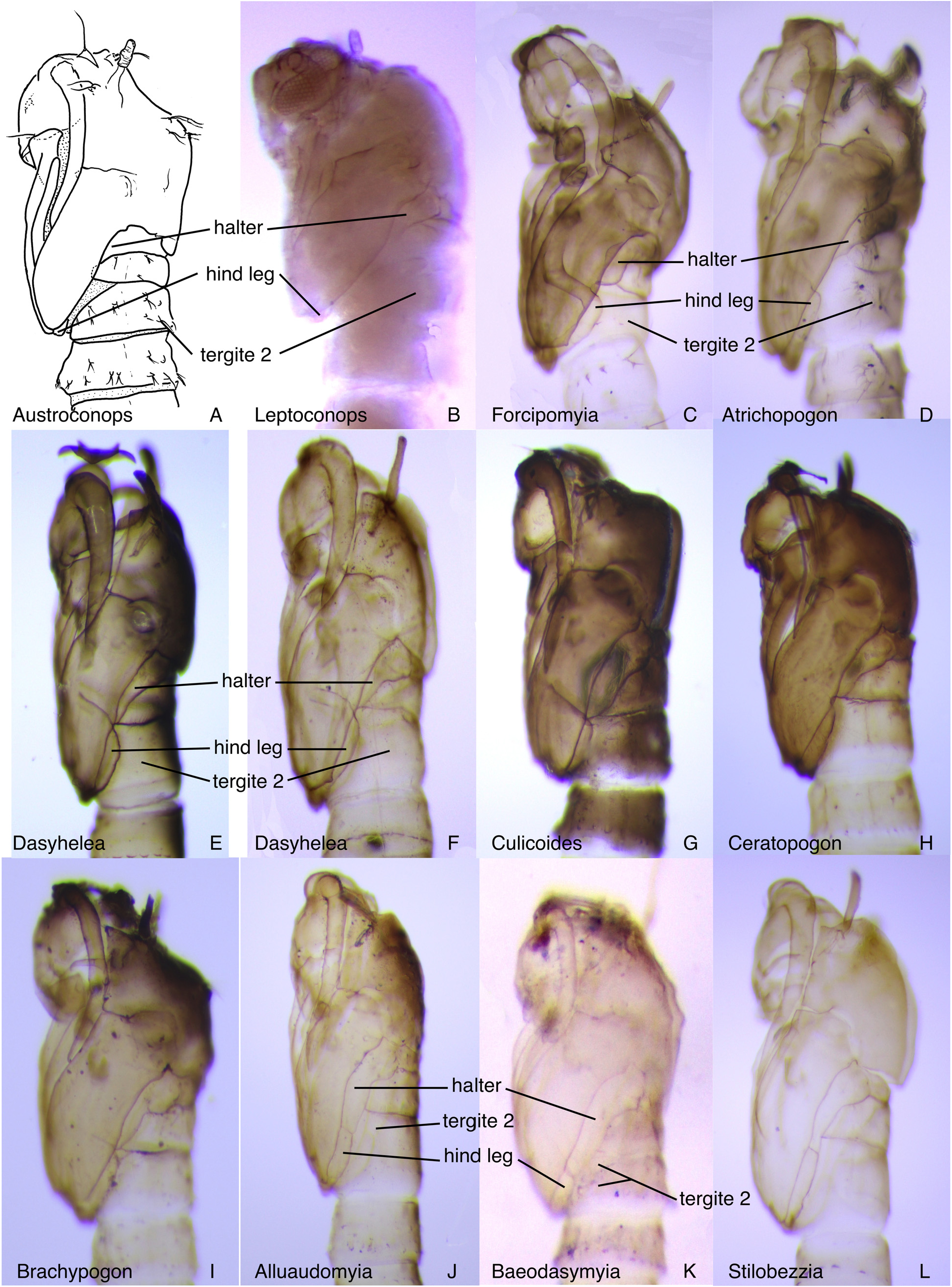
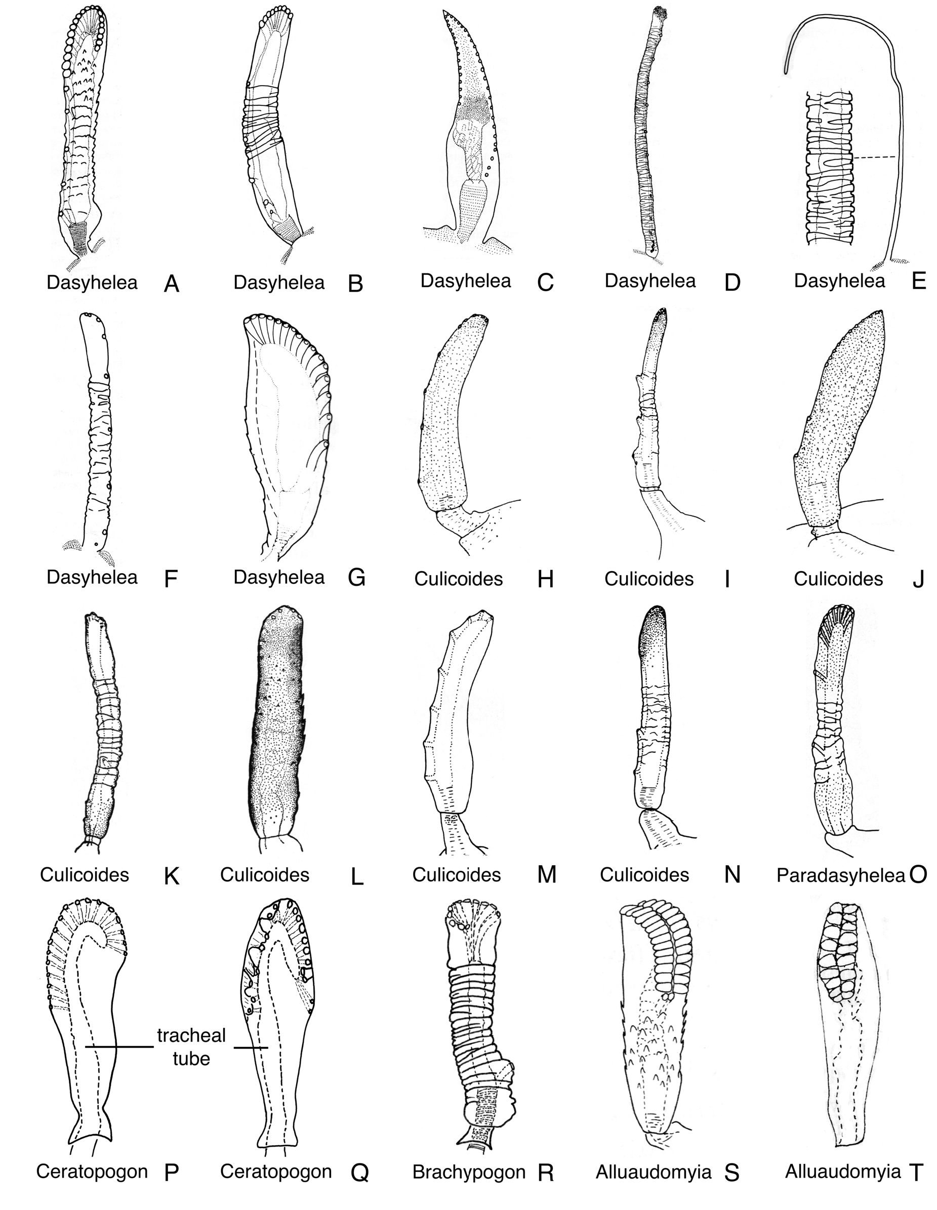
DESCRIPTION: Total length = 1.50–2.09 mm. Without larval exuviae retained on abdomen. Exuviae with flagellum appressed against lateral margin of face (as in Fig. 15A View FIGURE 15 ). Ecdysial tear medial to base of antenna (as in Figs. 15A View FIGURE 15 , 79D View FIGURE 79 ); along prothoracic extension. Head: Dorsal apotome ( Figs. 19E–G View FIGURE 19 ), without ventral line of weakness, without dorsomedial tubercle, without central dome; dorsolateral cephalic sclerite (as in Fig. 13B View FIGURE 13 ) separated from scutum by thin cuticle, separate from scutum upon emergence, each side separated medially by dorsal apotome in whole pupa; mouthparts ( Fig. 24B View FIGURE 24 ) with mandible, lacinia well-developed, overlapping; palpus extending equal to or just posterior to posterolateral margin of labium; labium separated medially by labrum, hypopharynx; apex of antenna ( Fig. 35A View FIGURE 35 ) anterior to posterior extent of midlength portion of midleg (portion lateral to mesosternum); sensilla: dorsal apotomals ( Figs. 19E–G View FIGURE 19 )—1 moderate to elongate seta, 1 campaniform sensillum; dorsolateral cephalic sclerite sensilla—2 setae, 1 campaniform sensillum; clypeal-labrals ( Fig. 24B View FIGURE 24 )—2 elongate setae; oculars ( Fig. 24B View FIGURE 24 )—2 elongate setae. Thorax: Prothoracic extension ( Fig. 24B View FIGURE 24 ) wide, welldeveloped, extending from palpus to antenna; mesonotum with short tubercles, extending posteromedially, completely dividing metathorax medially ( Fig. 48B View FIGURE 48 ); respiratory organ ( Fig. 43O View FIGURE 43 ) length/width = 6.50–7.06, elongate, slender, circular in cross-section, with pores closely abutting or slightly separated at apex of respiratory organ, arranged in single row, with additional, more basal pores, outer surface with annulations, without other surface modifications, with elongate, slender pedicel, base with short posteromedial apodeme, membranous base of respiratory organ short, tracheal tube straight to slightly curved along length, with spirals restricted to base, distally smooth or plates; wing ( Fig. 35A View FIGURE 35 ) with apical tubercle lateral to apex of hind leg, separated medially by fore-, midlegs; halter apex and hind leg (as in Fig. 32G View FIGURE 32 ) just separate; halter apex abutting anterolateral knob-like extension of tergite 2; legs ( Fig. 35A View FIGURE 35 ) with lateral margin of foreleg near midlength of wing evenly curved; hind leg visible at lateral margin of wing (as in Fig. 32G View FIGURE 32 ); with apex of foreleg moderately anterior to apex of midleg; apex of hind leg slightly dorsal to, partially abutting apex of midleg laterally; sensilla: anteromedials—1 seta; anterolaterals—2 setae, 1 campaniform sensillum; dorsal setae ( Fig. 29M View FIGURE 29 )—D-1-T, D-2-T, D-4-T, D-5-T setae, D- 3-T campaniform sensillum, D-3-T posterior to D-4-T; supraalar 2—campaniform sensillum; metathoracics ( Fig. 48B View FIGURE 48 )—1 seta, 2 campaniform sensilla; M-3-T near anterior margin of metathorax. Abdomen: without pigmentation pattern, segment 2 as wide or slightly wider than segment 3, segments with undivided, thin to thick setae, with rounded to pointed, short tubercles, tergites or sternites entire, each without membranous disc; segment 9 ( Fig. 73I View FIGURE 73 ) not strongly modified, terminal processes closely approximated basally, each projecting posterodorsolaterally, tapering to pointed apex; sensilla: tergite 1 ( Fig. 48B View FIGURE 48 ) with 7 setae, 1 or 2 campaniform sensilla, including 3 lateral sensilla, D-2-I, D-3-I closely approximated, D-7-I situated posteriorly near D-8-I; segment 4 ( Fig. 57B View FIGURE 57 )—D-2-IV, D-3-IV short to moderately elongate setae on short tubercles; D-5-IV, D-8-IV, D-9- IV short setae, D-5-IV, D-4-IV, D-7-IV, D-8-IV, D-9-IV on short, separate tubercles, posterior dorsal sensilla in transverse row, arranged medially to laterally: D-5-IV, D-4-IV, D-7-IV, D-8-IV, D-9-IV; L-1-IV short seta on short tubercle, well anterior of posterior lateral setae; L-2-IV, L-3-IV, L-4-IV moderately elongate setae, on short to pointed tubercles, V-5-IV, V-6-IV, V-7-IV short to moderately elongate setae on rounded tubercles; segment 8 without D-3-VIII, without L-1-VIII; segment 9 ( Fig. 73I View FIGURE 73 )—with D-5-IX, D-6-IX campaniform sensilla.
DISTRIBUTION AND HABITAT: The genus Paradasyhelea is known from 11 species in the Southern Hemisphere from New Caledonia, Australia, New Zealand, Chile and Argentina and (one species) in the Olympic Peninsula of the northwest USA ( Borkent 2014). Immatures have been found in the sandy, loamy or muddy margins of creeks or rivers, bogs, or swamps. There is one record from the muddy margin of a lake ( Kettle & Elson 1975a).
TAXONOMIC DISCUSSION: Three species of Paradasyhelea are known as pupae, all from Australia ( Tables 2–3 View TABLE 2 View TABLE 3 ). Elson-Harris & Kettle (1985a) provide a key to these species (republished by Elson-Harris & Murray (1992)) and suggested some features which would distinguish Paradasyhelea pupae from those of Culicoides but I could not confirm these differences as being consistently different between the two genera.
MATERIAL EXAMINED: P. albipunctata : 6 pupal exuviae (of paratypes), Kiandra, New South Wales, Australia, 19-XII-1956 (USNM); 1 pupal exuviae (of paratype), Oxford Falls, New South Wales, Australia, 6-XII- 1956 (USNM), 1 pupal exuviae, as previous locality, 10-XI-1956 (USNM); 1 pupal exuviae (of paratype), McCarr's Creek, New South Wales, Australia, 20-IX-1956 (USNM); 2 pupal exuviae (of paratype), as previous locality, 11-XI-1956 (ANIC); 1 pupal exuviae, Colo Vale, New South Wales, Australia, 17-I-1957 (USNM); 1 pupal exuviae (of paratype), Middle Creek, Narrabeen, New South Wales, Australia, 4-XI-1956 (ANIC); 1 pupal exuviae (of paratype), as previous locality, 8-IX-1956 (ANIC). P. minuta : 1 pupal exuviae (of paratype), South Creek, Deewhy, New South Wales, Australia, 27-IX-1956 (USNM); 2 pupal exuviae (of paratypes), Middle Creek, Narrabeen, New South Wales, Australia, 12-IX-1956 (USNM); 1 pupal exuviae (of paratype), Galston Gorge, New South Wales, Australia, 6-IX-1956 (USNM); 1 pupal exuviae, Colo Vale, New South Wales, Australia, 7-III-1957 (ANIC). P. sp: 18 pupal exuviae, S. Manjimup, Western Australia, Australia, 30-X-1985 (ANIC); 3 pupal exuviae, Gap Creek, Mittagong, New South Wales, Australia, 10-II-1966 (ANIC); 2 pupal exuviae, McCarr's Creek, New South Wales, Australia, 8-II-1966 (ANIC); 1 pupa (in glycerin), Borlase Stream, Lake Rotoiti, St. Arnaud, Nelson Lakes National Park, 41°48'52"S 172°51'55"E, South Island, New Zealand, 1–2-II-2000 (CNCI).


No known copyright restrictions apply. See Agosti, D., Egloff, W., 2009. Taxonomic information exchange and copyright: the Plazi approach. BMC Research Notes 2009, 2:53 for further explanation.
|
Kingdom |
|
|
Phylum |
|
|
Class |
|
|
Order |
|
|
Family |