
Paramesanophrys typica, Pan & Fan & Gao & Chen, 2016
|
publication ID |
https://doi.org/10.5852/ejt.2016.191 |
|
DOI |
https://doi.org/10.5281/zenodo.3852226 |
|
persistent identifier |
https://treatment.plazi.org/id/03812658-FFFF-6E65-FDA6-2C34FCDCFE36 |
|
treatment provided by |
Valdenar |
|
scientific name |
Paramesanophrys typica |
| status |
gen. et sp. nov. |
Paramesanophrys typica gen. et sp. nov.
urn:lsid:zoobank.org:act:
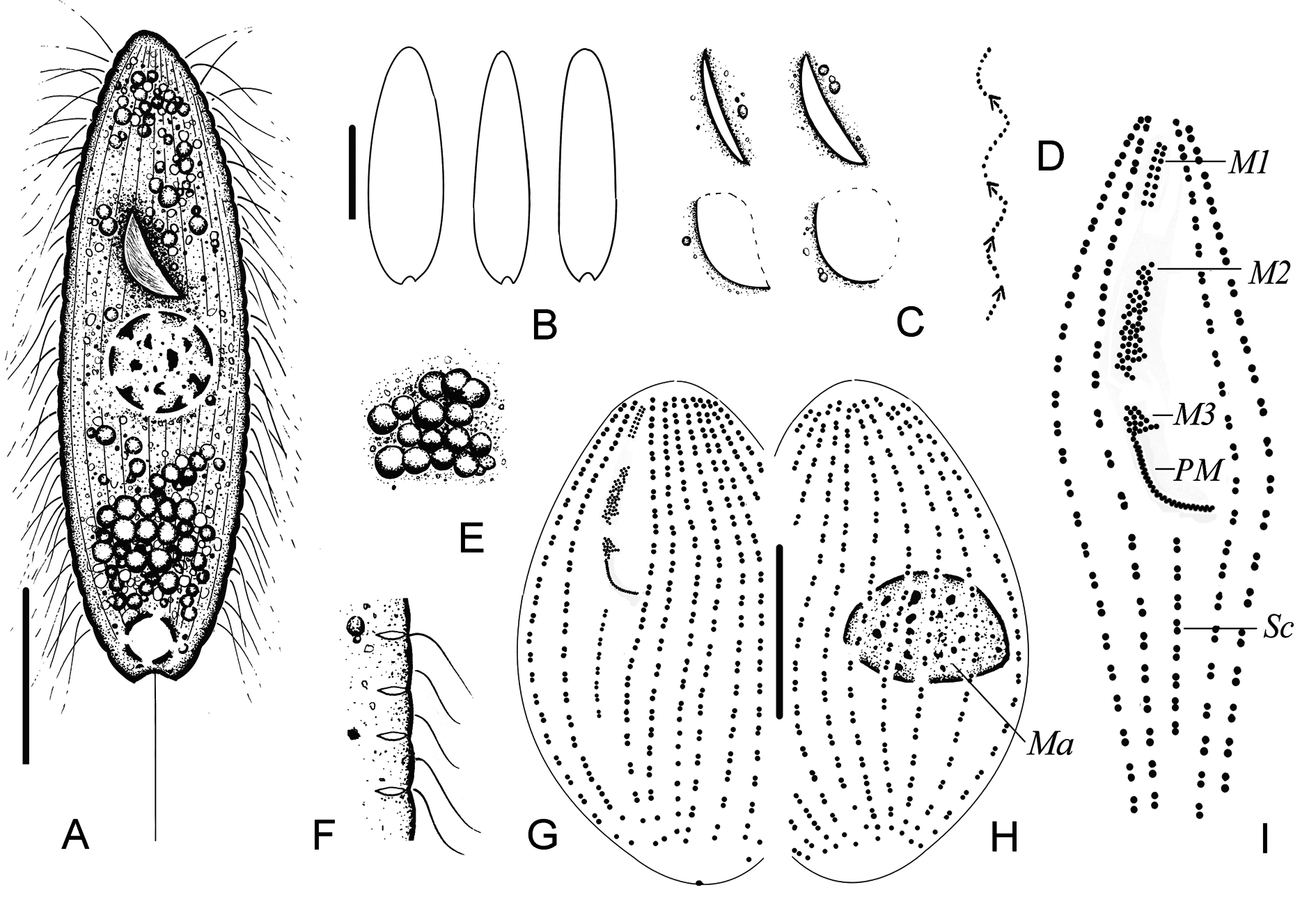
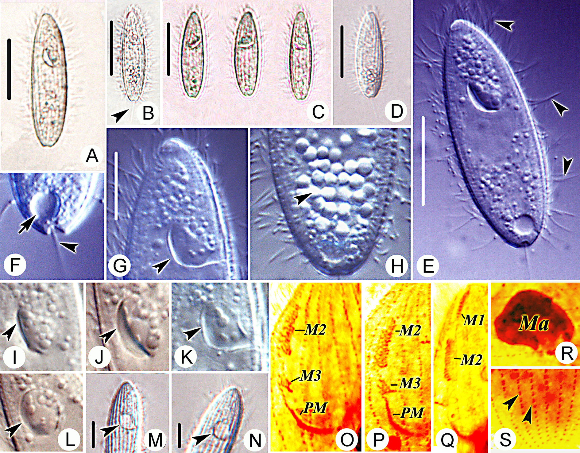
Figs 2–3 View Fig View Fig , 4A View Fig ; Table 2
Diagnosis
Size in vivo about 90–100 × 25–35 µm, elongate body, with pointed anterior end and narrowly rounded caudal end; posterior end distinctly depressed where caudal cilium located; buccal field approximately 40% of body length; 20 or 21 somatic kineties; M1 with 8–10 basal bodies in each kinety; M2 and M3 irregularly multi-rowed; scutica comprising c. seven kinetosome pairs; single macronuclear nodule; contractile vacuole caudally positioned; marine habitat.
Etymology
The epithet of this new species, typica (Greek, the type /typical, gender feminine), refers to the fact that it is the type of the new genus, Paramesanophrys gen. nov.
Type locality and ecological features
Coastal waters of Daya Bay (22°66'23'' N, 114°65'09'' E), Guangdong Province, China, with pH 8.0, salinity 31‰ and water temperature about 16 °C.
Type slides
A protargol slide with the holotype specimen encircled in black ink is deposited in the Laboratory of Protozoology, Ocean University of China (PXM-2011042101). A paratype slide is deposited in the Natural History Museum, London, UK (2016.3.10.1).
Description
Size 90–100 × 25–35 µm in vivo, body elongate, spindle-shaped, with pointed anterior end ( Figs 2 View Fig A–B, 3A–D). Posterior end narrowly rounded and distinctly depressed in middle of caudal margin at bottom of caudal cilium ( Figs 2A View Fig , 3A, F View Fig ). Buccal field approximately 40% of body length; shape of buccal cavity frequently changed from “falcate-shaped” to oval to circular, then conversed ( Figs 2C View Fig , 3G View Fig , I–N). Pellicle slightly indented at bases of cilia ( Figs 2F View Fig , 3E, H View Fig ). Extrusomes spindle-shaped, c. 2–4 µm long ( Fig. 2F View Fig ). Cytoplasm colourless to greyish, containing several to many large ( c. 5 μm across) food vacuoles filled with bacteria, often concentrated in anterior and posterior ends of body ( Figs 2A, E View Fig , 3E, H View Fig ). Single ellipsoid to spherical macronucleus, c. 15 µm across, no micronucleus observed ( Fig. 3R View Fig ). Contractile vacuole caudally located, approximately 8 μm across during diastole, pulsating at intervals of approximately 30 s ( Figs 2A View Fig , 3F View Fig ). Somatic cilia, approximately 10 μm long, densely arranged; single caudal cilium approximately 30 μm long ( Figs 2A View Fig , 3 View Fig E–F). Movement by swimming while rotating about long body axis without pause or by gliding on substrate ( Fig. 2D View Fig ).
Twenty or 21 somatic kineties, extending entire length of body and consisting of dikinetids in most of body and monokinetid in rest of body ( Figs 2 View Fig G–H, 3S). Buccal apparatus ( Figs 2I View Fig , 3 View Fig O–Q) consisting of PM and three Parauronema -like membranelles. M1 composed of two rows of kinetids with 8–10 basal bodies each ( Figs 2I View Fig , 3Q View Fig ). M2 and M3 irregularly multi-rowed. M3 much shorter than M2 ( Fig. 3 View Fig O–Q).
Abbreviations: CV = coefficient of variation in %; M = median; Max = maximum; Mean = arithmetic mean; Min = minimum; n = number of individuals examined; SD = standard deviation.
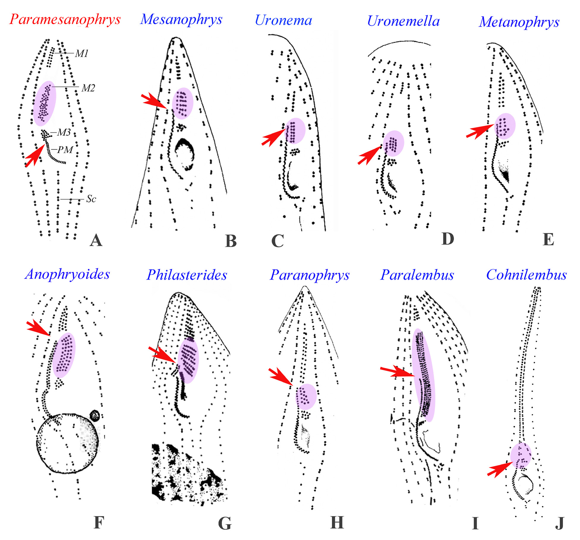
PM with paired basal bodies organized in zigzag pattern, extending anteriorly to posterior end of M3 ( Figs 2G, I View Fig , 3O, Q View Fig ). Scutica located at posterior end of PM, comprising c. seven or eight kinetosome pairs aligned in line parallel to somatic kineties ( Figs 2G, I View Fig ).
Mesanophrys carcini ( Grolière & Léglise, 1977) Small & Lynn in Aescht, 2001 Figs 4B View Fig , 5 View Fig ; Table 2
Small & Lynn in Aescht (2001) did not formally combine this species with Mesanophrys Small & Lynn in Aescht, 2001 . However, since they fixed it as the type species, they automatically produced the combination.
Some characteristics, e.g., a larger body size and fewer somatic kineties, were found in the Qingdao population. Hence, a description of the Qingdao population as well as a comparison between different populations are supplied.
Description of Qingdao population
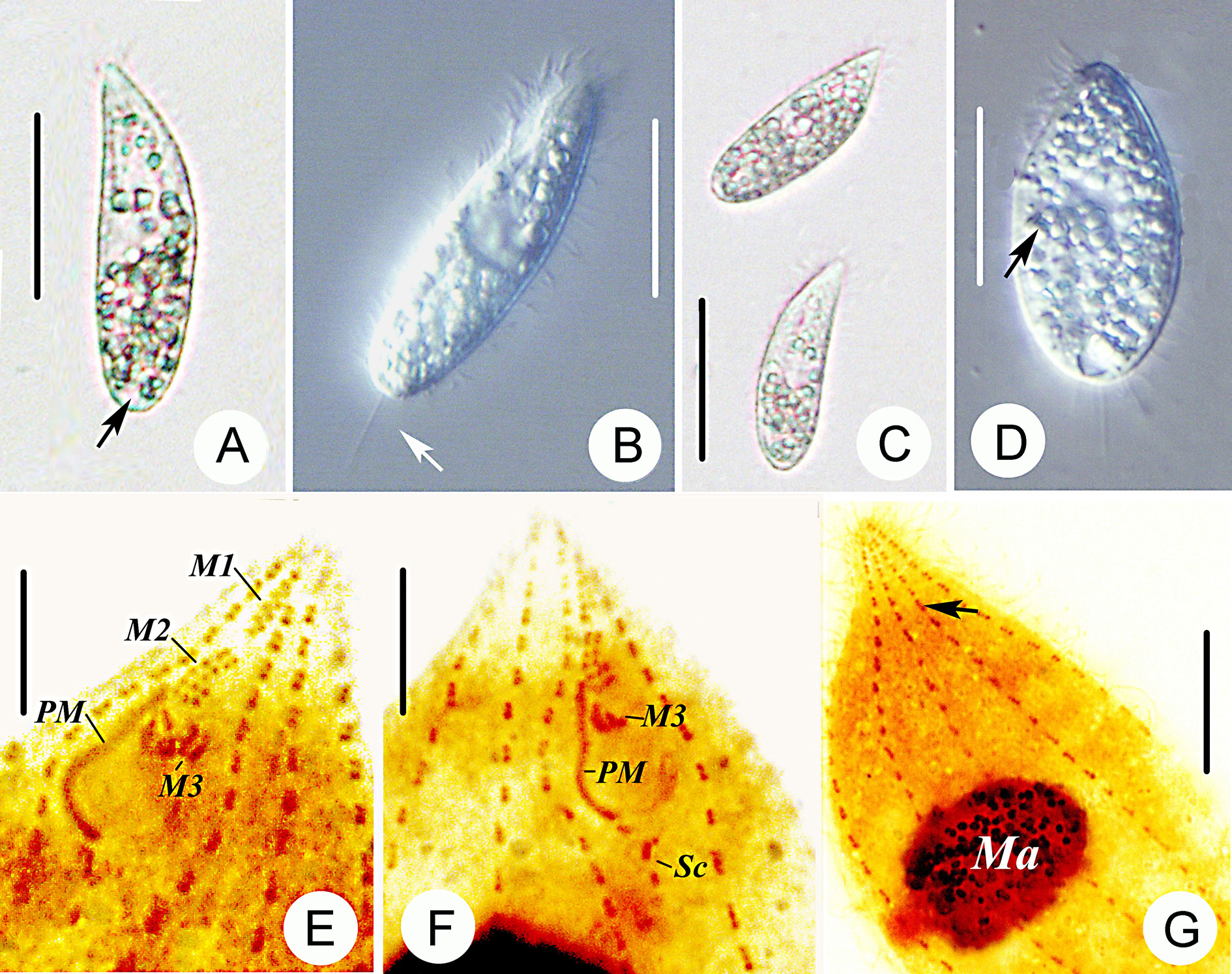
Body size 45–65 × 15–25 μm in vivo, spindle-shaped to long fusiform, with pointed anterior end and narrowly rounded caudal end ( Fig. 5 View Fig A–C). Body shape variable, likely due to nutritional conditions or stage in life cycle: from slender, spindle-like to pyriform ( Fig. 5 View Fig C–D). Buccal field short and narrow, with length of about 30% of body ( Fig. 5B View Fig ). Somatic cilia densely arranged and about 6–8 µm long ( Fig. 5B View Fig ). Pellicle thin and smooth, with no distinguishable extrusomes. Cytoplasm colourless to slightly greyish, containing several to many differently-sized (3–5 μm) refringent granules ( Fig. 5A, D View Fig ). Single caudal cilium about 15 µm in length ( Fig. 5B View Fig , arrow) and one large, spherical, centrally located macronucleus; one micronucleus closely associated with macronucleus. Contractile vacuole small (5 μm across), terminally positioned and pulsating at intervals of approximately 30 s ( Fig. 5A View Fig , arrow). Movement by continuous swimming in water without pause or gliding slowly on substrate.
Ten or 11 somatic kineties, consisting of dikinetids in anterior two-thirds and monokinetid in posterior third of body ( Fig. 5G View Fig , arrow). M1 slightly separated from apex, composed of two rows of kinetids with 7–9 basal bodies each ( Fig. 5 View Fig E–F). M2 composed of five or six longitudinal rows, each containing about 6–8 basal bodies ( Fig. 5 View Fig E–F). M3 located close to M2, much shorter than M2 and composed of three short, irregularly arranged rows of kinetosomes ( Fig. 5 View Fig E–F). PM extending anteriorly to posterior end of M2. Scutica Y-shaped, with c. four pairs of kinetosomes ( Fig. 5 View Fig E–F).
Ecological features
Salinity 32‰, pH 7.9 and water temperature about 11 °C.
No known copyright restrictions apply. See Agosti, D., Egloff, W., 2009. Taxonomic information exchange and copyright: the Plazi approach. BMC Research Notes 2009, 2:53 for further explanation.
|
Kingdom |
|
|
Phylum |
|
|
Class |
|
|
SubClass |
Scuticociliatia |
|
Order |
|
|
Family |
|
|
Genus |