
Alphocoris caudatus Rédei, Tsai & Jindra, 2018
|
publication ID |
https://doi.org/10.11646/zootaxa.4382.2.5 |
|
publication LSID |
lsid:zoobank.org:pub:3F5277F2-9FA5-4004-8BFF-4A1627AC6C07 |
|
DOI |
https://doi.org/10.5281/zenodo.5946448 |
|
persistent identifier |
https://treatment.plazi.org/id/03818784-FFC1-FFF9-C5D8-FE22FF68F986 |
|
treatment provided by |
Plazi |
|
scientific name |
Alphocoris caudatus Rédei, Tsai & Jindra |
| status |
sp. nov. |
Alphocoris caudatus Rédei, Tsai & Jindra , sp. nov.
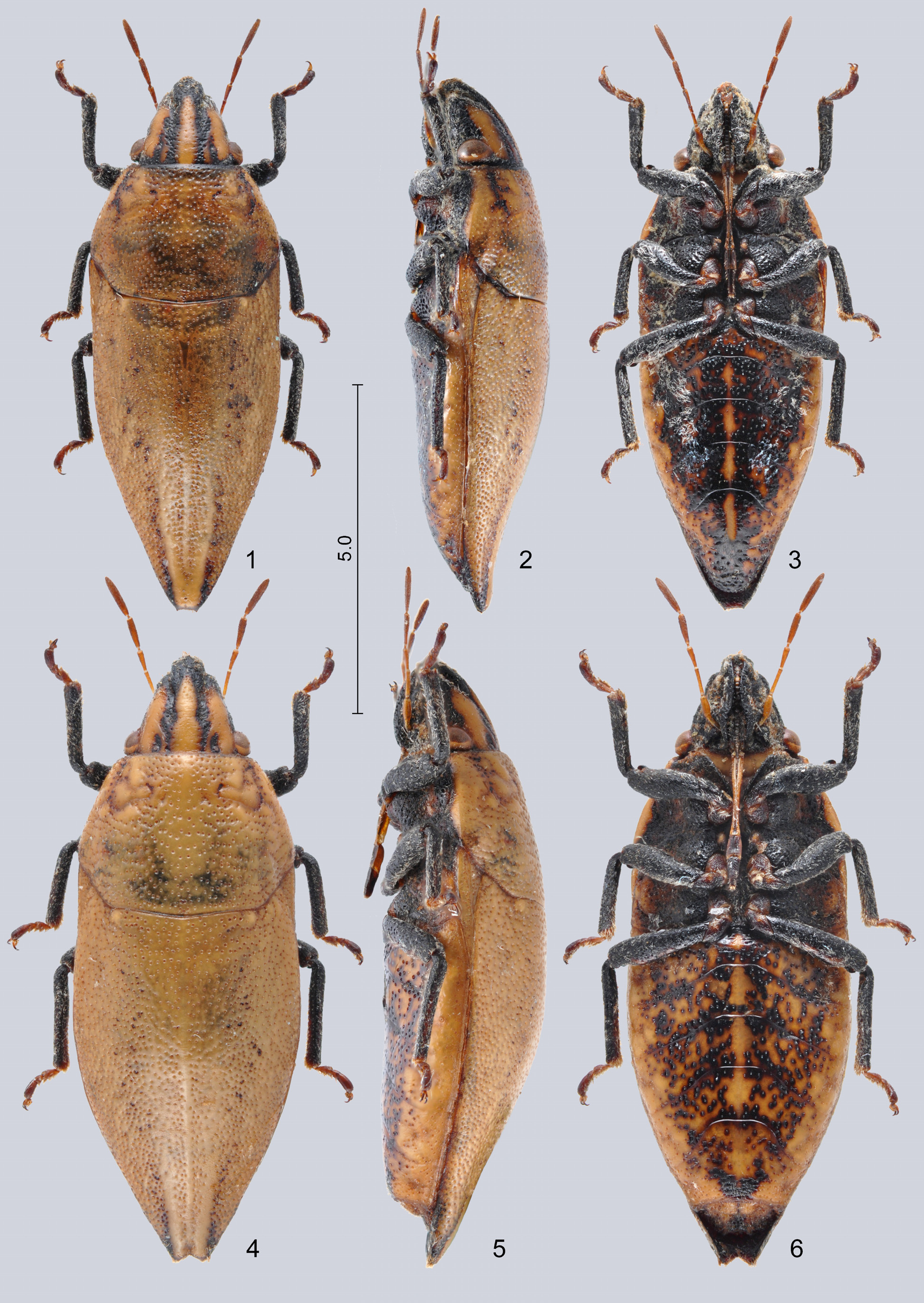
Figs. 1–6 View FIGURES 1–6 , 19–21, 28–29, 34–36, 44, 51–54, 59–62
Type material. Holotype: ♂, “ INDIA occ., Maharashtra state \ 7–11 October 2005 \ Mulshi enV., 40 km W of Pune \ F. Kantner lgt.”; mounted on card, genital capsule remoVed and glued to the same card separately but otherwise intact ( ZJPC). Paratypes: same label as in holotype ( 3 ♂♂ 1 ♀ ZJPC, 2 ♂♂ HNHM, 1 ♂ NMNS); “ India W, 30.ix.-2.x.2005 \ Maharastra state \ 40 km W of Pune \ Mulshi enV., J. BEZDĚK lgt.” ( 1 ♀ NMPC)
Diagnosis. Readily recognized among all congeners by its rather uniformly coloured dorsum lacking dark longitudinal Vittae ( Figs. 1, 4 View FIGURES 1–6 ), and the posterior portion of the scutellum and abdomen being strongly constricted in both sexes ( Figs. 1–6 View FIGURES 1–6 ) but particularly in males (Fig. 21).
Description. Colour, integument and vestiture. Dorsum rather uniformly stramineous with contrasting black markings on head but without dark longitudinal Vittae on scutellum, subshining; head stramineous to yellow, anterior part of clypeus, a longitudinal Vitta on mandibular plate laterally anteriad of eyes, mesal margin of mandibular plates together with adjacent lateral margins of clypeus continued in a pair of broad submedian Vittae extending to base of head, postocular portion laterally, and whole Ventral surface of head black; yellow areas of head with a few scattered, colourless, inconspicuous punctures, black areas densely and coarsely punctured; scape and pedicel of antenna dark yellowish or brown, flagellum rather dark brown, but occasionally whole antenna suffused with dark brown except of base of scape and basipedicellite; labium stramineous, segments III and IV suffused with dark brown; pronotum, scutellum, and exposed portions of fore wings rather uniformly stramineous, with small, not conspicuous dark patches formed by confluent dark punctures as follows: an irregular longitudinal streak laterad and a small dot immediately mesad of cicatrices, and a pair of submedian, longitudinal, anteriorly indistinctly delimited Vittae on posterior fourth to fifth of scutellum; pronotum and scutellum densely and rather uniformly and regularly punctured, but with a pair of unpunctured, slightly eleVated, somewhat callose surfaces between cicatrices and anterior margin of pronotum, furthermore midline of scutellum and a pair of submedian longitudinal bands in posterior third of scutellum also unpunctured, with Very fine and shallow punctures between and around them; middle and posterior portions of scutellum also frequently with a few irregularly scattered, Very large, black punctures; prothoracic hypomeron (= deflexed marginal portion of pronotum) stramineous, thoracic pleurites greatly black but paler laterally, coarsely punctured; coxae and trochanters dark brown, suffused with black, femora and tibiae black, tarsi brown, suffused with black towards apex; ground colour of abdominal Venter stramineous but densely coVered by coarse, greatly confluent black punctures therefore pale ground colour only Visible as a broad Vitta along lateral margin, a median Vitta usually interrupted at intersegmental sutures, and pale interspaces between punctures on disk; outer laterotergites stramineous, broadly suffused with black mesally, inner laterotergites and mediotergites black. Dense, tomentose pubescence of dorsum was lacking in the examined material (probably it was rubbed off), but its remnants were present on some specimens particularly on anterior portion of head, Ventral surface of body, and legs; scape and basipedicellite nearly glabrous, distipedicellite with a few scattered, fine hairs, flagellum with fine, dense, adpressed to semierect pilosity.
Structure. Body elongate, about 2.7 times as long as its humeral width ( ♂, ♀), more strongly constricted posteriorly than A. naso sp. nov. and A. asper sp. nov. therefore appearing more gracile. Head (Figs. 19–20) elongate subtriangular, about as wide as long, width across eyes about 1.35 ( ♂) / 1.25–1.3 ( ♀) times as long as interocular distance; eyes relatiVely small, semiglobose, strongly protruding laterally; distance from anterior margin of eye to leVel of apex of mandibular plate measured along longitudinal axis of body about 2.75 times as long as length of eye in dorsal View; apical, protruding portion of clypeus relatiVely short, less than one third as long as distance from anterior margin of eye to leVel of apex of mandibular plate measured along longitudinal axis of body; mandibular plates with a weak, obtuse longitudinal carina laterally anteriad of Ventral margin of eye; apex of buccula closely approaching apex of clypeus. Labium reaching bases of hind coxae. Thorax. Pronotum 1.4–1.45 times as broad as its median length; lateral margins smooth in their whole length, straight anteriorly, then gradually transform to broadly rounded humeri. Scutellum 1.55–1.65 times as long as its greatest width, posterior portion strongly tapering with conVerging lateral margins straight, apex conspicuously narrow (distance between paired protruding terminal angles shorter than width of anterior portion of clypeus) and considerably surpassing apex of abdomen, terminal part apically narrowly and almost transVersely truncate between a pair of minute lateral denticles ( ♂) or narrowly angularly excised ( ♀). Pregenital abdomen more strongly tapering apically than in A. naso sp. nov. and A. asper sp. nov., Ventrite VII of male conspicuously elongate (Fig. 21), posterolateral angles of abdominal Ventrites III–IV not produced.
External male genitalia. Genital capsule ( Figs. 28–29 View FIGURES 28–33 , 34 View FIGURES 34–43 ) highly elongate oVal in posterior View ( Fig. 34 View FIGURES 34–43 ), Ventral rim broadly conVex in dorsal View ( Fig. 28 View FIGURES 28–33 ), posterior portion relatiVely strongly produced ( Fig. 29 View FIGURES 28–33 ). Paramere as in Figs. 35–36 View FIGURES 34–43 . Phallus ( Figs. 44 View FIGURES44–46 , 51 View FIGURES 51–58 ): second pair of conjunctiVal processes (cp-II) subdiVided into 2+2 branches, dorsal branch ( Fig. 44 View FIGURES44–46 : cp-II1) membranous, greatly enlarged, surpassing apex of aedeagus; Ventral branch ( Fig. 44 View FIGURES44–46 : cp-II2) sclerotized, fin-shaped, broad; aedeagus weakly dilated subapically, distalmost portion narrow, tubular.
External female genitalia. Terminalia as in Figs. 52–53 View FIGURES 51–58 . Ovipositor: dorsomesal margin of laterotergite IX ( Figs. 53 View FIGURES 51–58 , 59: lt9) deeply emarginate around postgenital abdomen; ValVifer IX (Fig. 59: Vf9) with a relatiVely small, oVal median portion prolonged into a pair of elongate, obliquely transVerse rod-like arms joining rami of ValVulae IX without moVeable articulation; ValVulae VIII (Figs. 59: Va8) and IX well deVeloped, each with distinct sclerotized rami (Fig. 59: ra8), rami of ValVulae VIII weakly sclerotized towards its base, distinct, sclerotized gonangulum lacking. Inner ectodermal genital tracts: gynatrium saccular, elongate, proVided with a pair of long anterolateral pouches (Fig. 60: algp); ring sclerites could not be traced in the single examined specimen; base of spermatheca associated with a broad, plate-like fecundation sclerite (Fig. 60: fec). Spermatheca (Figs. 61–62): proximal duct (Figs. 61–62: pd) greatly broadened, thin-walled, saccular, joining to gynatrium through a broad opening; dilation (Figs. 61–62: dil) thick-walled, outer surface irregularly wrinkled, capable of inVagination into proximal duct as shown in Fig. 64 for A. naso sp. nov.; distal duct (Figs. 61–62: dd) Very short and thin, membranous, dorsoVentrally flattened; intermediate part ( Fig. 54 View FIGURES 51–58 ) with a small proximal flange, lacking distal flange, thin-walled portion (flexible zone) occupying its distal two-thirds, septum and fretum could not be traced in the single examined specimen; apical receptacle ( Fig. 54 View FIGURES 51–58 ) with a cup-shaped, strongly curVed proximal portion and a Voluminous, globose apical portion.
Measurements (in mm; N = 3 ♂♂ / 2 ♀♀). Body length 8.00–8.72 / 8.82–9.10; length of head 1.62–1.75 / 1.75–1.86, width across eyes 1.75–1.76 / 1.81–1.91, interocular distance 1.28–1.32 / 1.43–1.47; lengths of scape 0.37–0.44 / 0.43–0.44: basipedicellite 0.32–0.39 / 0.39–0.43: distipedicellite 0.25–0.27 / 0.27–0.29: basiflagellum 0.43–0.44 / 0.49: distiflagellum 0.62–0.63 / 0.68–0.69; median length of pronotum 2.08–2.16 / 2.21–2.35, humeral width 2.93–3.04 / 3.23–3.35; median length of scutellum 4.73–5.00 / 5.20–5.39, greatest width 2.88–3.12 / 3.33–3.37.
Etymology. The specific epithet caudatus , - a, - um (meaning ‘caudate’, ‘haVing or proVided with a tail’) is an adjectiVe formed from the Latin noun cauda (‘tail’) by adding the suffix - atus; it refers to the posteriorly strongly produced and tapering scutellum.
Distribution. Only known from the type locality near Pune in Maharashtra State, western coastal region of India.


No known copyright restrictions apply. See Agosti, D., Egloff, W., 2009. Taxonomic information exchange and copyright: the Plazi approach. BMC Research Notes 2009, 2:53 for further explanation.
|
Kingdom |
|
|
Phylum |
|
|
Class |
|
|
Order |
|
|
Family |
|
|
Genus |