
Anoplodactylus nanus, Krapp, Franz, Kocak, Cengiz & Katagan, Tuncer, 2008
|
publication ID |
https://doi.org/ 10.5281/zenodo.180521 |
|
DOI |
https://doi.org/10.5281/zenodo.6234205 |
|
persistent identifier |
https://treatment.plazi.org/id/038287D5-FFF7-FFB6-10FC-C3422C063937 |
|
treatment provided by |
Plazi |
|
scientific name |
Anoplodactylus nanus |
| status |
sp. nov. |
Anoplodactylus nanus View in CoL n. sp.
( Figures 3–6 View FIGURE 3 View FIGURE 4 View FIGURE 5 View FIGURE 6 )
Material: 1 ovigerous male from St. 3a., 0 3.03.1995, Gencelli Cove, Turkey, Cystoseira crinita , 1.5 m. (Figure 1). Transferred to Berlin Museum, repository number: Pycnogonida ZMB Nr. 367.
Diagnosis: A tiny yet compact Anoplodactylus species (about half size of A. angulatus ) with ventral angular proboscis tip, further distinguished from this morphologically similar species by the ventral suture between the ventral proboscis antimeres, more robust and thick-set legs, a greater number (6 in the holotype male vs. 3–4 in angulatus ) of femoral cement gland pores. The latter character may be compared with fig. 6d in Krapp (1973). The larger species A. angulatus lacks the distal-most and slenderer spine on the propodal heel. The (sub-) terminal robust setae on both tibiae present in many Anoplodactylus species were not found in A. nanus , although such a spine was present in the typical situation on its dorso-distal femur end.
Description: Body small (length 0.4 mm), compact, trunk and especially cephalon appearing relatively stout, tapering from segment 2 to 4 in width and length. Dorsally two sutures only, segments 3 and 4 fused; ventrally there appears a partly obliterated furrow between these two segments. No apparent ornamentation (setae or spines) on trunk or crurigers under light microscope ( Fig. 3 View FIGURE 3 a–c).
Ocular tubercle low, close to anterior margin of cephalon, its anterior outline convex in lateral view, posterior outline concave and slowly merging into dorsal segment surface. Four round and feebly pigmented eyes in normal configuration, lateral sense organs inconspicuous in glycerol or lactophenol ( Fig. 3 View FIGURE 3 b,c).
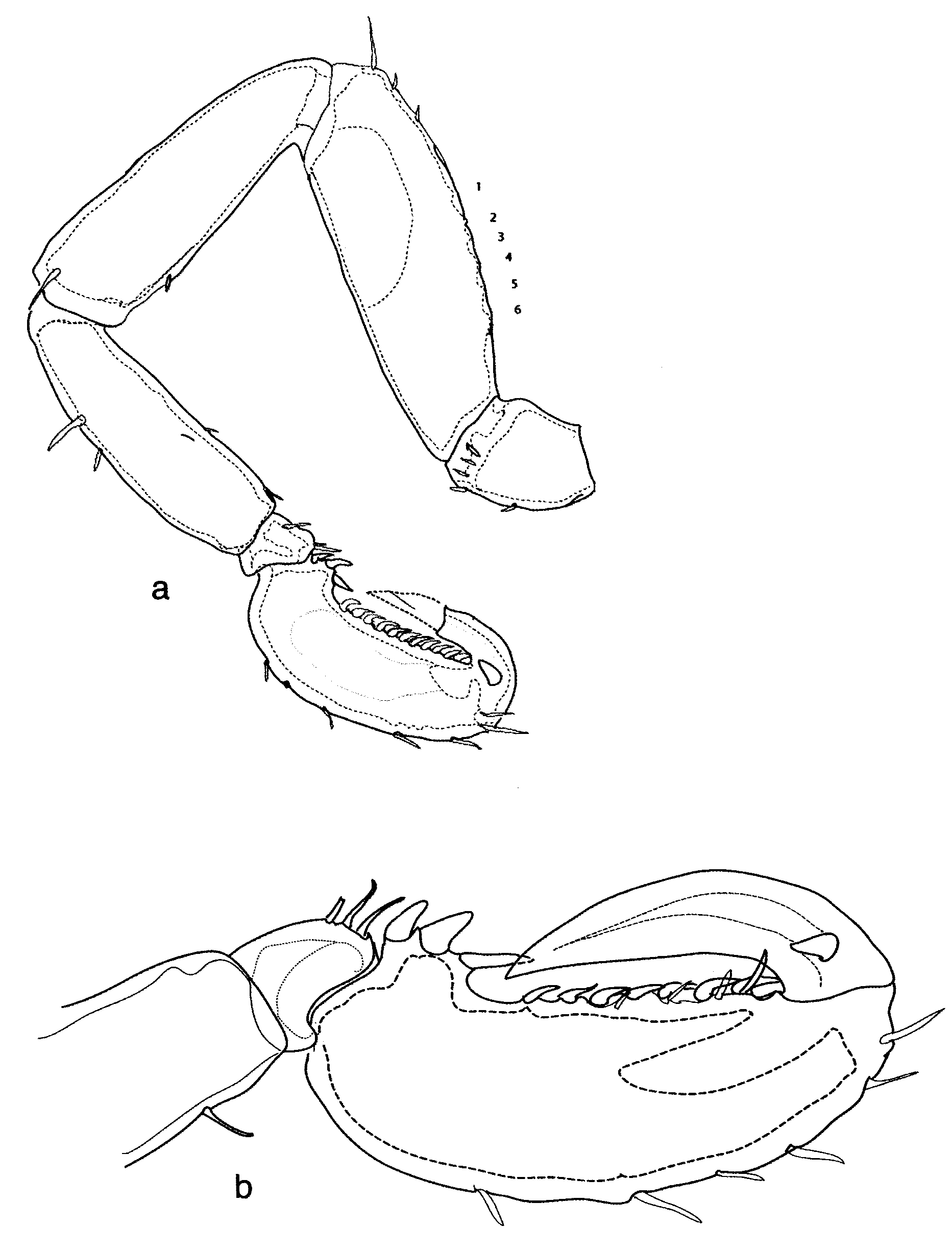
Proboscis cylindrical in dorsal view, in lateral view somewhat bulging ventrally. In ventral view tip with two protruding angular extremities of ventral antimeres (reminiscent of Anoplodactylus angulatus or robustus). Long median suture apparent in ventral view between ventral antimeres ( Fig.6 View FIGURE 6 b).
Chelifore scape long and slender, overhanging proboscis, very feebly curved, distally slightly swollen, bearing chela which is bent ventrally and towards midline. Chela fingers of equal length, movable finger with about 3 short teeth, immovable finger apparently toothless, “cutting" surfaces curved like minute ice-tongs, tips crossing ( Fig. 6 View FIGURE 6 b).
Ovigers 6-articled, 3rd article longest, 1st article a broad cone with dorsal extremity truncated (formed like a frustrum), its base almost as broad as 1st lateral process, article 4 about as long as article 2, terminal (6th) article shortest, thorn-like. On inner surface of article 5 minimally 2 tiny spinules, directed backwards, apparently as a retinaculum for the egg masses ( Fig. 6 View FIGURE 6 a).
Legs relatively long ( Fig. 3 View FIGURE 3 a). Coxae 1 and 3 subequal, coxa 2 longer by a fifth ( Fig. 5 View FIGURE 5 b). Second coxae on legs 3 and 4 of male holotype without genital spur. Femur and tibia 1 subequal, tibia 2 shorter than either. ( Fig. 4 View FIGURE 4 a). Cement glands in femur dorsal of intestine, about six pores opening level with dorsal femur surface, without any protruding ducts ( Fig. 5 View FIGURE 5 a). Tarsus short, almost triangular in outline, bearing 3–4 setae ventrally, dorsal aspect shortened and without setae ( Fig. 4 View FIGURE 4 b). Propodus compact, ectally bulging and bearing 6–8 robust setae. Heel distinct, three robust spines, distal-most spine slenderest. Sole straight, bearing about 9–11 distally-inclined robust spines, most proximal spine much more slender and separated from remainder by distinct gap; robust spines flanked on both sides by some setae; no trace of a cutting lamina. Main claw heavy, almost as long as sole, auxiliary claw short (less than one third of proximal claw diameter) ( Fig. 4 View FIGURE 4 b).
Remarks: Owing to the paucity of material (only the ovigerous male holotype available) we refrained from measuring trunk length, as we did not dare dissect more than the third and first legs (see figs. 4 and 5). The left oviger has been drawn still attached to the first lateral process. The right oviger with two cemented egg masses has been left attached.
Measurements: Length of coxa 3: 0.019, femur: 0.53, tibia 1 0.50, tibia 2: 0.42, tarsus: 0.008, propodus: 0.37 mm.
Etymology: Due to its dwarfish size — one of the tiniest Anoplodactylus species known — we chose to call it nanus , i. e. of dwarf size (Latin); adjective, gender masculine as that of the genus.
The species belongs to a group of species which share the following characters:
1) proboscis in ventral view angular, due to the extremities of the paired ventral antimeres (except Anoplodactylus virescens {Hodge, 1864} which probably belongs to this group)
2) presence of stout yet short auxiliary claws
3) no cutting sole lamina
4) cement gland pores on dorsal surface of femur, absence of cement gland ducts, only a series of openings on outer side of femur
5) compact build, lateral processes separated by less than half their diameter
6) cephalon compact
7) ocular tubercle a very low „cone“ at the anterior extremity of the cephalon
8) no male genital spurs on coxae 3 of legs 3 and 4.
The composition of this Anoplodactylus group was briefly outlined in Krapp (1996), more comparative studies of the species pertaining to it are urgently needed.


| ZMB |
Museum für Naturkunde Berlin (Zoological Collections) |
No known copyright restrictions apply. See Agosti, D., Egloff, W., 2009. Taxonomic information exchange and copyright: the Plazi approach. BMC Research Notes 2009, 2:53 for further explanation.
|
Kingdom |
|
|
Phylum |
|
|
Class |
|
|
Order |
|
|
Family |
|
|
Genus |