
Rhizophysa tricornuta, Pugh, P. R., 2019
|
publication ID |
https://doi.org/10.11646/zootaxa.4669.1.1 |
|
publication LSID |
urn:lsid:zoobank.org:pub:6E2F8FE4-4524-44B1-B5F8-BCC58D4FDF8E |
|
DOI |
https://doi.org/10.5281/zenodo.3796976 |
|
persistent identifier |
https://treatment.plazi.org/id/0384A837-826B-FF9C-FF37-F90FFD267E61 |
|
treatment provided by |
Plazi |
|
scientific name |
Rhizophysa tricornuta |
| status |
|
Bedot and Bathyphysa View in CoL
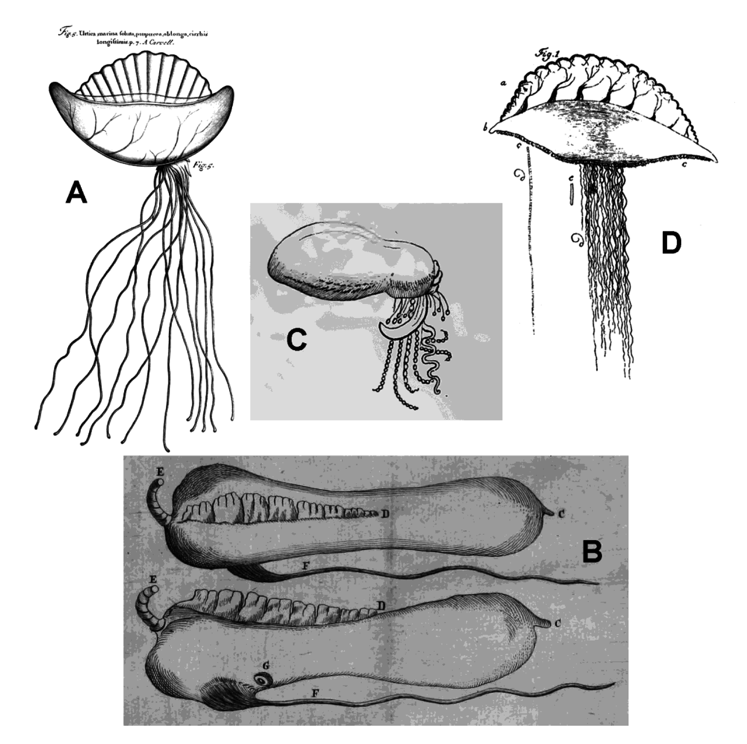
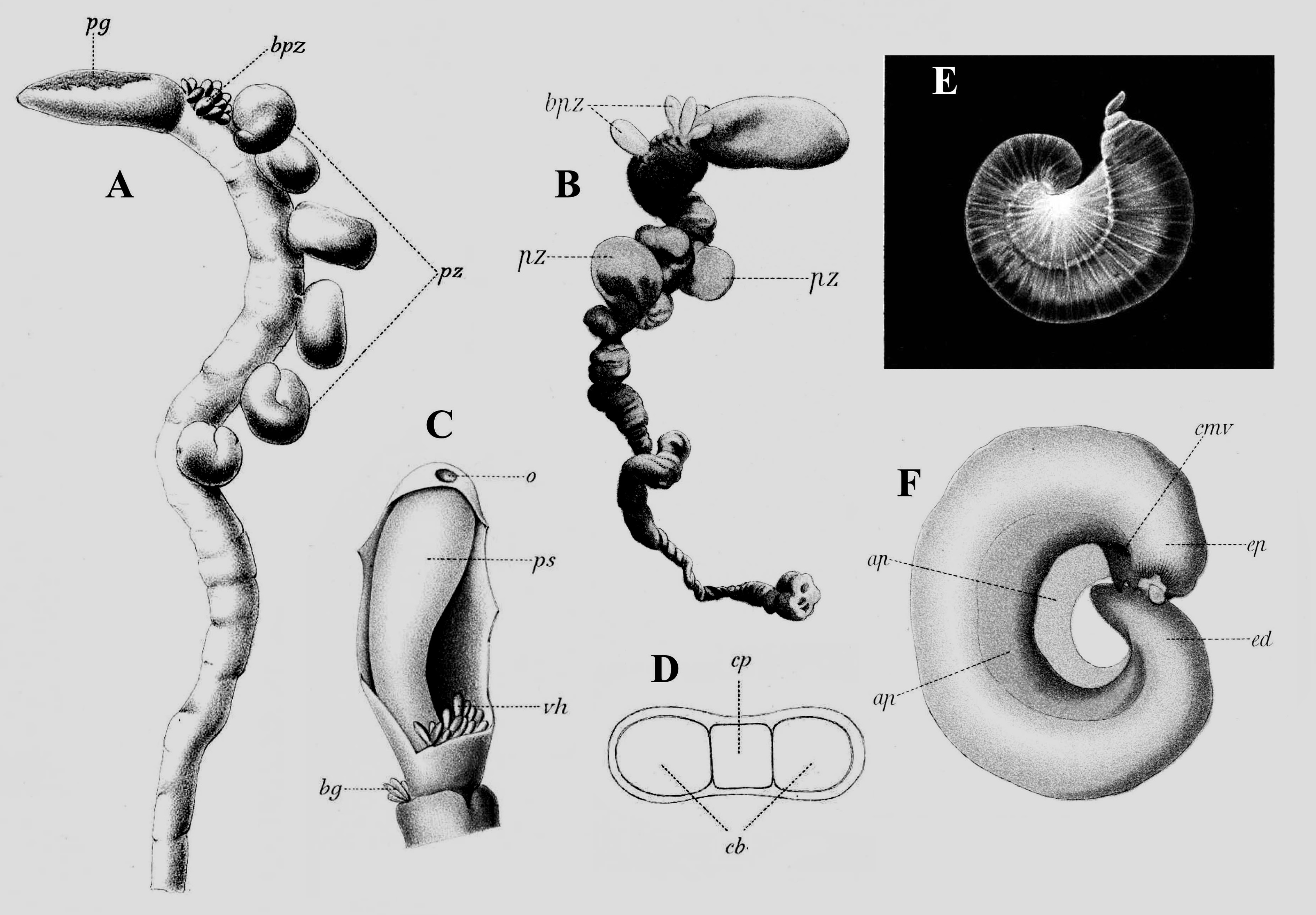
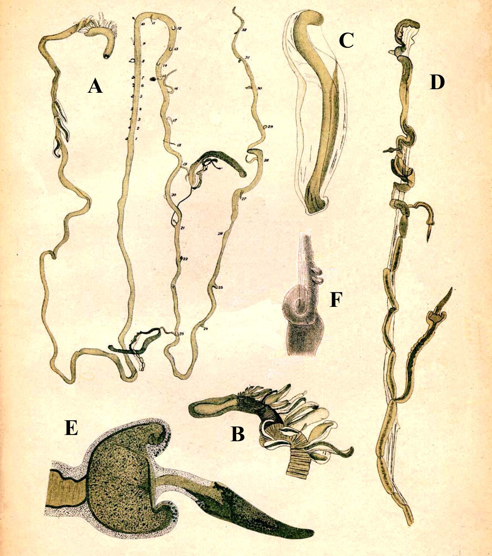
Having brought the history of Physalia physalis up to the present, let us now turn to the rhizophysid cystonects. As noted above we got away lightly with Haeckel’s (1888b) interpretation of the genus Bathyphysa as belonging to the family Forskaliidae . However, this interpretation caused great confusion to Bedot (1893) when he described a bathyphysid species under the name Bathyphysa Grimaldii Bedot. Firstly, Bedot adopted Haeckel’s terminology, dividing the stem into the anterior nectosome and posterior siphosome, and he noted that for his specimens the former was reduced to just the pneumatophore, which had hypocystic villi at its base (see Figure 26C View FIGURE 26 ). Instead of nectophores, Bedot believed that the upper part of the stem bore peculiar and specialised zooids, which he name pneumatozoïdes (see Figure 26A, B View FIGURE 26 , pz), which were said to be borne on short peduncles. These zooids were de- scribed as consisting of a tube, closed at both ends and lined by endodermal cells, and curved into a C-shape by a transparent ectodermal membrane united to its walls ( Figure 26 E, F View FIGURE 26 ). Bedot considered that the endodermal tube was filled with air, attributing to them a hydrostatic function; hence their name.
Posterior to these so-called pneumatozoïdes Bedot (1893) described the siphosome with a great number of gastrozooids, although it would seem that all the latter were detached. The gastrozooids were said to be similar to those described by Studer (1878a) for Bathyphysa abyssorum , i.e. with two longitudinal replis or ptera, and borne on long peduncles, which bore small swellings filled with nematocysts. Bedot rejected Haeckel’s idea that these swellings represented the sites of attachment of bracts, and considered that bracts were totally absent, noting that the so-called bract that Studer described for B. abyssorum was nothing more than a small gastrozooid. Tentacles were also present, which had exactly the same structure as the peduncles of the gastrozooids. Bedot concluded that his B. Grimaldii was actually more closely related to Rhizophysa than to Forskalia , but he considered that there were sufficient differences, particularly the presence of pneumatozoïdes, to warrant the establishment of a new family, the Bathyphysidae , as Haeckel (1888b) himself had suggested; but in Bedot’s case within the Cystonectae . Bedot considered that the presence of ptera on the gastrozooids was an important taxonomic character and thus included Studer’s B. abyssorum and Fewkes’s B. ( Pterophysa) grandis within the genus. However, Bedot, like Fewkes, considered that there was only one genus and he chose Studer’s original genus Bathyphysa , as opposed to Fewkes’ substitute one Pterophysa . Nonetheless, although Bedot also noted the presence of ptera on the gastrozooids of Studer’s R. conifera , for some unexplained reason he excluded this species not only from the genus Bathyphysa , but also from his family Bathyphysidae . He did not mention Studer’s R. inermis at all.
With regard to bathyphysid species Schneider (1898) was remarkably brief. To him they were all cystonect siphonophores and he placed them all in the genus Pterophysa . He separated off P. conifera from his other species on the basis that its gastrozooids were not borne on long peduncles, and its tentacles were simple, without tentilla. However, he did not explain why he considered the gastrozooids as sessile. He also recognised P. abyssorum , with gastrozooids borne on long peduncles, but without tentacles, and suggested that P. grandis and Bathyphysa grimaldii were most probably junior synonyms it. With regard the so-called pneumatozoïdes, described by Bedot (1893), he correctly identified them as nothing more than young gastrozooids.
Unfortunately, Schneider’s (1898) contention goaded Bedot (1904) into publishing a further paper on his Bathyphysa grimaldii . Most of this paper is taken up with detailed descriptions of the anatomy of the so-called pneumatozoïdes, which Bedot believed contained a specialised organ of unknown function. However, Bedot changed his mind with regard to two features of the pneumatozoïdes. Firstly, what he had previously thought was a short peduncle, he now interpreted as a short filament, said to be on the dorsal side, and that the pneumatozoïde was, thus, attached directly to the stem. Secondly, he decided that he was wrong to consider the endodermal cavity as having contained “air”. He also changed his mind with regard to the presence of a peduncle on the mature gastrozooid, now regarding it as a tentacle such that the gastrozooid was attached directly to the stem. Attention was also draw to the fact that this tentacle was positioned on the gastrozooid in exactly the same position as the filament on the pneumatozoïde. But he was still quite adamant that the two structures were entirely different. Thus he took issue with Schneider stating ( ibid. p. 22) 66: “If the pneumatozoïdes were young gastrozooids, they should have smaller dimensions than the latter. However, this is not the case and I studied a great number of pneumatozooids whose length varied between 30 and 40 mm, which is the size of the largest gastrozooids. In addition - and this is the most important point - the pneumatozooids are never open at the distal end, while it is easy to distinguish the mouth of the gastrozooids even when they are very young”. The question of size of a zooid appears to be similar to the question – “how long is a piece of string?”, but he was entirely wrong with regard to his believe that young gastrozooids should always have a mouth opening, because it is now clear that the so-called pneumatozoïdes are indeed the developing gastrozooids, and the youngest ones have yet to develop a mouth opening. As for the strange organ that these zooids were supposed to contain, Lens & van Riemsdijk (1908, p. 107): “looked in vain for the apparatus ... We made microscopical sections of the base of siphons in different specimens but there was not the slightest indication of any such structure”. However, photographs of B. conifera in a tank, taken by Rob Sherlock clearly show that a small white sphere usually is present at the distal end of the gastrovascular canal, just below the tapered tip of the young gastrozooid, which has a mauve tinge (see below). This may be what Bedot was referring to.
So, returning to Schneider’s (1898) classification, the species Bathyphysa grimaldii would now appear to be more closely allied with Pterophysa conifera , as both had now been described with gastrozooids without, or at least only short, peduncles, but with simple tentacles, without tentilla. As Fewkes’s (1886) specimen of P. grandis consisted of a denuded stem and pneumatophore, and some young detached gastrozooids with ptera, but without tentacles and, presumably, a peduncle, then it is impossible to categorise the species on this basis. This leaves B. abyssorum as the only species to have been described with gastrozooids borne on long peduncles, and possibly without tentacles. However, as discussed above, it is extremely likely that what have been interpreted as peduncles are more likely to be tentacles, and that a peduncle, if present, is likely to be quite short. The presence/absence of a peduncle seems, therefore, to be a complete red herring.
Mayer, Chun and Schneider
Mayer (1894, p. 239) considered that: “Dr. Fewkes [1879] gives a good description and a very poor sketch, which is apparently intended to represent R. Eysenhardtii . In our opinion it would be better to adopt the nomenclature of Haeckel, and give to this species the name of Cannophysa, as it is evidently very closely allied to, if not identical with, Cannophysa Murrayana from the Canary Islands ”. This appears to be a slightly arrogant on Mayer’s part as a) Fewkes’s paper predates Haeckel’s monograph; b) Fewkes described and, admittedly poorly, illustrated the tentilla of R. filiformis not R. eysenhardtii ; and c) Mayer should have synonymised C. Murrayana with R. eysenhardtii , and not the other way round. Mayer then goes on to give a description of specimens of what he calls C. Eysenhardtii,
66 Original quote: “Si les pneumatozoïdes étaient de jeunes gastrozoïdes, ils devraient avoir des dimensions inférieures à celles de ces derniers. Or, cela n’est pas le cas et j’ai étudié un grand nombre de pneumatozoïdes dont la longueur variait entre 30 et 40mm, ce qui est la dimension des plus gros gastrozoïdes. En outre - et c’est là le point le plus important – les pneumatozooids n’ont jama s d’ ou erture à leur extr m t d stale, tandis que l’on distingue facilement la bouche des gastrozoïdes lors même qu’ils sont très jeunes.”
which contains virtually no characters of any worth, together with three poor illustrations. With regard to the tentilla all he said was ( ibid. p. 240): “The secondary filaments are fine hair-like structures terminating in a bunch of nematocysts (see Plate III, Fig. 4 View FIGURE 4 )”. However, that figure (see Figure 27 View FIGURE 27 ) clearly shows that distally they are divided into three lobes, from which one would conclude that they are very young tentilla of the tricornuate type of R. filiformis . Thus, clearly Mayer appears not to know his eysenhardtii from his filiformis . Also included in his Plate III was a figure (see Figure 27 View FIGURE 27 ) of what he referred to, in the legend, as C. filiformis , although Mayer made no reference whatsoever to it in the text. The specimen clearly is deformed as the stem is highly twisted, but it gives the impression that the tentacles bore extremely large tentilla of a type not previously described. One wonders, therefore, why Mayer gave it the specific name filiformis , presumably likening it to the well-known filiformis or perhaps, from the above, eysenhardtii . We will return to this figure shortly.
Chun (1897, p. 83) 67, for once did not agree with Haeckel, and considered, with regard to the family Rhizophysidae , that the two basic characters that Haeckel had used to subdivide the family, i.e. the positioning of the gonodendra and the structure of the tentilla, were untenable. Regarding the former he said: “A principle of classification, which applies to not only numerous genera, but also to two subfamilies, in my opinion should be based on structural features that in all cases allow a clear and unambiguous decision to be made.” Chun believed that the positioning and number of the gonodendra was more related to the age of the specimen. With regard to the number of types of tentilla present he said ( ibid.) 68: “Whomever now attached generic value to the structure of the tentilla would have to split [specimens of] R. filiformis , dependant on their age, into no less than three genera, characterised by mono-, tri- and bimorphic tentilla … [thus] the structure of tentacles and stinging buttons should not be given too high a systematic value”. Finally, he split his Order Rhizophysaliae into two sub-orders, the Rhizoidea, including the families Epibulidae and Rhizophysidae , and the Physaloidea, for the family Physalidae .
Among the Plankton Expedition samples Chun (1897) found only two Rhizophysa species, which he called R. eysenhardtii and R. murrayana . But he also listed all the siphonophore species previously found in the Atlantic Ocean and which apparently he considered to be valid. For the family Rhizophysidae be included the following species:
Rhizophysa Eysenhardtii Gegenbaur.
Rhizophysa (Cannophysa) Murrayana Haeckel.
Rhizophysa clavigera Chun (= Cannophysa filiformis Mayer ).
Rhizophysa (Linophysa) conifera Studer.
Rhizophysa filiformis Forsskål.
Salacia polygastrica (= S. uvaria Fewkes ?) Haeckel.
Although Chun (1897, p. 84) 69 recognised that there was a close similarity between the tentilla of Rhizophysa eysenhardtii and R. murrayana , he believed that there were some differences as he said, with regard to R. murrayana : “As I will describe them in more detail at another time, I only remark that in the case of the latter species, in the angle at the base of the weaker [side] branches are muscle fibres which run along the middle of the main branch to the base these branches.” However, this description apparently never appeared in print and, as noted above, the structure referred to had already been illustrated by Fewkes (1879), and is now known to be characteristic of the mature tricornuate tentilla of R. filiformis .
We have already dealt with most of the species in the list above. However, Chun (1897) did not describe Rhi- 67 Original quote: “Ein Eintheilungsprincip, welches nicht nur zur Aufstellung zahlreicher Gattungen, sondern auch zweier Unterfamilien Verwerthung findet, sollte meines Erachtens an Strukturverhältnisse anknüpfen, welche in allen Fällen einen klaren und unzweideutigen Entscheid gestatten.”
68 Original quote: “Wer nun der Gestaltung der Seitenfäden generischen Werth beilegt, müsste die Rh. filiformis je nach ihrem Alter in nicht weniger denn drei Genera zerspalten, welche durch monomorphe, trimorphe und bimorphe Seitenfäden charakterisirt wären … der Struktur von Tentakeln und Nesselknöpfen keinen allzu hohen systematischen Werth beizulegen.” 69 Original quote: “Da ich dieselben an einer anderen Stelle noch eingehender schildern werde, so bemerke ich nur, dass ebenso wie bei der letzteren Art in dem Winkel zwischen dem Ansatz der schwächeren Seitenäste Muskelfasern entwickelt sind, welche längs des mittleren Hauptastes bis zur Basis der Seitenäste verstreichen.”
zophysa clavigera (= Cannophysa filiformis Mayer ), and one can only presume that he considered it to be a new species, distinct from R. filiformis . He had earlier rejected Mayer’s placement of it within the genus Cannophysa as it was impossible to use Haeckel’s key to identify that genus since no gonodendra were present. Bigelow (1911, p. 318) also provided a very reasonable explanation with regard to this species when he said: “Whether R. clavigera is really a distinct species can hardly be determined from Mayer’s very confused account, or from his figure which was evidently drawn from a fragmentary specimen. My opinion is that it was probably R. filiformis , with siphons and tentacles twisted together”. Thus the suppose tentilla are actually gastrozooids. Although I am not sure what Bigelow means by “very confused account” for, as mentioned above, no description of this species was actually given. However, like many of Mayer’s new siphonophore species, this one is best ignored and, as Bigelow (1911) suggested, treated as a synonym of R. filiformis .
Of course, Schneider (1898, p. 164) 70 rejected Chun’s classification of his order Rhizophysaliae when he said: “In my view, the relationships of all the relevant forms are so close that a division of the same family into 3 and even 2 Suborders (the whole group of Cystonects being considered only as a Suborder, as the Siphonophora as a whole was considered only as an Order) does not appear justified”. However, with regard to the genus Rhizophysa, Schneider entirely agreed with Chun that care must be taken for specific identifications based on the structure of the tentilla but, based on this, he expressed his surprise, correctly in our opinion, that Chun (1897) had retained the species R. murrayana and R. gracilis . Nonetheless, Schneider’s (1898) choice of species that he believed belonged to the genus Rhizophysa is itself rather strange. Firstly, he considered ( ibid. p. 170) 71 that: “The type of this genus is generally considered to be Gegenbaur’s 53 R. filiformis from Messina”. He noted that Forsskål’s (1776) illustration of R. filiformis (see Figure 3B View FIGURE 3 ) showed no side branches to the tentacles, but contemplated that they might have been lost, or simply overlooked.
However, Schneider (1898) then did just that for his second rhizophysid species, which was Rhizophysa Mertensii Brandt, 1835 . He considered the tentacles of this species to bear simple tentilla, and thereby likened it to Gegenbaur’s (1859) R. eysenhardtii and Haeckel’s (1888b) Nectophysa Wyvillei. As Bigelow (1911, p. 318) pointed out: “Schneider (’98) used the name R. mertensii Brandt to replace eysenhardtii Gegenbaur , evidently supposing that Brandt’s (’35, p. 33[233]) description of the tentilla as “ Tentacula composita ramulis, i.e. tentaculis porpriis, simplicibus” meant that they were filiform. But Haeckel (’88b, p. 329) who examined Mertens’ unpublished figures of this species expressly states that it “exhibits distinctly two different kinds of branched tentilla””. Thus Bigelow ( ibid.) concluded that: “there seems to be only one course open, namely, to consider mertensii a synonym of filiformis , on the strength of its having two kinds of tentilla”. In fact, what Bigelow quoted was Brandt’s description of the genus Epibulia , while that for the species E. ( Macrosoma) Mertensii itself, Brandt stated ( ibid. p. 233): “ Corpus longissimum , pallidissime fuscescens, Vesica natatoria ovata corpori concolor. Tentacula composita plurima, alterna, corpori concolora. Proboscides suctoriae plures in trunco secundae, pallide roseae, apice in peltam dilatabiles ”. In addition, Brandt’s ( ibid.) description of the species also stated: “ Corpus longissimum , pallidissime fuscescens ”. So the long stem was said to be of the palest brown colour, while the gastrozooids were pale pink, thus indicating that Mertens’ specimen could have been R. eysenhardtii . However, as we only have Brandt’s description, without illustration, to go by it really is best to consider the species name Mertensii as a nomen nudum
The third species that Schneider (1898) included in the genus Rhizophysa was R. uvaria Fewkes, 1886 .As noted above, this species, supposedly had polygastric “cormidia” and, thus, would belong in Haeckel’s (1888b) genus Salacia - the sheer inventiveness of which we have already considered. As for the genus Epibulia , we have already dealt with the views of Schneider.
Lens and van Riemsdijk (1908)
This last point was discussed further by Lens & van Riemsdijk (1908), who reviewed all the descriptions of rhizophysid and bathyphysid species. They included two genera in the family Rhizophysidae , and with regard to Rhizophysa species, they said ( ibid. 100): “So far as we can judge by the litterature [sic] ... we can only accept two species, Rhizophysa filiformis Forsk. 1775 and Rhizophysa Eysenhardtii Ggbr. 60 [59]. We cannot admit the validity of any
70 Original quote: “Meiner Auffassung nach sind die Verwandtschaftsbeziehungen aller hierher gehörigen Formen so enge,
dass eine Auseinanderreissung derselben zu 3 Familien und gar 2 Unterordnungen (die ganze Gruppe der Cystonecten ist nur
als Unterordnung zu betrachten, wie die Siphonophoren insgesammt nur als Ordnung) nicht berechtigt erscheint.” 71 Original quote: “Als Typus dieser Art betrachtet man allgemein die R. filiformis Gegenbaur’s 53 von Messina.”
of the new genera which Haeckel has proposed ( Cannophysa, Linophysa , Aurophysa , Pneumophysa , Nectophysa ): their characteristics are based on differences too slight to permit us to look upon them as more than two species of the same genus˝.
Lens & van Riemsdijk (1908, p. 100) stated: “Haeckel also often repeats that he will describe the specimens in a future work (see 88b Pneumophysa p. 328). His descriptions of Aurophysa and Linophysa are quite insufficient. Cannophysa and Nectophysa have been treated somewhat better, but we cannot find any difference from the original Rhizophysa Eysenhardtii , looking through the description of Cannophysa Murrayana, from Rhizophysa filiformis or comparing it with Nectophysa Wyvillei. Haeckel writes simply of the latter (88b p. 327) “Another closely allied species seems to be Rhizophysa Eysenhardtii described by Gegenbaur””. They then gave quite detailed descriptions of both R. filiformis and R. eysenhardtii ( Figure 28 View FIGURE 28 ) of which they had several specimens.
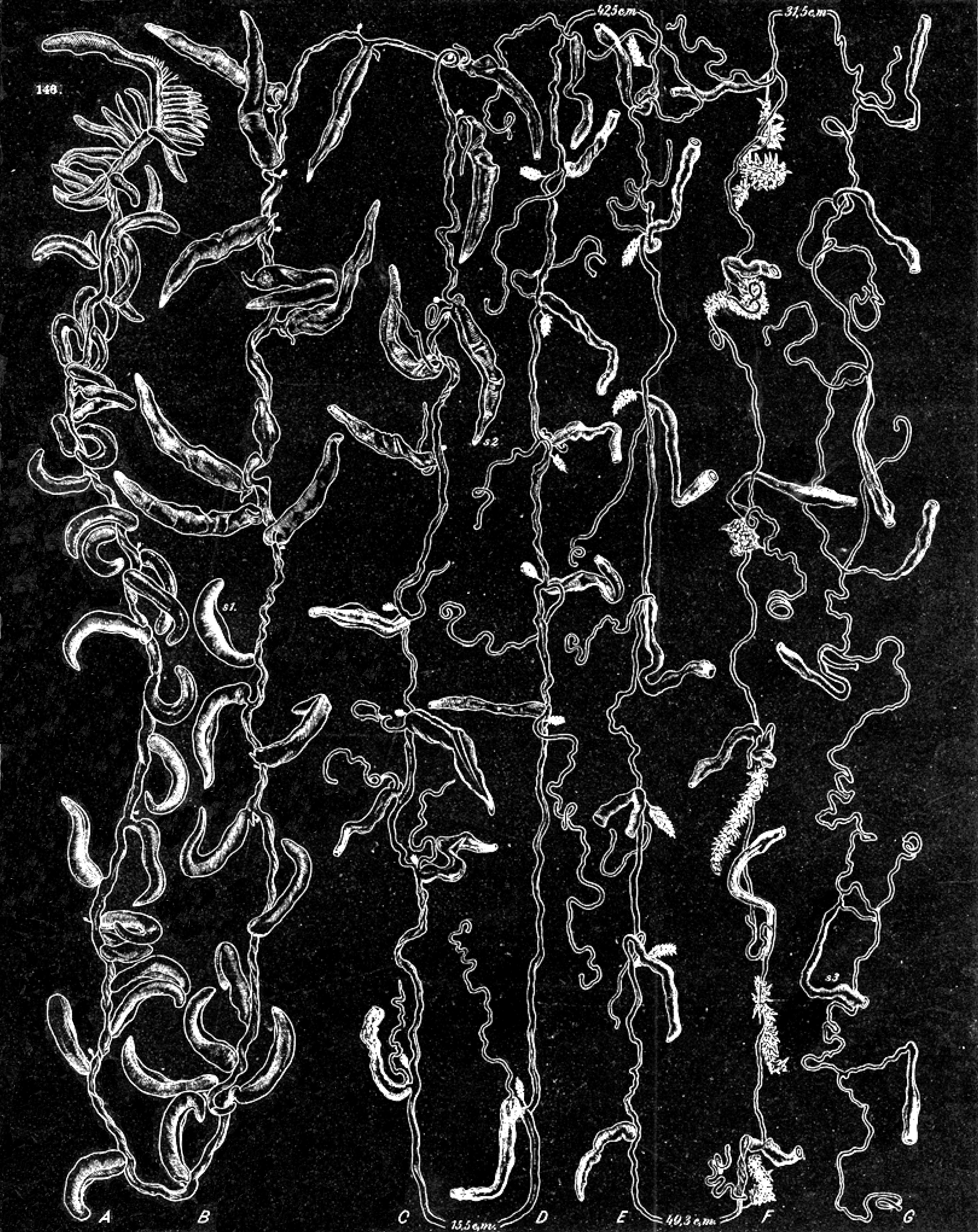
For the bathyphysid species, Lens & van Riemsdijk (1908) gave a detailed summary of what had been published to date, but they fell into the trap with regard to the presence/absence of a peduncle on the gastrozooids. With regard to Bathyphysa abyssorum they noted that in Studer’s (1878a) Plate III figures 38-39 (see Figure 16B View FIGURE 16 ) the so-called peduncles to the gastrozooids appeared to be wound around the stem rather than connecting with it. Thus, they reached the preliminary conclusion that these filaments were more likely to be tentacles than peduncles. They attempted to resolve this point by looking at the original and still extant material but apparently, because of the large size of the specimen, they were only able to re-examine one of the original gastrozooids from the original specimen. Clearly, this was insufficient to resolve the point and so they ultimately decided against this option as they interpreted Studer’s (see Figure 16A View FIGURE 16 ) illustration as clearly showing that the gastrozooids were attached to the stem by long peduncles. As has already been discussed, this conclusion is almost certainly wrong.
Thus, Lens and van Riemsdijk (1908) divided the known bathyphysid species among the genera Pterophysa , with sessile, and Bathyphysa , with pedunculate gastrozooids. They agreed with Schneider that Rhizophysa conifera and P. grandis were closely related, but maintained them as separate species. However, they never referred to the former species as P. conifera presumably because they considered that the gastrozooids were borne on short peduncles, while those on P. grandis were sessile. They reached the latter conclusion based on the examination of some additional material that Fewkes had subsequently identified as that species. This uncertainty as to the exact status of the species conifera , also applies to one of Lens & van Riemsdijk’s new species, as is discussed below. However, they also moved Bedot’s B. grimaldii into the genus Pterophysa , and considered that it was probably identical with P. grandis .
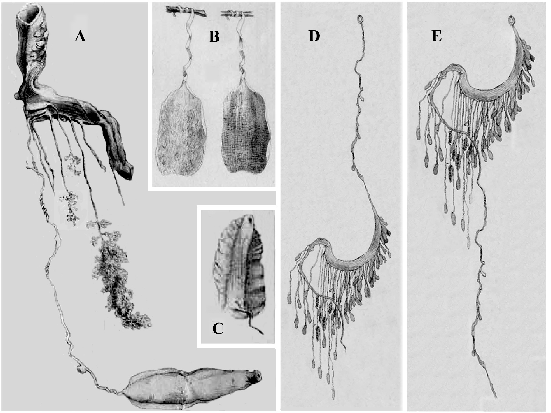
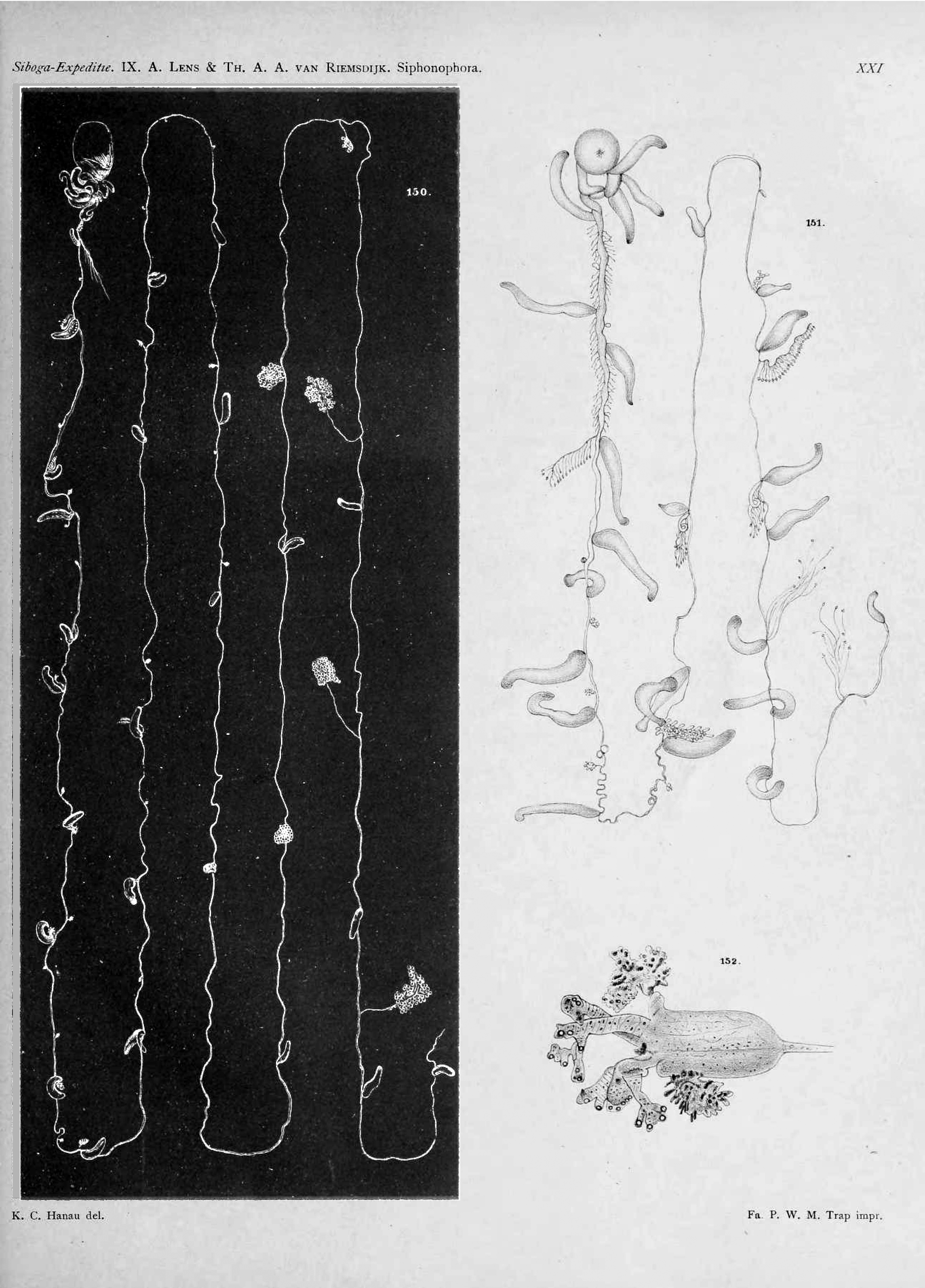
Lens & van Riemsdijk (1908) then gave an extensive re-description of Pterophysa grandis (see Figures 29 View FIGURE 29 , 30A View FIGURE 30 ) based on their own material. They could not find any hypocystic villi at the base of the pneumatophore, but stated that such might have been obscured by the opacity of the walls. However, they noted that the young gastrozooids did not possess tentacles, and that this structure did not begin to develop until the c. 60 th gastrozooid from the anterior end of the siphosome. Gonodendra also were not present between the most anterior gastrozooids, but buds of them, apparently just anterior to a gastrozooid, began to appear well before the tentacle began to be developed on the gastrozooid. All the gastrozooids, at whatever degree of maturity, were attached directly to the stem.
A small part of the large specimen from Siboga St. 52, and the smaller specimens from Sts. 185 and 284 have been re-examined by the present author, thanks to the kindness of Elly Beglinger (Zoological Museum, University of Amsterdam). All of Lens and van Riemsdijk’s specimens were initially fixed in formalin and later, perhaps after they had examined them, transferred into alcohol. The portion from the St. 52 specimen, although in poor condition, clearly came from toward the posterior end as it included many mature gastrozooids and a well-developed gonodendron. However, what was most striking was that many of the long, tubular gastrozooids were borne on short peduncles, up to 2.5 mm in length. The other two specimens were more contracted so that often the gastrozooids appeared to be attached directly to the stem, but evenso a short peduncle occasionally could be discerned. The only conclusion that could be reached was that the gastrozooids did possess a short peduncle, but that preservation may have resulted in it becoming severely contracted so as to give the appearance that it did not exist. With regard to the gonodendron, there were approximately forty side branches from the central gonostyle, each of which bore a terminal gonopalpon, a primordial asexual nectophore and about eight gonophores. The exact distribution of the gonophores with respect to the asexual nectophore could not be assessed accurately due to the poor state of preservation.
Next, Lens & van Riemsdijk (1908) described their two “new” bathyphysid species. For Pterophysa ( Bathyphysa) Studeri Lens & van Riemsdijk (see Figure 30 B View FIGURE 30 ) they described the presence of hypocystic villi at the base of an enormous ( 35 mm long) pneumatophore, and they devoted a large part of their description to their structure. Only three gastrozooids remained attached to the stem, situated at 28, 52 and 64 mm from the anterior end of the siphosome. They were all considered to be mature, but it is clear that all of them were young as they possessed obvious ptera, but no tentacles, and only one was said to have a mouth opening. Another three gastrozooids and: “a vast amount of tentacles, and stems and other appendages, impossible to determine” ( ibid. p. 114) were also found with the specimen. The authors were unable to ascribe the species to either of the genera discussed above, because they believed that the gastrozooids were borne on short peduncles, and thereby, like Rhizophysa (P.) conifera , possessed a character that was intermediate between the two genera as they had earlier defined them. Although they noted that their specimen closely resembled Studer’s B. abyssorum : “To call our specimen Bathyphysa we thought too daring” ( ibid. p. 113). However, if one compares their illustration of one of the attached gastrozooids (see Figure 30 B View FIGURE 30 , cen- tre), with those of young gastrozooids of P. grandis (see Figure 30 A View FIGURE 30 ) it is clear that in both the ptera continue right to the base of the gastrozooid. Since Lens & van Riemsdijk considered the gastrozooids of the latter species to be attached directly to the stem, it is unclear why they considered those of P. ( B.) studeri to have a short peduncle.
What remains of Lens & van Riemsdijk’s material of Pterophysa (Bathyphysa) studeri from Siboga St. 126 has been re-examined by the present author, but it consists of only a few pieces of denuded siphosomal stem, some of which are flattened and ribbon-like, as noted by Lens & van Riemsdijk. Thus, we can only rely on their description for further consideration of this species.
Finally, Lens & van Riemsdijk (1908) described Bathyphysa sibogae Lens & van Riemsdijk , based on two specimens that bore mature gastrozooids with distinct peduncles (see Figure 31 View FIGURE 31 ). Small hypocystic villi were present around the base of the pneumatophore. The young gastrozooids, at the anterior end of the siphosome, clearly bore ptera ( Figure 30A View FIGURE 30 ) and were attached directly to the siphosomal stem. The shape of these gastrozooids was said to; “resemble more or less a foliaceous bract of Physonecta ” ( ibid. p. 115). The first bud of a gonodendron was found at some distance from the budding zone. Most of the siphosomal stem was denuded of gastrozooids, but the authors found one mature one that was borne on a long peduncle, with a small gonodendron attached just anterior to it. From the base of the gastrozooid arose an annulated tentacle with a tentillum attached at each internode. The tentillum had a very distinctive shape (see Figure 31 D, E View FIGURE 31 ).
In summary, Lens & van Riemsdijk (1908) considered that two bathyphysid genera should be distinguished: Bathyphysa characterised by the mature gastrozooids being borne on long peduncles ( B. abyssorum and B. sibogae ); and Pterophysa with “sessile” gastrozooids attached directly to the siphosomal stem ( P. grandis and P. grimaldii ). On this basis Lens & van Riemsdijk were unable to place two other species, Studer’s Rhizophysa conifera and their own Pterophysa ( Bathyphysa) studeri , into either genus as they believed that for these species the gastrozooids were borne on short peduncles. Nonetheless, as noted above, some of the gastrozooids on Lens & van Riemsdijk’s specimens of P. grandis have been found to have short peduncles, while the supposed peduncles to the young gastrozooids on their P. (B.) studeri are considered to have been imagined, since the ptera stretched down to the very base of the gastrozooid, although it was still uncertain as to whether the mature gastrozooids of this species were borne on peduncles. From this it would seem that there is very little, if anything, that separates the species R. conifera , P. grandis , P. grimaldii or P. (B.) studeri .
The Last 110+ Years
We owe a debt of gratitude to Bigelow (1911), in his Monograph on the Siphonophorae , for his attempts to sort out the disastrous state of the taxonomy of this group in the aftermath of Haeckel. Unfortunately, he was not always successful and, indeed, introduced further confusion in the case of certain genera, such as the physonect Athorybia . We have already discussed his thoughts regarding species of Physalia . For the rhizophysids and bathyphysids, Bigelow (1911) largely followed the views of Lens & van Riemsdijk (1908) and others. For the former, the only genus he recognised was Rhizophysa and he included in it just R. filiformis and R. eysenhardtii . For the latter, he retained both the genera Bathyphysa and Pterophysa . He concluded (ibid. p. 321): “In Pterophysa there are P. ( Rhizophysa) conifera Studer , P. ( Bathyphysa) grimaldi Bedot , and P. grandis Fewkes. These three are so closely allied that I doubt whether they can be distinguished”. However, he did not include the species studeri in this category, noting only that Lens & van Riemsdijk were unable definitively to place it in either genus. Within the genus Bathyphysa Bigelow retained the two species, B. abyssorum and B. sibogae .
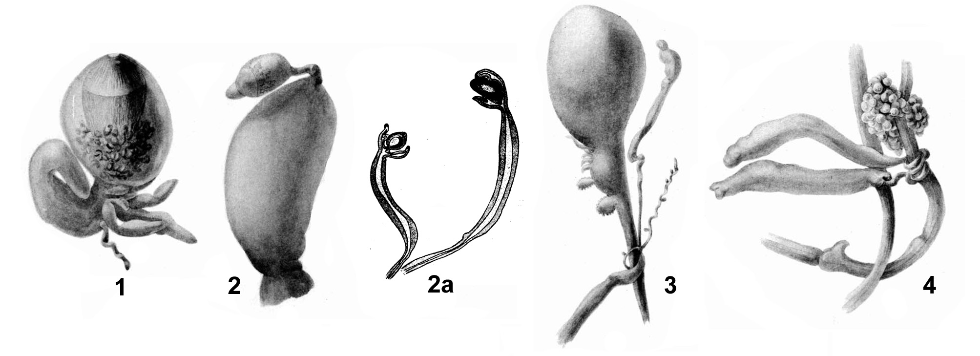
A huge monograph concerning the siphonophores caught by the Gauss during the Deutsche Südpolar-Expedition 1901-1903 was eventually published by Moser (1925). Within the collections there were a few fragments of rhizophysids, including what Moser considered to be a larval form (see Figure 32 View FIGURE 32 , 1 View FIGURE 1 ), consisting of a relatively small pneumatophore, 5 mm in length, and a very short fragment of the siphosome to which were attached some gastrozooids, one of which was considered to be at the terminal end. Moser noted the presence of hypocystic villi and also claimed that an apical pore was absent; speculating that it might appear later in development. Whether this really is a post-larval form of a Rhizophysa species or just the extreme anterior end of a larger specimen remains uncertain; but we need not considered it any further as it is totally unrecognisable.
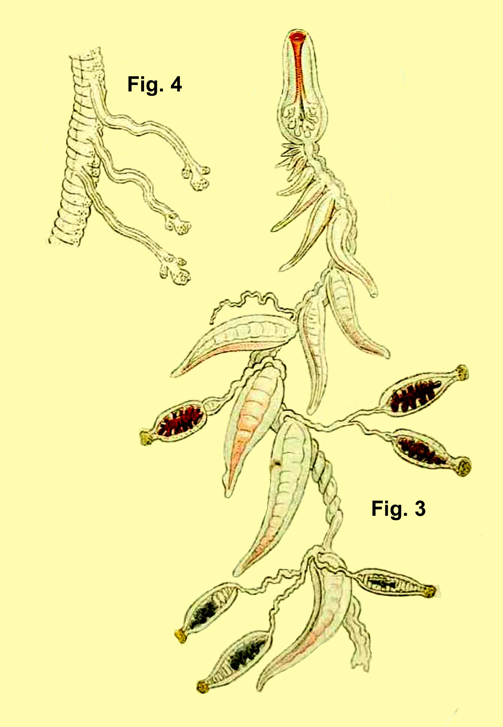
Moser (1925) also found fragments of two rhizophysid specimens that she considered had sufficient characters to warrant a specific designation, provisionally in the genus Rhizophysa . The first of these she named R. (?) tricornuta Moser (see Figure 32 View FIGURE 32 , 2, 2a View FIGURE 2 ) that was collected in the vicinity of the Cape Verde Islands. There is very little detail in the general description of this species, and it is difficult reconcile Moser’s fig. 2 (see Figure 32 View FIGURE 32 , 2 View FIGURE 2 ) with a pneumatophore, as she described it. The main portion is clearly a gastrozooid, but it is uncertain as to what the attached part might be. Apart from the detail regarding the tentilla, the only other character that Moser mentions is that the gastrozooids were borne on long peduncles, in contrast to the arrangement for R. filiformis . However, it seem, likely, as was the case for Cannophysa filiformis described by Mayer (1894) that the so-called peduncles were actually the tentacles of the detached gastrozooids that have become entangled around the stem. Moser spent much time comparing the structure of the tentilla, firstly with the “birds head” like ones described by Gegenbaur (see above) and then the tentilla of Athorybia ; noting that the central, terminal ampulla was devoid of nematocysts. However, she decided that in both cases there sufficient differences to distinguish her species. She stated that she would describe the tentilla in more detail in a later publication, but this never appeared. So, given the lack of detail, its seems best to consider this species as another nomen nudum.
The other species, of which Moser (1925) had three specimens, she named Rhizophysa (?) megalocystis Moser (see Figure 32 View FIGURE 32 , 3–4 View FIGURE 3 View FIGURE 4 ). The pneumatophore was relatively large, up to 7–8 mm in diameter, and had an apical pore, but no mention was made of hypocystic villi. Immediately below the pneumatophore there were five buds, increasing in size posteriorly, arranged in a single sequence. Moser considered that the siphosomal stem divided, which is extremely unlikely (see the discussion below regarding Bathyphysa japonica Kawamura ). The remainder of her description is very confusing and lacking in detail, although it does appear that Moser considered that, as well as gastrozooids with their tentacles, and gonodendra, there was the possibility that palpons were present. A close examination of Moser’s Plate XXXIII ( Figure 32 View FIGURE 32 , figs. 3-4) appears to show that she fell into the same trap as Studer (1878) in that the long narrow peduncles of the gastrozooids actually might be their tentacles that have become wrapped around the siphosomal stem and which might explain the division in the stem that she described. Nonetheless, there is so little detail that, again, it is felt that this species must be considered as a nomen nudum.
The only subsequent authors that have commented on these two species of Moser appear to be Bigelow & Sears (1937, pp. 64–65). They remarked that: “ R. tricornuta like R. filiformis , has “vogelkopfartigen Tentakelknopfen” [bird-headed tentacle buttons] ( Moser, 1925, p. 445). According to Moser, the terminal median beak of her new species corresponded more closely to the terminal ampulla in the young tentillum of Anthophysa ( Bigelow, 1911, P1. 20, Fig. 8 View FIGURE 8 ), or of Agalmidae before the cnidoband has become spirally coiled, than to the terminal median structure in the beak-like tentilla of R. filiformis . But comparison of Haeckel’s (1888b, P1. 24, Fig. 9 View FIGURE 9 ) figure of the trifid tentillum of R. filiformis , and of the ‘Thor’ examples of the latter, with Moser’s (1925, Textfig. 59) illustration of tentilla of tricornuta fails to show any essential difference: the terminal structure is, in fact, only slightly more dilated in Moser’s figure than it is shown by Haeckel, or than in the ‘Thor’ example ... And the lateral filaments show the same indication of incipient subdivision. We are, therefore, inclined to refer tricornuta to the synonymy of filiformis .
“Moser’s R. megalocystis is set apart by the possession of palpons (“Tentakeltaster”, Moser, 1925, p. 447) bearing giant nematocyst capsules. As no corresponding structures have ever been described for Rhizophysa , it cannot be referred to that genus, but may belong to the genus Salacia , in which the polygastric cormidia are described by Haeckel (1888b, p. 332) as bearing thin spindle shaped tubes, “perhaps palpons which remain permanently mouthless”, as well as siphons”. The present author does not agree with this conclusion, as noted above.
From henceforth the number of Rhizophysa species was stabilised at two, R. filiformis and R. eysenhardtii , but Leloup (1936) gave an extensive review of the genus Bathyphysa and the six species that Bigelow (1911) had included in his sub-family Bathyphysinae. Unfortunately, Leloup started badly by resurrecting Bedot’s (1893, 1904) contention regarding the presence of pneumatozoïdes. Leloup agreed with Bedot (1904) that the term pneumatozoïde was inappropriate, as the zooids had no hydrostatic function, but still considered that they were not the first stages of the development of the gastrozooids proper, but had a totally different function. He reached this conclusion because he contended that no intermediary stages between the pneumatozoïdes and full grown gastrozooids had been found. We now know (see Biggs & Harbison, 1976) that the young gastrozooids, with ptera, but without tentacles, do gradually transform into the mature gastrozooid, with a well tentacle, but with the ptera reduced or totally resorbed. Fortunately, however, this erroneous assumption did not greatly affect the remainder of Leloup’s discussion.
For Studer’s Rhizophysa conifera Leloup (1936) believed that as the gastrozooids matured they came to be borne on small peduncles, and possessed a tentacle without tentilla. He likened the apparent disappearance of the ptera on these mature gastrozooids to that which Lens & van Riemsdijk (1908) had described for the gastrozooids of Pterophysa grandis . With regard to Studer’s other species, Bathyphysa abyssorum, Leloup was able to examine numerous detached gastrozooids from the original specimen. These led him to conclude that the filaments that appeared to attach the gastrozooids to the stem were actually tentacles that had become wrapped around the latter, as Lens & van Riemsdijk had initially suggested. He was able to discern the scar left from where the gastrozooid had been attached to the stem, although he described this as lying at the distal end of the gastrozooid. Leloup, therefore, concluded that the gastrozooids were sessile, being attached directly to the stem and bearing a simple tentacle, without tentilla. Unfortunately the present author has been unable to ascertain whether Studer’s specimens are still in existence.
For Pterophysa grandis Leloup (1936) noted that, from Lens & van Riemsdijk’s (1908) redescription, all the gastrozooids were attached directly to the stem and bore simple tentacles, without tentilla. For both Bathyphysa grimaldi and P. ( B.) studeri Leloup was able to examine some of the original material. Although he considered that the presence of the special organ that Bedot (1904) had described at the distal end of the pneumatozoïdes set these structures apart, he considered that the mature gastrozooids in both species were both attached in the same way, presumably directly to the stem or perhaps on a small bud-like excrescence. Finally he was able to re-examine the material of B. sibogae and concluded that Lens & van Riemsdijk were wrong to describe the gastrozooids as being borne on long peduncles, having found numerous loose gastrozooids, bearing a tentacle with tentilla, but without any sign of a peduncle. Thus, he concluded that for all species the gastrozooid was attached directly to the stem and that Lens and van Riemsdijk’s (1908) division of the bathyphysid species into two genera was not justified. He, therefore, retained the older generic name, Bathyphysa and, based only on the presence or absence of tentilla on the tentacles, he recognised just two species, namely B. conifera , with simple tentacles, and B. sibogae , with tentacles bearing tentilla.
Although most recent authors, with the notable exceptions of Stepanjants (1967) and Carré & Carré (1995), concurred with this conclusion, despite Leloup’s (1936) justifications for his conclusion that the gastrozooids of Bathyphysa sibogae were sessile being untenable.. This is because Biggs & Harbison (1976) have largely confirmed Lens & van Riemsdijk’s (1908) original description of B. sibogae and have shown that the mature gastrozooids are indeed borne on long peduncles, each bearing a tentacle with distinctive tentilla. With regard to B. conifera , including its four junior synonyms, it has been suggested that the mature gastrozooids may be borne on a short peduncles that, on preservation, strongly contract so as to give the appearance of their total absence. However, examination of numerous photographs of a living specimen of B. conifera taken by Rob Sherwood, who has kindly given the present author access to them, seem to indicate that there is no peduncle to the gastrozooid.
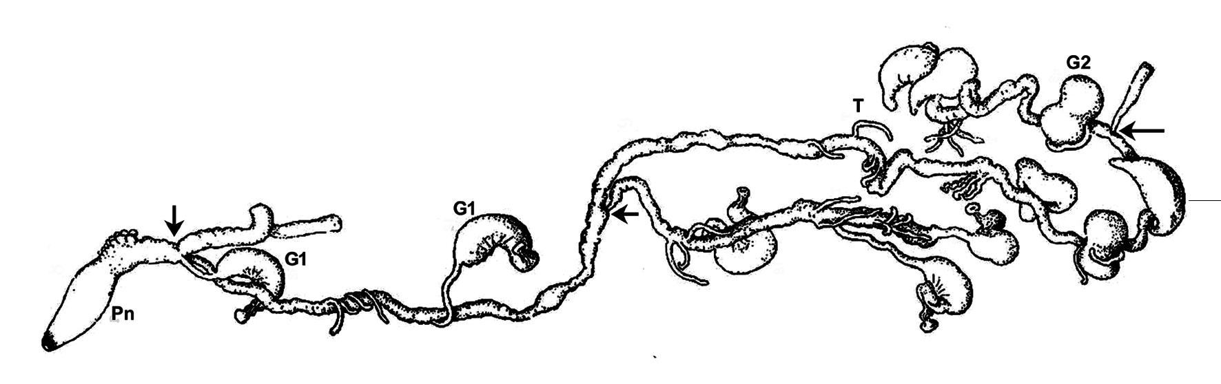
Subsequent to Leloup’s (1936) review another bathyphysid species, Bathyphysa japonica Kawamura 1947 , was described. The original description appeared in an obscure Japanese publications (see Pagès, 2002) but, fortunately, Kawamura (1954) published a further description in English. He took no account of Leloup’s (1936) review as he recognised six Bathyphysa species; but these are not the same as the six discussed by Leloup. He omitted Studer’s B. ( Rhizophysa) conifera and Lens & van Riemsdijk’s Pterophysa ( B.) studeri and replaced them with Haeckel’s (1888b) B. gigantae [sic] and B. sp. from Bigelow (1911), both of which are nomina nuda. As well as the description of the new species, Kawamura also gave details on a further specimen of B. grimaldii . Unfortunately, both descriptions are short in detail, and Kawamura described both species as having branched stems (see Figure 33 View FIGURE 33 ), which is, of course, theoretically impossible.
For Bathyphysa grimaldii Kawamura (1954) noted the absence of hypocystic villi at the base of the pneumatophore. He considered this to be a character distinguishing the genera Bathyphysa and Rhizophysa , but it is not in accord with previous descriptions and, indeed, is incorrect. He also described the presence of many “pneumatosiphons” or “pneumatozoides”, without specifically mentioning the presence of ptera, thereby adopting the erroneous terminology of Bedot (1893), but perhaps acknowledging that they were in fact siphons/gastrozooids, which Bedot did not. Like Bedot (1893), Kawamura ( ibid. p. 121) described these “bladder-like siphons” as having a short peduncle: “while the kidney-shaped gastric portion is converted into an airbladder-like structure, embracing a gas bubble in their interior”. However, as pointed out above, in his later paper Bedot (1904) corrected both of these interpretations, stating that the peduncle was actually a tentacle-like structure attached at the base of the sessile pneumatozoïde, and that the structure did not enclose an air bubble and, thus, was not a flotation device. Although Kawamura referred to Bedot’s paper in his references, he did not appear to have cited it anywhere in the text.
Kawamura (1954) also described “hook-shaped siphons”, presumably more mature ones, which he interpreted as being borne on long, slender peduncles. He also noted the presence on these of thin lateral membranes, i.e. ptera. Although Bedot (1904) observed greatly reduced ptera on his mature gastrozooids of Bathyphysa grimaldii he was unable to observe their mode of attachment, although he considered it most likely that they were attached directly to the stem. In addition, Kawamura mentioned two or three “zooids” that he believed were pairs of “pneumatosiphons” fused together. Finally, he illustrated a gonodendron apparently attached to a “pneumatosiphon”. There are thus many peculiarities in Kawamura’s description of B. grimaldii , not least the presence of a branching stem, many of which are not in accord with the redescription of the species by Bedot (1904). Unfortunately, according to Pagès (2002), the whereabouts of the specimen cannot be traced. However, the present author considers that Kawamura’s interpretations of various structures on this specimen were probably grossly inaccurate. The apparent branching of the stem must be a preservation artefact (see below) where various parts of the stem have become entangled with each other. The apparent fusion of the young gastrozooids (pneumatosiphons), again is a preservation artefact, while with regard to the attachment of the mature gastrozooids it can also been seen from Kawamura’s figures that if one interprets the singular structure at the proximal end of the gastrozooid as a peduncle, then a tentacle would be absent. As with many prior descriptions, it is most likely that what has been interpreted as a peduncle is in actuality a tentacle that has become wrapped around the stem. Thus there seems no reason not to consider Kawamura’s specimen as belonging to B. conifera .
For the other species, Bathyphysa japonica, Kawamura again believed that its stem was branched ( Figure 33 View FIGURE 33 ), and he made no mention of the presence of hypocystic villi at the base of the pneumatophore. Otherwise he gave little information of any descriptive value whatsoever, merely stating that “hook-shaped” and “bladder-shaped” gastrozooids, each with simple a tentacle, were present, as well as two fused siphons. However, it can be seen from his figure that the siphons closest to the pneumatophore are gastrozooids, not pneumatosiphons. Kawamura made no mention of the presence of ptera or the mode of attachment of the gastrozooid to the stem, and he distinguished his specimen from other Bathyphysa species on the basis of the smaller size and shape of the siphons, and the form of the pneumatophore. He then came to some strange generalised conclusions, which need not be considered here.
Totton (1965) agreed with Leloup (1936) that there were only two valid species, Bathyphysa conifera and B. sibogae , but he included Kawamura’s B. japonica as a species inquirenda because of the apparent presence of stem branches. Referring to both of Kawamura’s Bathyphysa descriptions Totton said ( ibid. p, 43): “My tentative interpretation is that these branches are gonodendra. The fact that they bear gastrozooids may perhaps be explained in the following way: Physonect gonodendra develop by successive budding of palpons from the bases of their predecessors. These palpons are reduced gastrozooids. In Bathyphysa gonodendra appear to develop from unreduced gastrozonoids [sic], though there is a terminal gonopalpon on the ultimate branchlets”.
Fortunately, the specimen of Bathyphysa japonica is still extant and was re-examined by Pagès (2002). Although Pagès did not mention whether hypocystic villi were present at the base of the pneumatophore, he was able to clearly show that the so-called branching of the stem was an artefact caused by the detachment of the posterior part of the stem, and its subsequent entanglement with the anterior part. Thus the stem part illustrated by Kawamura (see Figure 33 View FIGURE 33 ) between the most anterior and the next branching (arrowed) was actually two parts of the stem ravelled around each other. For the gastrozooids Pagès noted that the youngest ones, which included Kawamura’s fused siphons, bore ptera and were attached directly to the stem, but he made no mention of ptera on the older gastrozooids. Although the tentacles appeared simple, without tentilla, Pagès was uncertain as to whether this could be a preservation artefact, in that tentilla had been lost. This argument could, of course, be applied to all previous descriptions of B. conifera . In addition, as no mature gastrozooids actually were attached to the stem, he was uncertain as to whether they were borne on a peduncle or not, noting that although it was known that B. sibogae had mature gastrozooids borne on long peduncles the young gastrozooids were sessile. However, he erroneously stated that Lens & van Riemsdijk (1908), with regard to B. grandis , considered that peduncles were present on all gastrozooids although, as noted above, Lens & van Riemsdijk statement that the gastrozooids of this species were all sessile itself is incorrect.
Although Pagès (2002) was unsure as to whether Kawamura’s (1954) of Bathyphysa japonica could, with certainty, be associated with either B. conifera or B. sibogae , the information that he provided was quite sufficient enough to show that there were no specific characters by which one could continue to consider B. japonica as a valid species. For my part, it seems that there is every reason to consider the species as a junior synonym of B. conifera , based on the general construction of the preserved young gastrozooids, the absence of any tentilla on the tentacles (as even if they are lost some remnant of their attachment often remains), and the probability that the mature gastrozooids were attached to the stem by, at most, short peduncles.
Thus the ultimate conclusion is that of the seven species, excluding Haeckel’s Bathyphysa gigantea as it was never described, that have previously been considered for inclusion in the genus Bathyphysa , only two are valid, namely conifera and sibogae . The two species are, if the material is good enough, easily distinguished by the fact that the mature gastrozooids of B. sibogae are borne on long peduncles and bear tentacles with tentilla, while in B. conifera the gastrozooids have, at most, short attachment peduncles and their tentacles do not bear tentilla. In both species hypocystic villi are present at the base of the pneumatophore, although Totton (1965, p. 42) mistakenly said, in his diagnosis of the genus, that they were absent.


No known copyright restrictions apply. See Agosti, D., Egloff, W., 2009. Taxonomic information exchange and copyright: the Plazi approach. BMC Research Notes 2009, 2:53 for further explanation.
|
Kingdom |
|
|
Phylum |
|
|
Class |
|
|
Order |
|
|
SubOrder |
Cystonectae |
|
Family |
|
|
Genus |