
Polana ( Declivella ) maculosa, Gonçalves & Takiya & Mejdalani, 2018
|
publication ID |
https://doi.org/10.11646/zootaxa.4457.1.7 |
|
publication LSID |
lsid:zoobank.org:pub:39481728-AB63-4818-9D9D-20FFB99358DD |
|
DOI |
https://doi.org/10.5281/zenodo.5967975 |
|
persistent identifier |
https://treatment.plazi.org/id/656233E8-26AD-4A33-B49E-59E22B35F0BC |
|
taxon LSID |
lsid:zoobank.org:act:656233E8-26AD-4A33-B49E-59E22B35F0BC |
|
treatment provided by |
Plazi |
|
scientific name |
Polana ( Declivella ) maculosa |
| status |
sp. nov. |
Polana ( Declivella) maculosa View in CoL sp. nov.
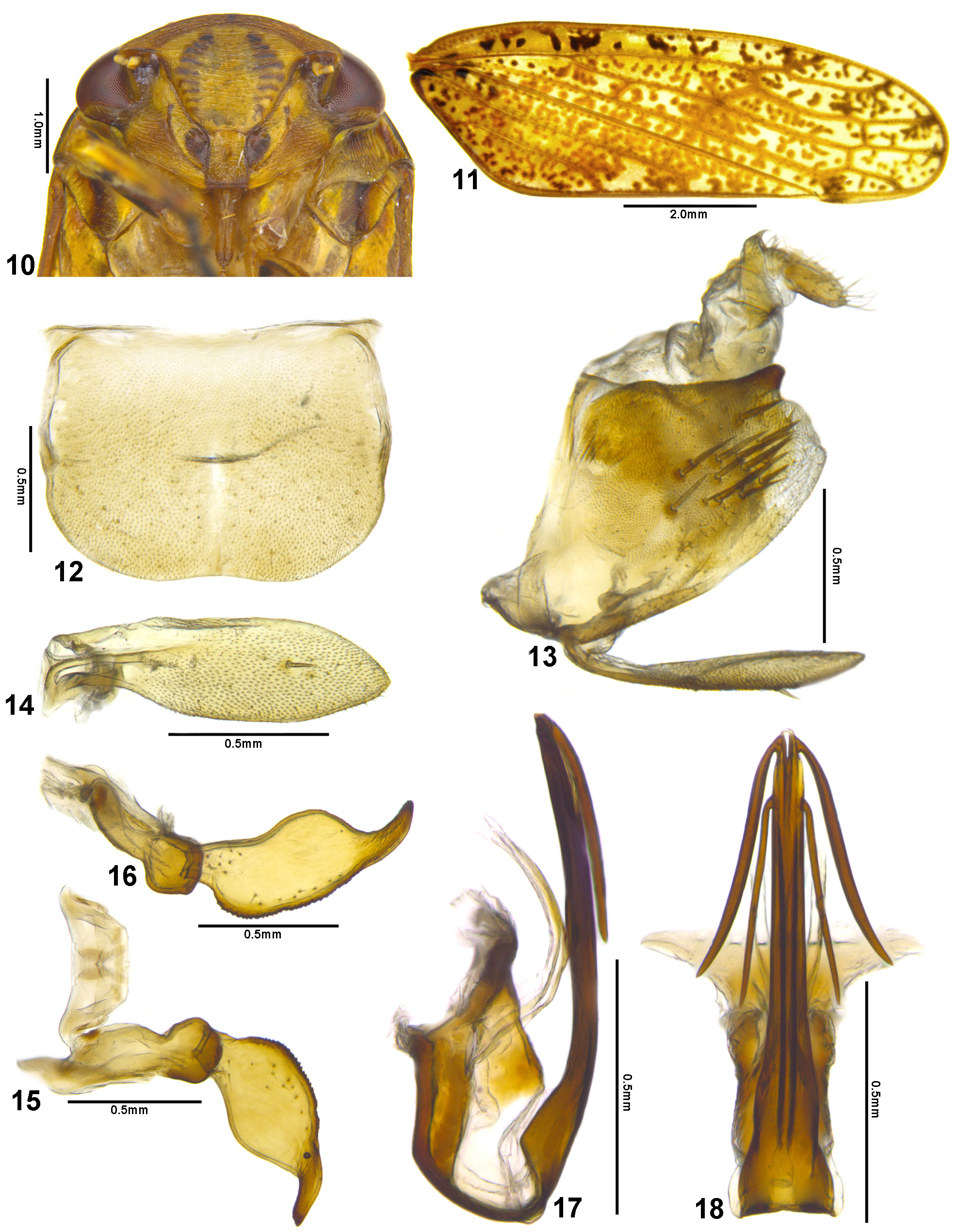
( Figs 10–18 View FIGURES 10–18 , 44, 45 View FIGURES 40–47 )
Diagnosis. Male pygofer ( Fig. 13 View FIGURES 10–18 ) with dorsoapical, rounded sclerotized small process; style ( Figs 15, 16 View FIGURES 10–18 ) broad medially and with ventral margin serrate; aedeagal shaft ( Figs 17, 18 View FIGURES 10–18 ) with two pairs of long and narrow lateral processes, one apical and one preapical.
Description (male holotype). Length 10.9 mm. Head ( Fig. 44 View FIGURES 40–47 ), in dorsal view, short, just slightly produced anteriorly; slightly narrower than pronotum; transocular width about five-sixths of humeral width of pronotum. Crown ( Fig. 44 View FIGURES 40–47 ) with median length approximately one-third of interocular width; anterior and posterior margins approximately parallel; surface slightly depressed on outer side of ocelli; texture with numerous punctures and transverse superficial striations; coronal suture indistinct. Ocellus large; equidistant between anterior and posterior margins of crown; closer to adjacent compound eye than to midline. Transition between crown and face ( Fig. 45 View FIGURES 40–47 ) indistinct and rounded. Face ( Fig. 10 View FIGURES 10–18 ) wider than high. Frontogenal suture not extending upwards beyond antennal ledge. Antennal ledge, in frontal view, approximately transverse; extending inwardly beyond frontogenal suture. Frons ( Fig. 10 View FIGURES 10–18 ) slightly swollen; texture with transversal superficial striations; lateral margin separated from eye by distance slightly larger than maximum width of clypeus. Epistomal suture distinct, complete. Clypeus ( Fig. 10 View FIGURES 10–18 ) higher than wide, expanded apically.
Pronotum, in dorsal view ( Fig. 44 View FIGURES 40–47 ), with lateral margins convergent anteriorly; posterior margin approximately straight; disk transversely striated and covered by numerous dark punctures; in lateral view ( Fig. 45 View FIGURES 40–47 ), strongly declivous anteriorly. Forewing ( Fig. 11 View FIGURES 10–18 ) subhyaline; venation distinct, not reticulated; apex rounded; appendix very narrow, extending only to second apical cell. Forefemur with AD, AM, and PD rows reduced and poorly defined, AD1, AM1, and PD1 indistinct; AV row restricted to basal half, formed by eight or nine setae, increasing in length towards apex, slightly spaced apart, seta AV1 indistinct; IC row formed by slightly arched comb of fine setae beginning at distal half of femur; PV row formed by three basal setae and one apical seta. Hind leg with femoral setal formula 2:2:1; tibial AD row with long spiniform setae with prominent bases, without intercalary microsetae; first tarsomere with two parallel rows of setae on ventral surface, pecten with seven platellae medially; apex of second tarsomere with four apical platellae.
Coloration. Mottled with brown ( Figs 44, 45 View FIGURES 40–47 ). Crown ( Fig. 44 View FIGURES 40–47 ) with dark brown to black median line, extending from anterior to posterior margin, faint medially; with black macula posterior to each ocellus. Frons ( Fig. 10 View FIGURES 10–18 ) with nine pairs of aligned black markings over muscle impressions. Lorum ( Fig. 10 View FIGURES 10–18 ) with large brown marking adjacent to clypeus. Pronotum ( Fig. 44 View FIGURES 40–47 ) mottled with dark brown on anterior half; disk with numerous dark brown punctures. Mesonotum ( Fig. 44 View FIGURES 40–47 ) with basolateral angles dark brown; with pair of elongate, oblique brown marks anterior to scutellar suture. Forewing ( Fig. 11 View FIGURES 10–18 ) covered by numerous brown maculae throughout surface. Legs ( Fig. 45 View FIGURES 40–47 ) mostly pale brown with yellow areas and scattered dark brown maculae; tarsi dark brown.
Male terminalia. Sternite VIII ( Fig. 12 View FIGURES 10–18 ) wider than long, median length approximately three-fourths of maximum width; posterolateral corners broadly rounded; posterior margin slightly excavated medially. Pygofer ( Fig. 13 View FIGURES 10–18 ) approximately as long as high; dorsal margin with small, preapical sclerotized bulbiform process; apex broadly rounded and slightly sclerotized; macrosetae distributed on median portion of apical half. Subgenital plate, in lateral view ( Fig. 13 View FIGURES 10–18 ), as long as pygofer; in ventral view ( Fig. 14 View FIGURES 10–18 ), ligulate, 3.1 times longer than wide; apex subacute; ventral surface with one median macroseta. Connective ( Fig. 15 View FIGURES 10–18 ) somewhat U-shaped, stem absent; total length about one-seventh of style length; dorsal surface with two parallel longitudinal keels. Style ( Figs 15, 16 View FIGURES 10–18 ) with outer lobe developed and rounded; in lateral view ( Fig. 16 View FIGURES 10–18 ), blade strongly expanded medially and slightly curved dorsally; ventral margin serrated, except base and apex; apex digitiform and strongly sclerotized. Aedeagus ( Figs 17, 18 View FIGURES 10–18 ) with preatrium very short; dorsal apodeme developed and expanded laterally; atrium elongated and bearing pair of narrow, elongated, fragile processes, directed posterodorsally; shaft tubular and directed dorsally; with two pairs of elongate lateral processes, directed ventrally, apical pair slightly more robust than preapical pair.
Female unknown.
Etymology. The name of the new species alludes to the mottled aspect of the forewings ( Figs 11 View FIGURES 10–18 , 44, 45 View FIGURES 40–47 ).
Material examined. Male holotype: “ Brasil, Roraima, Caracarai\ Serra da Mocidade,\ 600m., 1°36’N \ 61°54’W, bandeja amarela\ + septo, 15–26.i.2016;\ F.F.Xavier F°; R. Boldrini\ & P. Barroso”; “DNA voucher\ Entomologia DZRJ \ ENT 3816” ( INPA).
Remarks. Polana ( Declivella) maculosa sp. nov. is similar to P. ( D.) danesa in appearance and color ( Figs 46, 47 View FIGURES 40–47 ). However, P. danesa differs in having the forewing with a lower density of maculae per cell, the style with a thinner blade that is strongly bent dorsally, and the aedeagus with stem expanded apically and bearing three pairs of processes.
Additional material examined. Polana danesa , male holotype: “ Tocumen , Panama \ 6.I.1953, F.S.Blanton ” ( USNM).

No known copyright restrictions apply. See Agosti, D., Egloff, W., 2009. Taxonomic information exchange and copyright: the Plazi approach. BMC Research Notes 2009, 2:53 for further explanation.
|
Kingdom |
|
|
Phylum |
|
|
Class |
|
|
Order |
|
|
Family |
|
|
SubFamily |
Iassinae |
|
Tribe |
Gyponini |
|
Genus |