
Polana ( Striapona ) desela DeLong, 1979
|
publication ID |
https://doi.org/10.11646/zootaxa.4457.1.7 |
|
publication LSID |
lsid:zoobank.org:pub:39481728-AB63-4818-9D9D-20FFB99358DD |
|
DOI |
https://doi.org/10.5281/zenodo.5967977 |
|
persistent identifier |
https://treatment.plazi.org/id/03856B70-6306-7B0B-D5F8-FC13FC8F011B |
|
treatment provided by |
Plazi |
|
scientific name |
Polana ( Striapona ) desela DeLong, 1979 |
| status |
|
Polana ( Striapona) desela DeLong, 1979 View in CoL
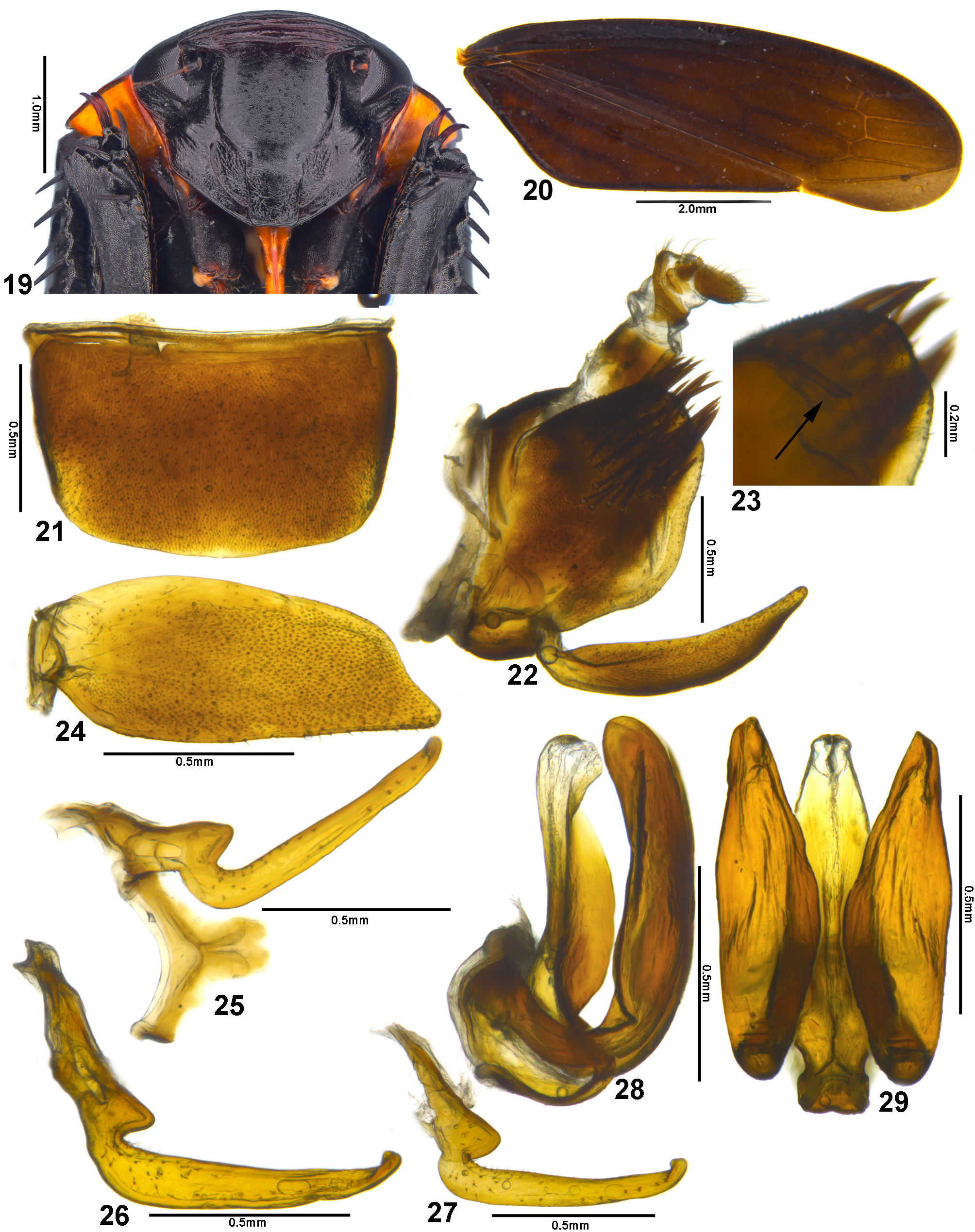
( Figs 19–39 View FIGURES 19–29 View FIGURES 30–39 , 48–54 View FIGURES 48–54 )
Diagnosis. Male sternite VIII ( Fig. 21 View FIGURES 19–29 ) about twice wider than long and with posterior margin broadly convex; male pygofer ( Fig. 22 View FIGURES 19–29 ) with apex slightly bilobed, with long and narrow inner basodorsal process ( Fig. 23 View FIGURES 19–29 ); style ( Figs 25–27 View FIGURES 19–29 ) long and narrow, with conspicuous outer lobe on basal half, apex curved dorsally and subacute; aedeagus ( Figs 28, 29 View FIGURES 19–29 ) with pair of long and broad processes of dorsal apodeme, positioned posteriorly to shaft, latter strongly flattened dorsoventrally.
Redescription (based on additional specimens). Length of male 8.7–9.7 mm (n = 4); female 9.9 mm (n = 1). Head ( Figs 48, 50, 51, 53, 54 View FIGURES 48–54 ), in dorsal view, slightly or moderately produced anteriorly; distinctly narrower than pronotum; transocular width about three-fourths of humeral width of pronotum. Crown ( Figs 48, 50, 51, 53, 54 View FIGURES 48–54 ) with median length approximately half of interocular width; anterior margin broadly rounded; surface flattened; texture with numerous transverse striae; coronal suture distinct or not. Ocellus small, closer to anterior crown margin than to posterior margin; closer to adjacent compound eye than to midline. Transition between crown and face ( Figs 49, 52 View FIGURES 48–54 ) defined, broad; transversely striated. Face ( Figs 19 View FIGURES 19–29 , 30 View FIGURES 30–39 ) slightly wider than high. Frontogenal suture extending dorsally beyond antennal ledge. Antennal ledge, in frontal view, slightly oblique downward in relation to frons; extending inwardly beyond frontogenal suture. Frons ( Figs 19 View FIGURES 19–29 , 30 View FIGURES 30–39 ) flattened; texture shagreen; lateral margin separated from lateral margin of eye by distance shorter than maximum width of clypeus. Epistomal suture indistinct. Clypeus ( Figs 19 View FIGURES 19–29 , 30 View FIGURES 30–39 ) higher than wide; with parallel lateral margins.
Pronotum, in dorsal view ( Figs 48, 50, 51, 53, 54 View FIGURES 48–54 ), with lateral margins convergent anteriorly; posterior margin slightly concave; disk transversely striated; in lateral view ( Figs 49, 52 View FIGURES 48–54 ), moderately declivous anteriorly. Forewing ( Figs 20 View FIGURES 19–29 , 31 View FIGURES 30–39 ) subhyaline; venation distinct, not reticulated; apex rounded; appendix broad, extending only to second apical cell. Forefemur with AD, AM, and PD rows reduced and poorly defined, except for apical setae AD1, AM1, and PD1; AV row formed by five long setae, slightly spaced apart and restricted to basal half of femur; IC row formed by slightly arched comb of fine setae beginning at apical half of femur and extending to AM1; PV row formed by four to five setae spaced apart and occupying entire femur, proximal seta distinctly smaller than others. Hind leg with femoral setal formula 2:2:1; tibial AD row with long spiniform setae with prominent bases and with two or more intercalary microsetae distributed along entire length; first tarsomere with one row of setae on ventral surface, external row indistinct; pecten with four platellae medially flanked by longer tapered lateral setae; second tarsomere with two apical platellae.
Coloration (based on holotype). Mostly dark brown ( Figs 48, 49 View FIGURES 48–54 ). Pronotum ( Figs 48, 49 View FIGURES 48–54 ) and mesonotum pale brown; pronotum with large, dark brown, transverse spot on disk. Forewing ( Figs 48, 49 View FIGURES 48–54 ) dark brown, without maculae.
Male terminalia (based on additional specimens compared to holotype). Sternite VIII ( Fig. 21 View FIGURES 19–29 ) wider than long, median length approximately half of maximum width; posterolateral corners slightly rounded; posterior margin broadly rounded. Pygofer ( Fig. 22 View FIGURES 19–29 ) approximately as long as high; dorsal margin slightly convex; ventrocaudal margin broadly rounded; macrosetae numerous and distributed throughout dorsoapical half; apex slightly bilobed ( Fig. 23 View FIGURES 19–29 ); inner basodorsal process ( Fig. 23 View FIGURES 19–29 ) present, weakly sclerotized, narrow and long, extending to apical third of pygofer, apex rounded. Subgenital plate, in lateral view ( Fig. 22 View FIGURES 19–29 ), as long as pygofer; in ventral view ( Fig. 24 View FIGURES 19–29 ), ligulate; about two times longer than maximum width; apex obtuse. Connective ( Fig. 25 View FIGURES 19–29 ) Yshaped; stem slightly shorter than arms; total length about one-fourth of style length. Style ( Fig. 25 View FIGURES 19–29 ), in dorsal view, with conspicuous outer lobe at basal half; in lateral view ( Fig. 26 View FIGURES 19–29 ), blade narrowing progressively towards apex; latter dorsally curved and subacute. Aedeagus ( Figs 28, 29 View FIGURES 19–29 ) with preatrium reduced; dorsal apodeme developed and expanded laterally, bearing pair of processes directed posteriorly and bent dorsally at right angle, located posteriorly to shaft; each process approximately tubular at basal third, strongly expanded laterally and flattened dorsoventrally at apical two-thirds, ligulate, long, slightly exceeding apex of shaft; atrium elongated; shaft with basal fourth tubular and strongly folded anteriorly, bending over itself, apical three-fourths directed dorsally, strongly expanded laterally, dorsoventrally flattened, lateral margins curved posteriorly; apex slightly sclerotized and rounded.
Female terminalia. Sternite VII ( Fig. 32 View FIGURES 30–39 ), in ventral view, about two times wider than long; posterolateral corners rounded and slightly produced posteriorly; posterior margin with broad median lobe, slightly shorter than posterolateral corners. Sternite VIII membranous. Pygofer ( Fig. 33 View FIGURES 30–39 ), in lateral view, short, about 1.5 times higher than long; macrosetae sparse on apical half; apex subacute. First valvifer ( Fig. 34 View FIGURES 30–39 ), in lateral view, subrectangular, slightly higher than long. First valvula ( Figs 34–36 View FIGURES 30–39 ) about six times longer than its maximum height; in lateral view ( Fig. 34 View FIGURES 30–39 ), almost straight; basal portion produced anterad and rounded; apical half with dorsal sculptured area strigate; ventral portion of apical half slightly sclerotized and with superficial longitudinal striae ( Fig. 35 View FIGURES 30–39 ); apical portion with lateral carina bearing 11–13 small teeth ( Fig. 36 View FIGURES 30–39 ); apex acute. Second valvula ( Figs 37–39 View FIGURES 30–39 ) robust; ventral margin expanded, forming flap; dorsal margin mostly straight; apical fourth narrowed and slightly curved dorsally; in dorsal view ( Fig. 37 View FIGURES 30–39 ), with apical portion triangular; apex acute. Gonoplac ( Fig. 33 View FIGURES 30–39 ) expanded at apical fourth; dorsoapical margin straight; apex rounded.
Material examined. Male holotype: “Peru,/ Squitos, [?], XI.[19]20”; “Holotype\ Polana \ ( Striapona )\ desela \ DeLong”; “OSUC 0158535” (OSU). Additional material: 1 male, “Peru, M[adre de] D[ios], Albergue\ Refugio Amazonas\ 12°52’30”[S]/ 69°24’35”[W]\ 231m, 06.xi.2016 \ D.Couceiro”; “Wired Amazon\ Project\ pan trap” (MUSM); 2 males and 1 female, “Brasil, Amazonas, Tapauá,\ Rio Ipixuna, porto Cotinha\ 13–18.x.2013, Terra Firme\ malaise, D.M.M.Mendes”, 1m (INPA), 1m, 1f (DZRJ); 1 male, “Brasil, Ro[ndônia], Porto Velho\ Rio Madeira\ 9°35’29,50”S; 65°2’57,60”W\ 09–12-. ix.2010, malaise\ R.R.Silva & R.M.Albuquerque” (INPA).
Morphological variation. We have observed some variation among additional studied material that is worth mentioning. Body lengths (8.7–9.7 mm) of male specimens studied are a little smaller than the holotype ( 10 mm). The color pattern is quite variable among localities and between sexes. Male specimens from Tapauá ( Amazonas, Brazil) have the mesonotum black and pronotum with anterior third black and remainder yellow ( Fig. 50 View FIGURES 48–54 ), whereas the female has the mesonotum yellow and pronotum yellow with a black transverse median spot ( Figs 51, 52 View FIGURES 48–54 ); both have a black crown. The male specimen from Porto Velho (Rondônia, Brazil) has a black mesonotum and the crown and pronotum completely orange-yellow ( Fig. 53 View FIGURES 48–54 ). Finally, the specimen from Madre de Dios ( Peru) has the crown and anterior third of pronotum reddish-brown, the remainder of pronotum red, and the mesonotum black ( Fig. 54 View FIGURES 48–54 ). The holotype and Amazonas specimens have the median length of the crown approximately half of the interocular width ( Figs 48, 50, 51 View FIGURES 48–54 ), the specimen from Rondônia has the crown length about one-third of the interocular width ( Fig. 53 View FIGURES 48–54 ), while the Peruvian specimen has the shortest crown, approximately one-fourth of the interocular width ( Fig. 54 View FIGURES 48–54 ). The male terminalia of the Brazilian specimens are indistinguishable from those of the holotype. However, slight differences in the style shape were found in the Peruvian male, which has the outer lobe rounded and the blade narrow ( Fig. 27 View FIGURES 19–29 ), whereas other specimens have the outer lobe more acute and blade wider ( Fig. 26 View FIGURES 19–29 ).
Notes. Only a few published papers have provided detailed descriptions of the female terminalia in Gyponini :
Engel & Takiya (2012), Da-Silva et al. (2014), Domahovski et al. (2014, 2015), Da-Silva & Coelho (2015), and Domahovski & Cavichioli (2015, 2017a, b). The difficulty of associating males and females and the apparently low morphological variation in the structures of the female terminalia are among the possible reasons for the small number of studies on this subject.
Da-Silva & Coelho (2015) described the female terminalia of Polana ( Polanana) amapaensis Coelho, 1991 and Domahovski & Cavichioli (2017a) described those of three species of Polana ( Varpulana) : P. ( V.) grossi Domahovski & Cavichioli, 2017 , P. ( V.) naja Domahovski & Cavichioli, 2017 , and P. ( V.) sapitanduva Domahovski & Cavichioli, 2017 . The structures of the female terminalia of all these species have characteristics similar to those of P. ( S.) desela , which are not known to occur in other genera of the tribe. These include: (1) first valvula with basal portion produced anterad and rounded ( Fig. 34 View FIGURES 30–39 ); (2) first valvula with apical portion with lateral carina bearing small teeth ( Fig. 36 View FIGURES 30–39 ); and (3) second valvula with ventral margin expanded, forming flap ( Fig. 38 View FIGURES 30–39 ). However, P. ( S.) desela differs from the aforementioned species in having the second valvula with the apical fourth narrowed and the dorsal margin without teeth ( Figs 38, 39 View FIGURES 30–39 ).


No known copyright restrictions apply. See Agosti, D., Egloff, W., 2009. Taxonomic information exchange and copyright: the Plazi approach. BMC Research Notes 2009, 2:53 for further explanation.
|
Kingdom |
|
|
Phylum |
|
|
Class |
|
|
Order |
|
|
Family |
|
|
SubFamily |
Iassinae |
|
Tribe |
Gyponini |
|
Genus |