
Tridensius, Jałoszyński, 2020
|
publication ID |
https://doi.org/10.11646/zootaxa.4755.2.3 |
|
publication LSID |
urn:lsid:zoobank.org:pub:8D3A61EC-17A4-4824-9D67-CAF1DECEDB36 |
|
DOI |
https://doi.org/10.5281/zenodo.3812777 |
|
persistent identifier |
https://treatment.plazi.org/id/038587BC-C06B-4C6B-3C8E-FED8DE63F88C |
|
treatment provided by |
Carolina |
|
scientific name |
Tridensius |
| status |
gen. nov. |
Tridensius View in CoL gen. n.
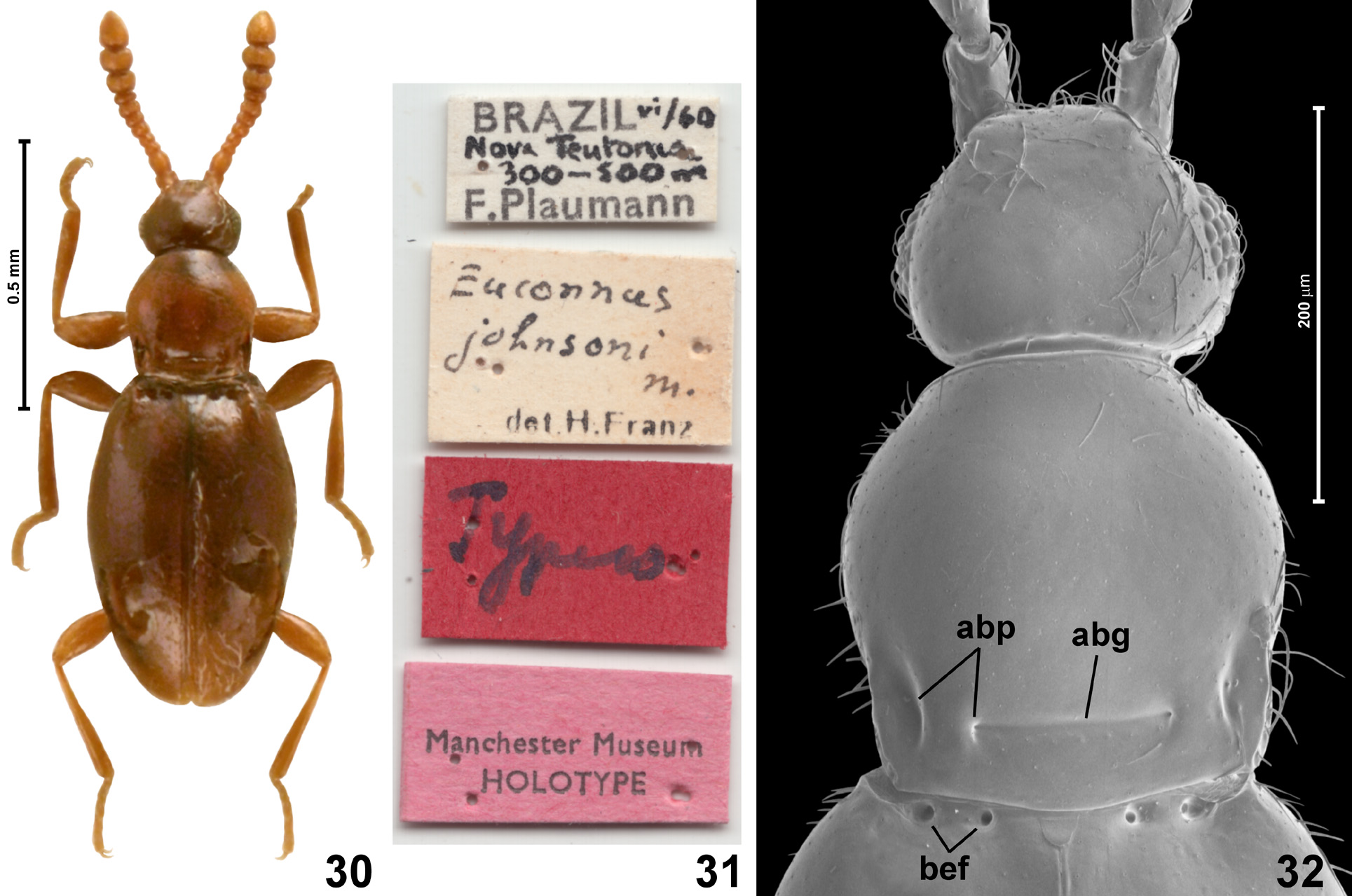
Type species: Euconnus brunneus Franz, 1988: 75 (here designated).
Diagnosis. Tridensius differs from all genera of Glandulariini in one apomorphy: mesoventral intercoxal process anteriorly (but behind procoxal rests of mesoventrite) forming a massive trident-shaped structure with lateral arms extending laterad and bent anterad; and in a set of synapomorphies, which, separately or in a different combination, are known in other Glandulariini : antenna gradually thickened; head in male with modified vertex; eyes closer to mandibular bases than to occipital constriction; thick bristles absent on head but present on sides of prothorax; submentum lacking lateral sutures; hypostomal ridges complete, connected just in front of small and circular posterior tentorial pits; the latter situated just in front of transverse impression demarcating ‘neck’ region; occipital constriction about as broad as frons between eyes, distinctly, but not much narrower than vertex; maxillary palpomere I strongly elongate; palpomere III swollen, about twice as long as broad and almost as broad as 1/3 width of head; palpomere IV relatively short, subconical, not rod-like; pronotum bell-shaped, broadest near middle, with two pairs of antebasal pits, inner pair connected by transverse groove, sublateral and lateral pronotal carinae absent; prosternum shorter than 1/3 of prothorax, with vestigial basisternal portion; prosternal process present, carinate, weakly elevated, sharply demarcated laterally; notosternal sutures complete, prosternum fully demarcated from hypomera both in front of and behind procoxal cavities, which are closed by overlapping (but not fused) postcoxal lobes of prosternum and hypomeron; hypomeral ridges complete, inner (adcoxal) portion of hypomeron divided by oblique ‘suture’ into broad and subtriangular posterior and strongly elongate, narrow anterior parts, hypomera anteriorly strongly expanding mesad and forming lateromesal subtriangular lobes in front of anterior prosternal margin; mesoscutellar shield not exposed between elytral bases; mesoventrite with shallow and diffuse anterolateral impressions functioning as procoxal rests, separated at middle by weakly elevated subtriangular median projection, the latter posteriorly touching, but not fused with, anterior portion of mesoventral intercoxal process, which is carinate, moderately elevated, nearly parallel-sided between mesocoxae; dorsolateral fovea present and deep; metaventral intercoxal process not separating metacoxae, composed of two moderately long pointed spines separated by narrow fissure; each elytron with two vestigial setose basal foveae; aedeagus with symmetrical median lobe, nearly symmetrical endophallic structures, ventral diaphragm, and slender, free parameres.
Description. Body ( Fig. 1 View FIGURES 1–3 ) relatively stout, with shallow constrictions between head and prothorax and between prothorax and elytra; sparsely setose.
Head capsule ( Figs 2–5 View FIGURES 1–3 View FIGURES 4–7 ) divided into ‘neck’ region retracted into pronotum and short, transverse anterior part; occipital constriction distinctly, but not much, narrower than vertex; vertex and frons confluent on sides, weakly convex and together about as long as broad, anterior margin of frons in dorsal view ( Fig. 3 View FIGURES 1–3 ) concave, supra-antennal tubercles large, diffuse, moderately elevated. Posterior portion of frons and vertex in male modified, with convexity of complex shape ( Fig. 4 View FIGURES 4–7 ), posterior margin of vertex lacking transverse ridge and not projecting posterodorsad. Eyes closer to mandibular bases than to occipital constriction, relatively coarsely faceted; antennal insertions broadly separated; tempora shorter than eyes. Head lacking thick bristles. Gular plate ( Fig. 5 View FIGURES 4–7 ; gp) large, with indistinct gular sutures; posterior tentorial pits situated slightly in front of transverse impression demarcating ‘neck’ region ventrally, round and small; submentum ( Fig. 5 View FIGURES 4–7 ; smn) not demarcated by lateral sutures, strongly transverse; hypostomal ridges ( Fig. 5 View FIGURES 4–7 ; hr) distinct, complete, connected at middle just in front of posterior tentorial pits. Mentum subtrapezoidal; prementum small, with narrowly separated bases of labial palps, labial palps poorly visible in the studied specimen, short. Maxillae generalized, as in most Glandulariini ; maxillary palpomere I conspicuously long, about 3 × as long as broad; II long, clavate and curved, III distinctly swollen, about twice as long as broad, nearly as broad as 1/3 of head width, palpomere IV slender but not rod-like, narrowing distad.
Antennae ( Fig. 1 View FIGURES 1–3 ) long and slender, gradually and weakly thickened distad; all antennomeres covered with sparse, long setae.
Pronotum ( Fig. 3 View FIGURES 1–3 ) in dorsal view bell-shaped, anterior margin arcuate, anterior corners broadly rounded, sides sinuate, slightly concave in posterior half, posterior corners blunt but well marked, posterior margin bisinuate. Pronotum with two pairs of antebasal pits ( Fig. 3 View FIGURES 1–3 ; abp), inner pair connected by transverse antebasal groove ( Fig. 32 View FIGURES 30–32 ; abg), lacking sublateral and lateral carinae. Sides of pronotum with thick bristles, especially dense on anteroventral portions of hypomera ( Fig. 5 View FIGURES 4–7 ).
Prosternum ( Figs 5–6 View FIGURES 4–7 ) with basisternal portion ( Figs 5–6 View FIGURES 4–7 ; bst) much shorter than coxal part; prosternal process developed as well-defined parallel-sided and weakly elevated carina, not separating procoxae; notosternal sutures ( Figs 5–6 View FIGURES 4–7 ; nss) complete; procoxal cavities closed by posterolateral lobes of prosternum that are overlapped with (but not fused to) postcoxal expansions of hypomera; hypomeral ridges ( Figs 5–6 View FIGURES 4–7 ; hyr) complete; inner (adcoxal) region of hypomeron divided by oblique ‘suture’ into broad subtriangular posterior portion and long, narrow anterior area; additionally posterior portion partly divided longitudinally by fine impressed line.
Mesonotum (not shown) with subtriangular scutellar shield about as long as broad, not exposed between elytral bases.
Elytra ( Figs 1, 3 View FIGURES 1–3 ) together oval, each with two vestigial, asetose basal elytral foveae ( Fig. 3 View FIGURES 1–3 ; bef); humeral calli and basal impressions present, apices of elytra rounded.
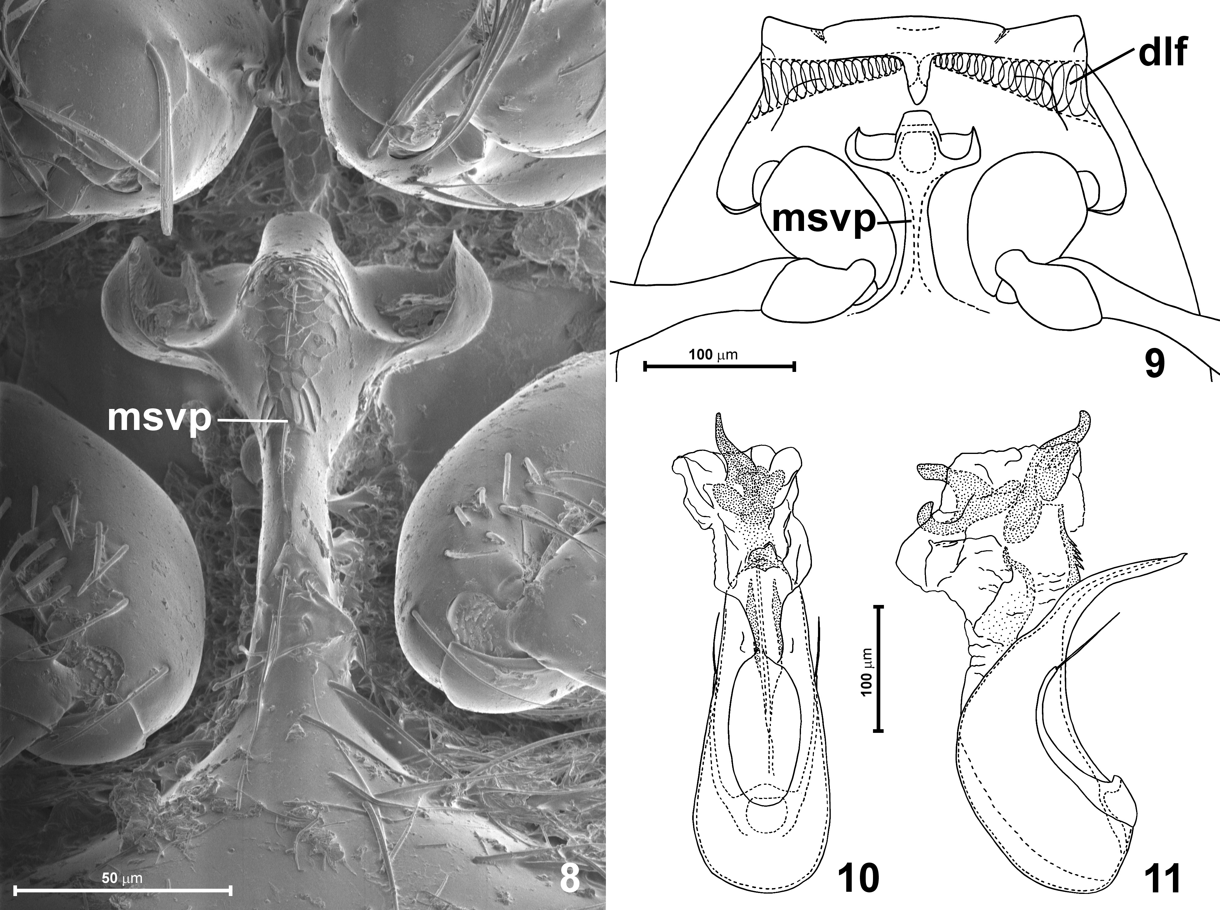
Mesoventrite ( Figs 7–9 View FIGURES 4–7 View FIGURES 8–11 ) with mesoventral intercoxal process ( Figs 7–9 View FIGURES 4–7 View FIGURES 8–11 ; msvp) carinate between mesocoxae, posteriorly fused with metaventrite, anteriorly with curved lateral arms forming a massive trident-like structure ( Fig. 8 View FIGURES 8–11 ), mesoventral process not extending to anterior mesoventral margin, but with separated subtriangular portion between diffuse and shallow anterolateral mesoventral impressions (= procoxal rests). Mesoventrite with deep dorsolateral foveae ( Fig. 9 View FIGURES 8–11 ; dlf), and with prominent mesocoxal projections.
Hind wings present.
Metaventrite ( Fig. 7 View FIGURES 4–7 ) slightly transverse, slightly broadening posteriorly; posterior margins of mesocoxal rests not carinate, but covered with dense setae; posterior metaventral margin strongly concave at each metacoxa, with metaventral intercoxal process ( Fig. 7 View FIGURES 4–7 ; mtvp) composed of two slender spines not separating metacoxae. Metanepisterna and metepimera narrow, largely covered by elytra.
Metendosternite (= metafurca) (not illustrated) with short and broad stalk and divergent lateral furcal arms with adjacent bases (nearly V-shaped).
Legs long and slender; pro- and mesocoxae oval, metacoxae strongly transverse and laterally reaching margins of metaventrite; all trochanters short and subtriangular; all femora distinctly clavate; tibiae slender; tarsi relatively short and robust.
Abdominal sternites unmodified, suture between sternites VII and VIII indistinct.
Aedeagus ( Figs 10–11 View FIGURES 8–11 ) elongate, with symmetrical median lobe and nearly symmetrical, complicated endophallus, parameres not fused with median lobe, slender, with apical setae.
Distribution and composition. Tridensius is represented by one nominal species known to occur in Venezuela.
Etymology. The name Tridensius refers to the trident-like mesoventral intercoxal process; from the Latin tridens, = three-toothed (English ‘trident’ originated from Latin noun use of adjective tridentem, nominative tridens). Gen- der masculine.
Remarks. Euconnus brunneus cannot be maintained as a member of Euconnus because of the metaventral intercoxal process developed as a pair of narrow spines that do not separate metacoxae (metacoxae separated and the process lacking spines in Euconnus ). The unique, trident-like structure of the mesoventral intercoxal process and a unique set of synapomorphies justify placing this species in a separate genus. Further comments on identification of Tridensius are given in the Discussion, including identification key to genera. Tridensius is morphologically similar to Protandroconnus and Trichocircus , all these genera have cephalic modifications in males, a conspicuously elongate maxillary palpomere I, a short prosternum with its anterior margin either strongly concave or shifted posterad in relation to the anteromesal margins of hypomera, the mesoventral intercoxal process carinate between mesocoxae, and contiguous metacoxae.


No known copyright restrictions apply. See Agosti, D., Egloff, W., 2009. Taxonomic information exchange and copyright: the Plazi approach. BMC Research Notes 2009, 2:53 for further explanation.
|
Kingdom |
|
|
Phylum |
|
|
Class |
|
|
Order |
|
|
Family |
|
|
SubFamily |
Scydmaeninae |
Tridensius
| Jałoszyński, Paweł 2020 |
Euconnus brunneus
| Franz, H. 1988: 75 |