
Sibylloconnus, Jałoszyński, 2020
|
publication ID |
https://doi.org/10.11646/zootaxa.4755.2.3 |
|
publication LSID |
urn:lsid:zoobank.org:pub:8D3A61EC-17A4-4824-9D67-CAF1DECEDB36 |
|
DOI |
https://doi.org/10.5281/zenodo.3812781 |
|
persistent identifier |
https://treatment.plazi.org/id/038587BC-C06F-4C6E-3C8E-FAE9DE49FCF8 |
|
treatment provided by |
Carolina |
|
scientific name |
Sibylloconnus |
| status |
gen. nov. |
Sibylloconnus View in CoL gen. n.
Type species: Euconnus sibyllensis Franz, 1984: 23 (here designated).
Diagnosis. Sibylloconnus differs from all genera of Glandulariini in one apomorphy: prosternum subtrapezoidal, with its anterior margin about half as wide as posterior margin, and consequently anterior regions of hypomera strongly expanding mesad; and in a set of synapomorphies, which, separately or in a different combination, are known in other Glandulariini : antenna with indistinctly delimited, pentamerous club, with antennomere VIII smaller than VII; head in male with modified frons and vertex; eyes closer to mandibular bases than to occipital constriction; thick bristles absent on head but present on sides of prothorax; submentum lacking lateral sutures; hypostomal ridges complete, connected far in front of small and circular posterior tentorial pits; the latter situated clearly in front of transverse impression demarcating ‘neck’ region; occipital constriction much narrower than vertex; maxillary palpomere I strongly elongate; palpomere III swollen, slightly more than twice as long as broad and almost as broad as 1/3 width of head (In Fig. 17 View FIGURES 15–18 palpomere III is collapsed dorsally and therefore distorted, it appears narrower than it truly is); palpomere IV relatively short, subconical, not rod-like; pronotum bell-shaped, broadest near middle, with one lateral pair of antebasal pits and with transverse groove not deepened laterally, sublateral carinae present, but not sharply marked, shifted laterad and obscured by bristles; prosternum shorter than 1/3 of prothorax, with basisternal portion distinctly shorter than coxal portion, but not vestigial; prosternal process marked as diffuse and weakly elevated longitudinal ridge not separating procoxae; notosternal sutures complete, prosternum fully demarcated from hypomera both in front of and behind procoxal cavities, which are closed by overlapping (but not fused) postcoxal lobes of prosternum and hypomeron; hypomeral ridges incomplete, obliterated shortly in front of procoxal cavities; inner (adcoxal) portion of hypomeron broad and subtriangular, hypomera anteriorly strongly expanding mesad and forming lateromesal subtriangular lobes behind anterior prosternal margin; mesoscutellar shield not exposed between elytral bases; mesoventrite with shallow and diffuse anterolateral impressions functioning as procoxal rests, separated at middle by narrow carina; mesoventral intercoxal process narrow, carinate, moderately elevated and parallel-sided between mesocoxae; dorsolateral fovea present and deep; metaventral intercoxal process not separating metacoxae, composed of two long pointed spines separated by narrow fissure; each elytron with two vestigial setose basal foveae; aedeagus with symmetrical median lobe, nearly symmetrical endophallic structures and slender, free parameres.
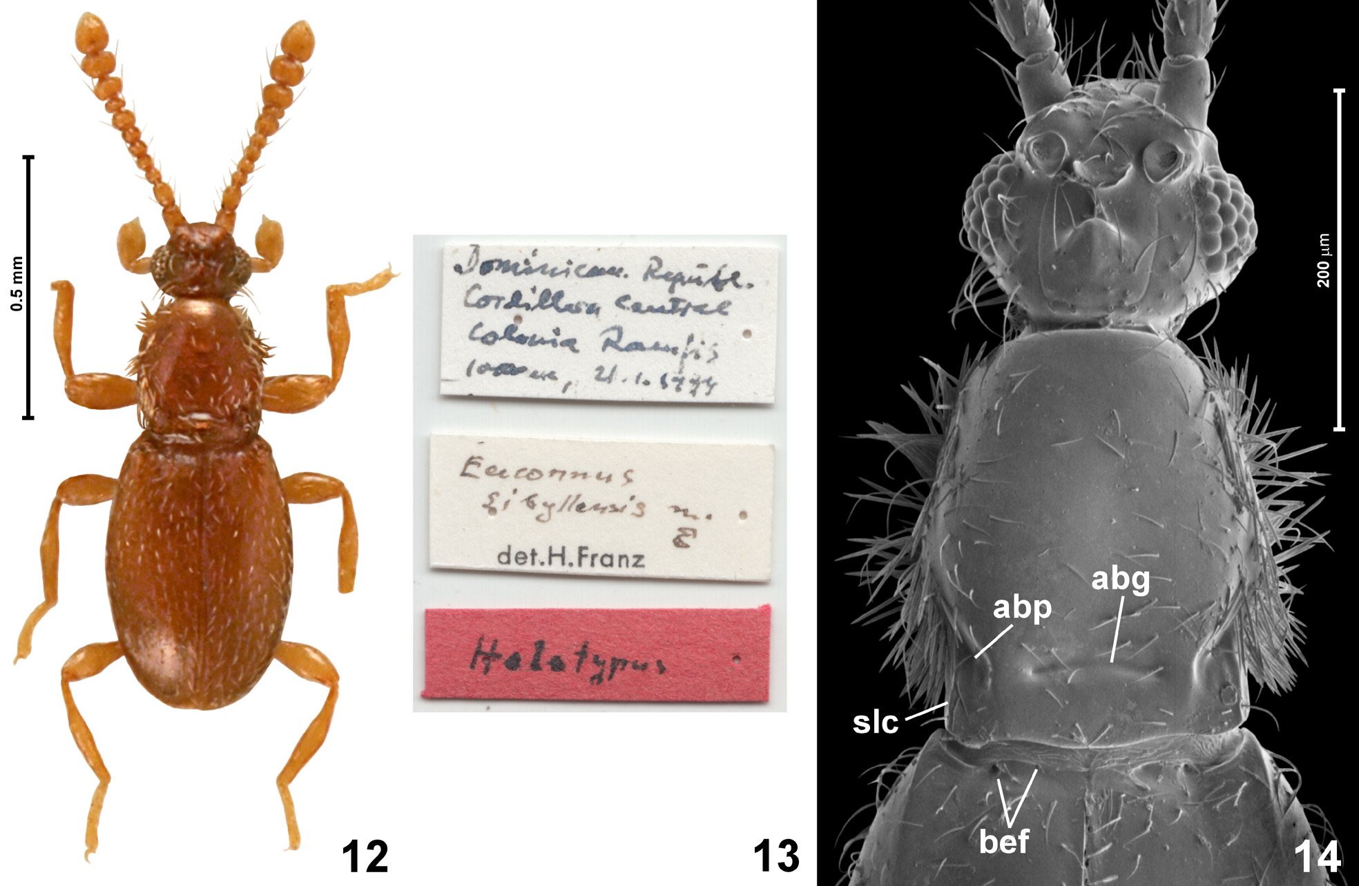
Description. Body ( Fig. 12 View FIGURES 12–14 ) moderately slender, with deep constriction between head and prothorax and shallow between prothorax and elytra; sparsely setose.
Head capsule ( Figs 14–17 View FIGURES 12–14 View FIGURES 15–18 ) divided into ‘neck’ region retracted into pronotum and short anterior part which is about as long as broad; occipital constriction much narrower than vertex; vertex and frons confluent, weakly convex and together about as long as broad, supra-antennal tubercles weakly marked. Frons and vertex in male modified ( Figs 15–16 View FIGURES 15–18 ), with median tubercles and lateral impressions, posterior margin of vertex not projecting posterodorsad, with rounded transverse ridge demarcating it from ‘neck’ region. Eyes closer to mandibular bases than to occipital constriction, relatively coarsely faceted; antennal insertions broadly separated; tempora indistinctly shorter than eyes. Head lacking thick bristles. Gular plate ( Fig. 17 View FIGURES 15–18 ; gp) large, with indistinct gular sutures; posterior tentorial pits ( Fig. 17 View FIGURES 15–18 ; ptp) situated distinctly in front of transverse impression demarcating ‘neck’ region ventrally, round and small; submentum ( Fig. 17 View FIGURES 15–18 ; smn) not demarcated by lateral sutures, strongly transverse; hypostomal ridges ( Fig. 17 View FIGURES 15–18 ; hr) distinct, complete, connected at middle far in front of posterior tentorial pits and forming one continuous arcuate ridge. Mentum ( Fig. 17 View FIGURES 15–18 ; mn) subtrapezoidal with anterior margin slightly longer than posterior margin; prementum small, with narrowly separated bases of labial palps, labial palps short, with palpomere II the longest. Maxillae generalized, as in most Glandulariini ; maxillary palpomere I about twice as long as broad; II long, clavate and curved, III distinctly swollen, slightly less than twice as long as broad, nearly as broad as 1/3 of head width, palpomere IV slender but not rod-like, narrowing distad.
Antennae ( Fig. 12 View FIGURES 12–14 ) long and slender, with indistinctly delimited pentamerous club, but antennomere VIII small- er than VII.
Pronotum ( Fig. 14 View FIGURES 12–14 ) in dorsal view bell-shaped, anterior margin weakly arcuate, anterior corners poorly marked, broadly rounded, sides sinuate, distinctly concave in posterior half, posterior corners blunt but well marked, posterior margin bisinuate. Pronotum with one lateral pair of antebasal pits ( Fig. 14 View FIGURES 12–14 ; abp) and transverse antebasal groove ( Fig. 14 View FIGURES 12–14 ; abg); sublateral carinae ( Fig. 14 View FIGURES 12–14 ; slc) short and weakly elevated, rounded and indistinct, shifted close to lateral pronotal margins. Sides of pronotum with thick bristles, especially dense on anteroventral portions of hypomera ( Fig. 17 View FIGURES 15–18 ).
Prosternum ( Figs 17–18 View FIGURES 15–18 ) with basisternal portion ( Fig. 18 View FIGURES 15–18 ; bst) much shorter than coxal part; prosternal process developed as diffuse and weakly elevated longitudinal ridge, not separating procoxae; notosternal sutures ( Fig. 18 View FIGURES 15–18 ; nss) complete; procoxal cavities closed by posterolateral lobes of prosternum that are overlapped with (but not fused to) postcoxal expansions of hypomera; hypomeral ridges ( Fig. 18 View FIGURES 15–18 ; hyr) incomplete, obliterated shortly in front of procoxal cavities; inner (adcoxal) region of hypomeron broad and subtriangular, posteriorly partly divided longitudinally by fine impressed line.
Mesonotum (not shown) with subtriangular scutellar shield about as long as broad, not exposed between elytral bases.
Elytra ( Figs 12, 14 View FIGURES 12–14 ) together oval, each with two vestigial, asetose basal elytral foveae ( Fig. 14 View FIGURES 12–14 ; bef); humeral calli and basal impressions present, apices of elytra rounded.
Mesoventrite ( Figs 19–20 View FIGURES 19–22 ) with mesoventral intercoxal process ( Figs 19–20 View FIGURES 19–22 ; msvp) carinate, posteriorly fused with metaventrite, disrupted in front of mesocoxae, with its narrow anterior portion separating diffuse and shallow anterolateral mesoventral impressions functioning as procoxal rests ( Figs 19–20 View FIGURES 19–22 ; pcr). Mesoventrite with deep dorsolateral foveae ( Fig. 20 View FIGURES 19–22 ; dlf), and with prominent mesocoxal projections.
Hind wings present, long.
Metaventrite ( Fig. 19 View FIGURES 19–22 ) elongate, slightly broadening posteriorly; posterior margins of mesocoxal rests not carinate; posterior metaventral margin weakly concave at each metacoxa, with metaventral intercoxal process ( Fig. 19 View FIGURES 19–22 ; mtvp) composed of two slender spines not separating metacoxae. Metanepisterna and metepimera narrow, largely covered by elytra.
Metendosternite (= metafurca) (not illustrated) with short and broad stalk and divergent lateral furcal arms with adjacent bases (nearly V-shaped).
Legs long and slender; pro- and mesocoxae oval, metacoxae strongly transverse and laterally reaching margins of metaventrite; all trochanters short and subtriangular; all femora distinctly clavate; tibiae slender; tarsi relatively short and robust.
Abdominal sternites ( Fig. 19 View FIGURES 19–22 ) unmodified, suture between sternites VII and VIII indistinct.
Aedeagus ( Figs 21–22 View FIGURES 19–22 ) elongate, with symmetrical median lobe and nearly symmetrical, simple endophallus, parameres not fused with median lobe, slender, with apical setae.
Distribution and composition. Sibylloconnus is represented by one nominal species known to occur in the Dominican Republic.
Etymology. Combination of sibyllo - taken from the type species name (which was dedicated to Sibylle Klap- perich) and - connus, as in Euconnus . Gender masculine.
Remarks. Euconnus sibyllensis cannot be maintained as a member of Euconnus because of the metaventral intercoxal process developed as a pair of narrow spines that do not separate metacoxae (metacoxae separated and the process lacking spines in Euconnus ). The uniquely shaped prosternum and a unique set of synapomorphies justify placing this species in a separate genus. Further comments on identification of Sibylloconnus are given in the Discussion, including identification key to genera. Relationships of Sibylloconnus remain unknown until a conclusive phylogenetic analysis has been carried out.


No known copyright restrictions apply. See Agosti, D., Egloff, W., 2009. Taxonomic information exchange and copyright: the Plazi approach. BMC Research Notes 2009, 2:53 for further explanation.
|
Kingdom |
|
|
Phylum |
|
|
Class |
|
|
Order |
|
|
Family |
|
|
SubFamily |
Scydmaeninae |
Sibylloconnus
| Jałoszyński, Paweł 2020 |
Euconnus sibyllensis
| Franz, H. 1984: 23 |