
Geoplana excelentissima, Negrete, Lisandro, Brusa, Francisco & Damborenea, Cristina, 2012
|
publication ID |
https://doi.org/ 10.5281/zenodo.281607 |
|
DOI |
https://doi.org/10.5281/zenodo.5628004 |
|
persistent identifier |
https://treatment.plazi.org/id/0387085C-3207-5704-52F3-779AFEECE479 |
|
treatment provided by |
Plazi |
|
scientific name |
Geoplana excelentissima |
| status |
sp. nov. |
Geoplana excelentissima sp. nov.
Study material. Holotype MLP 6535. C. Damborenea & F. Brusa col. Anterior region: horizontal sections on 40 slides (12 µm thick). Pre-pharyngeal region: transversal sections on 15 slides (10 µm thick). Pharynx: sagittal sections on 70 slides (12 µm thick). Copulatory apparatus: sagittal sections on 85 slides (12 µm thick).
Diagnosis. Body large, broad and flat (length: 165 mm, maximum width: 16 mm, maximum height: 2.5 mm); dorsal surface black with a white median strip, two paramedian white stripes, two lateral white stripes, and two marginal pinkish stripes with a blue inner surface; eyes initially marginal and uniserial encircling the anterior tip, backwards becoming dorsal and pluriserial with clear halos along the black lateral bands; intrabulbar prostatic vesicle with folded walls; penis papilla traversed by a straight and eccentric ejaculatory duct; female atrium wide and folded.
Type locality. Tropical rainforest near Quebrada Boca Piedras (09° 30.999’ S; 72° 47.891’ W) at 242 masl, in Department of Ucayali, Peru.
Etymology. The species is devoted to Eveline du Bois-Reymond Marcus, eminent turbellariologist, who carried out the greatest contribution on the diversity of land planarians of Peru. The specific name refers to the way in which Ernesto Marcus, her husband, referred to her at the beginning of his works on turbellarians (“ minha Esposa, Excelentíssima Snra. D. Eveline du Bois-Reymond Marcus ”).
Description. External features. The body of the land planarian is broad and flat, with the anterior end pointed and posterior end blunt. When crawling, the specimen reached 165 mm in maximum length, and 16 mm in width at pharynx level. After fixation, the flatworm is 149 mm long and 14 mm wide (9.4% width: length ratio). Maximum height is 2.5 mm at pre-pharyngeal level. Mouth and gonopore are located at 77.5 mm (52%) and 93 mm (62.4%) respectively, from the anterior end.
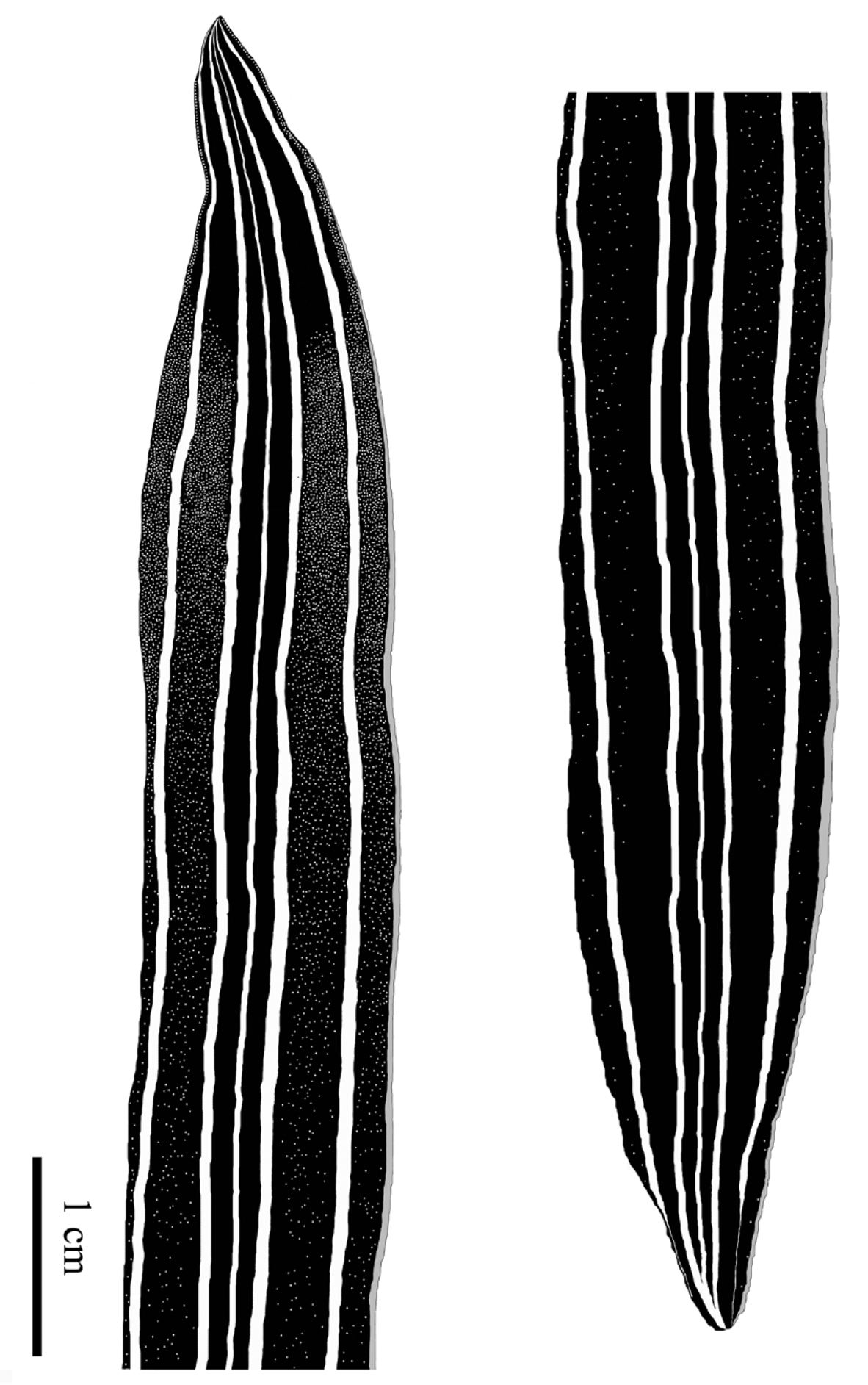
The dorsal side is black, with five white stripes, one thin median, two thicker paramedian and two lateral ones with the same width as the last ones. The margins of the body are pink. The dark colour defines five bands: one black median band (crossed by the median stripe), two black lateral bands (separated by the paramedian stripes) and two marginal bands (separated from the lateral bands by the lateral stripes). The marginal bands have a dark blue pigment toward the outer surface of the body, contacting with the pinkish margin ( Fig. 1 View FIGURE 1 A). Near the posterior tip the lateral stripes become very thin. The ventral surface is light orange-brownish, with a clearer midline. A very thin pinkish pigment is present in the margins of the ventral surface ( Fig. 1 View FIGURE 1 B).
The eyes are initially marginal and uniserial encircling the anterior tip, on the blue surface. Between 6-8 mm from the anterior end, they are biserial and between 8-14 mm tri-tetra serial. Posteriorly, the eyes become pluriserial. Beyond 18 mm they are dorsal on the black lateral bands of the body. At 50 mm, on the pre-pharyngeal region, the eyes are less numerous. At the level of the pharynx they are more isolated, and posterior to the copulatory apparatus the eyes become scattered along the marginal bands to reach the posterior tip ( Fig. 2 View FIGURE 2 ).
Epidermis, secretions and musculature at pre-pharyngeal region. The dorsal epidermis is 25 μm high and ventrally it is 27.5 μm high. The creeping sole reaches the glandular margin and it covers the entire ventral surface of the body. Rhabditogen cell bodies are below the cutaneous musculature. Their secretions open onto the whole epidermal surface.
In the dorsum and in the body margins rhabditogen secretion is abundant, and ventrally it occupies only the apical region of the epidermal cells. Below the rhabditogen cell bodies, in the parenchyma there are glandular secretions both cyanophil and erythrophil, the latter more abundant. Their secretions open principally at the dorsal surface. The glandular margin is composed of two types of secretory cells, cyanophil and erythrophil, and some rhabditogen cells ( Fig. 3 View FIGURE 3 A, D).
The cutaneous musculature is formed by the three layers typical of the Geoplaninae : an external circular layer, a diagonal layer, and an internal longitudinal layer which is arranged in bundles ( Fig. 3 View FIGURE 3 C). In the pre-pharyngeal region, circular and diagonal layers have both dorsally and ventrally similar thickness, 5-7.5 µm and 25-30 µm thick, respectively. The thickness of the longitudinal layer is approximately three times higher than the diagonal one, being 75-87.5 µm and 90-102.5 µm thick in the dorsal and ventral surface, respectively. Cutaneous muscular index (CMI) ranges between 9.1% and 10.5%.
The parenchymatic muscle fibres are arranged in three layers: (1) a dorsal layer with decussated fibres (75- 112.5 μm thick) located under the dorsal submuscular peripheral nerve net; (2) a well developed supra-intestinal transverse layer (200-225 μm thick); and (3) a sub-intestinal transverse layer (125-150 μm thick). Numerous oblique and dorso-ventral fibres run among the intestinal branches, and extend to the body margins.
A lot of nematode larvae were observed in the parenchyma of the ventral surface of the body, both in the prepharyngeal region and in the copulatory apparatus. Also, the larvae are located within the cutaneous musculature ( Fig. 4 View FIGURE 4 ).
Digestive system. The mouth is located in the middle of the pharyngeal pouch. The pharynx, collar type, is 15.9 mm long (~10% of body length) and strongly folded ( Fig. 5 View FIGURE 5 A).
The dorsal insertion is situated in the posterior third. The pharynx presents very abundant glandular erythrophil secretion and cyanophil in a less quantity; this secretion is abundant in the folds and at the level of the insertions. The external epithelium is cubic and ciliated (erythrophil), with three muscular layers; an external longitudinal one (5 µm thick), a circular one (15 µm thick) and a longitudinal one (5 µm thick). The internal epithelium is cubic ciliated (erythrophil) with a thick layer of longitudinal muscular fibres, with some circular fibres intermingled (135-150 µm thick). The oesophagus is extremely long (13.2 mm) and dorsal to the pharynx (oesophagus:pharynx ratio, 83%). It has a ciliated cubic epithelium with a strong longitudinal musculature (100-125 µm thick).
Male reproductive system. The testes are rounded and located beneath the rhabditogen glands and the dorsal parenchymatic musculature, interrupting the continuity of the supra-intestinal parenchymatic muscle fibres ( Fig. 3 View FIGURE 3 C). The testes are placed on 5 rows on both sides of the body; they are mature and charged with spermatozoa ( Fig. 3 View FIGURE 3 A, C). The relationship between the height of the testes and the height of the body varies between 7.4% and 7.8%.
The efferent ducts are internal and slightly dorsal to the ovovitelline ducts. They are inside the sub-intestinal parenchymatic muscular layer ( Fig. 3 View FIGURE 3 A, B). The epithelium of the efferent ducts is cubic and ciliated. Behind the pharynx the efferent ducts run latero-ventrally, they are broadened and charged with spermatozoa. They reach the proximity of the penis bulb and give on to tubular intrabulbar paired projections of the prostatic vesicle. These projections, which are the first portion of the prostatic vesicle, ascend slightly towards the sagittal plane. They present a ciliated cylindrical epithelium with a fine erythrophil secretion. The paired projections open into a sinuous unpaired ascending portion (1.1 mm long). The unpaired prostatic vesicle, with circular (15 µm thick) and longitudinal muscle layers (2.5 µm thick), continues as an expanded and irregular chamber (1.5 mm long) with folded walls ( Figs. 6 View FIGURE 6 , 7 View FIGURE 7 B). The epithelium is cylindrical, glandular and ciliated. It receives abundant erythrophil secretions, which is finer in its distal portion. Outside the penis bulb there are glands whose erythrophil secretion reaches the prostatic vesicle ( Figs. 5 View FIGURE 5 B, 6). The ejaculatory duct, whose epithelium is cylindrical, is a straight canal that goes through the penis papilla eccentrically. The cylindrical papilla is 1 mm long, blunt with a cylindrical epithelium. It occupies almost the whole male atrium (1.25 mm long). The atrium has an inner circular muscle layer and an outer longitudinal one (10 µm each). The musculature of the penis bulb is strong and compact, being formed by fibres arranged in different directions.
Female reproductive system. The ovaries were not observed in the slides of the anterior part of the body, but the rest of the female reproductive system is fully developed ( Figs. 5 View FIGURE 5 B, 7). The ovovitelline ducts are placed below the sub-intestinal parenchymatic muscular layer. The vitelline glands, placed dorsal and ventrally to the intestinal branches, give on to the oviducts along their run ( Fig. 3 View FIGURE 3 A-C). Behind the gonopore the ovovitelline ducts rise towards the sagittal plane. These ascending portions get scarce secretion from shell glands. As they ascend, they receive more secretion and in their final portion they are horizontal, receiving a great quantity of secretion ( Figs. 5 View FIGURE 5 B, 6, 7A). They join, forming a short glandular common ovovitelline duct just before they empty dorsally into a short vagina (250 µm long).
The epithelium of the glandular common duct is cubic and ciliated, while the vagina has a cylindrical non ciliated epithelium. The vagina is crescent-shaped and it opens dorsally in the atrium ( Fig. 7 View FIGURE 7 A). A muscular coat formed by circular (50 µm thick) and longitudinal fibres (75 µm thick) encircle the vagina and the atrium. The female atrium (4.25 mm long) has folded walls ( Figs. 5 View FIGURE 5 B, 6, 7A), with a cubic non ciliated epithelium apically erythrophil.


| MLP |
Museo de La Plata |
No known copyright restrictions apply. See Agosti, D., Egloff, W., 2009. Taxonomic information exchange and copyright: the Plazi approach. BMC Research Notes 2009, 2:53 for further explanation.